Cite as
Ercan M, Mungan S, Güzel I, et al. Serum asymmetric di-methylarginine and nitric oxide levels in Turkish patients with acute ischemic stroke. Adv Clin Exp Med. 2019;28(5):693–698. doi:10.17219/acem/78360
DOI
10.17219/acem/78360
Copyright
© 2019 by Wroclaw Medical University This is an article distributed under the terms of the Creative Commons Attribution Non-Commercial License (http://creativecommons.org/licenses/by-nc-nd/4.0/)
Address for correspondence
Işıl Güzel E-mail: [email protected] Funding sources None declared Conflict of interest None declared Acknowledgements
We are grateful to Roche and Beckman Diagnostics for providing reagent kits free of charge for this study.
Received on October 28, 2015 Reviewed on March 3, 2016 Accepted on October 6, 2017 Published online on August 29, 2018
Abstract
Background. Nitric oxide synthase (NOS) is present in the brain and cerebral arteries and it enables the synthesis of nitric oxide (NO), which plays a critical role in brain perfusion. Asymmetrical dimethylarginine (ADMA) is an endogenous NOS inhibitor.
Objectives. The aim of this study was to evaluate serum ADMA levels, which are an indicator of endothelial dysfunction of the renal functions in patients with acute ischemic stroke, and to determine whether there is a possible correlation between ADMA and NO levels and the l-arginine-to-ADMA ratio.
Material and methods. Fifty-two patients (22 male and 30 female; mean age: 75.2 ±10.1 years) with a diagnosis of acute ischemic stroke in the first 24 h post-stroke and 48 healthy individuals (controls; 13 male and 35 female; mean age: 60.1 ±7.92 years) were included in this study. The risk factors recorded and evalu-ated were age and gender of the patients, serum lipid levels, serum ADMA levels, nitrate-to-nitrite ratios, l-arginine, l-arginine-to-ADMA ratios, sedimentation rate, C-reactive protein (CRP), urea and creatinine levels, and glomerular filtration ratio (eGFR).
Results. The mean serum ADMA level was 0.48 ±0.23 µM for the patients and 0.36 ±0.18 µM for the controls. The mean NO level was 2.78 ±0.59 µM for the patient group and 4.49 ±2.84 µM for the controls. The ADMA levels for the patient group were significantly higher than for the control group (p = 0.011); the NO levels for the patients were significantly lower than for the controls (p < 0.001). The logistic regres-sion method demonstrated that ADMA and NO levels may be independent risk factors for the patient group, and the receiver operating characteristic (ROC) curve analysis showed that both of these variables were discriminative risk factors.
Conclusions. An increased serum level of the NOS inhibitor ADMA was found to be a possible independent risk factor for ischemic stroke.
Key words: nitric oxide, asymmetrical dimethylarginine, asymmetrical dimethylarginine/arginine levels, arginine, acute ischemic stroke
Serum asymmetric dimethylarginine and nitric oxide levels
in Turkish patients with acute ischemic stroke
Mujgan Ercan
1,A,D, Semra Mungan
2,B,E,F, Işıl Güzel
2,D–F, Huseyin Tugrul Celik
3,E,F, Ceylan Bal
4,B,F,
Sedat Abusoglu
5,C,F, Deniz Akbulut
6,D,F, Esra Firat Oguz
7,A,F, Fatma Meric Yilmaz
8,C,F1 Department of Biochemistry, Aydın Public Health Laboratory, Turkey
2 Department of Neurology, Ankara Numune Training and Education Hospital, Turkey 3 Department of Biochemistry, Faculty of Medicine, Turgut Ozal University, Ankara, Turkey 4 Department of Biochemistry, 4th Ankara Occupational Diseases Hospital, Turkey 5 Department of Biochemistry, Faculty of Medicine, Selçuk University, Konya, Turkey 6 Çukurova Dr. Aşkım Tüfekçi Hospital, Adana, Turkey
7 Department of Biochemistry, Ankara Numune Training and Education Hospital, Turkey 8 Department of Biochemistry, Faculty of Medicine, Yıldırım Beyazit University, Ankara, Turkey
A – research concept and design; B – collection and/or assembly of data; C – data analysis and interpretation; D – writing the article; E – critical revision of the article; F – final approval of the article
Introduction
Nitric oxide synthase (NOS), a requirement for the synthesis of nitric oxide (NO), which plays a critical role in the control of brain perfusion, is located in the brain and the walls of the cerebral arteries.1–3 Asymmetrical
dimethylarginine (ADMA) is a competitive endogenous inhibitor of endothelial NOS, which decreases endothelial NO synthesis and leads to the loss of NO bioavailability.3
Age, diabetes mellitus (DM), hypertension (HT), carotid arterial intima-media thickness, hyperlipidemia, hyper-homocysteinemia, obesity, inflammation, and sickle cell disease have all been found to be associated with increased ADMA blood levels.1,4 In addition, ADMA has been
sug-gested to be an independent marker of acute stroke and transient ischemic attack.5–7
Increased ADMA concentration has been found to be associated with a more rapid renal disease progression and mortality via reducing NO bioavailability, which re-sults in inflammation and oxidative stress – typical fea-tures of renal disease progression.8–10 Asymmetrical
di-methylarginine is also considered to be a uremic toxin, so it is also possible that ADMA contributes to progressive renal dysfunction by functionally impairing the integrity of the glomerular filtration barrier, promoting proteinuria, interstitial and glomerular fibrosis, and oxidative stress.11,12
Although ADMA is attributed to impaired renal function, it is not yet clear whether ADMA is a marker or a risk factor for renal disease progression.
In this study, we aimed to investigate the relationship be-tween serum levels of ADMA and NO and the l-arginine-to-ADMA ratio (indicators of endothelial dysfunction) in patients with acute ischemic stroke, and to discuss the possible confounding effect of renal function on ADMA and NO levels.
Material and methods
Ethics statement
This study was approved by the Ethical Committee of Ankara Numune Training and Research Hospital, Tur-key. Informed consent was obtained from each participant before the study.
Study population
A total of 52 patients who were admitted to the Depart-ment of Neurology of Ankara Numune Training and Re-search Hospital, Turkey, between December 2009 and May 2010 with a diagnosis of acute ischemic stroke in the first 24 h post-stroke and 48 healthy individuals were included in the study.
The diagnosis of ischemic stroke was based on the patient’s history, clinical examination, computed tomography (CT)
examination, and magnetic resonance imaging (MRI). Hy-pertension was defined according to the current World Health Organization (WHO) criteria and/or the use of an-tihypertensive drugs. Stroke severity on admission was as-sessed using the National Institutes of Health Stroke Scale/ Score (NIHSS). An NIHSS of 0–1 was considered mild, 2–8 was moderate, and ≥9 was severe.13 Patients with
hemor-rhagic stroke, coronary artery disease, mitral fibrillation, kidney or liver disease, rheumatological or collagenous disease, those with a diagnosis of cancer, fever or infection, those receiving anticoagulation treatment, and those who had undergone vascular surgery in the previous 3 months were excluded from this study.
Methods
Blood samples were drawn from the patients on the morn-ing of the 1st day of admission to the Department
of Neu-rology into 10 mm tubes with red caps, not containing gel (BD Vacutainer; Becton, Dickinson and Co., Franklin Lakes, USA). After at least 30 min of incubation, the specimen was centrifuged at 1,500 × g for 10 min, and then lipid profiles and urea, creatinine and C-reactive protein (CRP) levels were determined using commercial kits (Beckman Coulter DXC 800; Beckman Coulter, Inc., Brea, USA). A fibrinogen test was performed using a tube containing citrate and a Sie-mens CA 7000 device (Siea Sie-mens Healthcare Diagnostics, Marburg, Germany), and the erythrocyte sedimentation rate (ESR) was determined using a tube containing citrate and a Berkhun SDM 100 device (Blood Testing Equipments; Mediko Dardanel, Canakkale, Turkey). The serum samples to be used in determining ADMA, NO and arginine levels were stored at −80°C until analysis.
Serum ADMA and arginine levels were measured on the same day, after all the samples were collected by using an Applied Biosystems MDS SCIEX API 3200 LC-MS/MS system device (Applied Biosystems, Concord, Canada) in the ESI-positive mode and an Agilent Eclipse XDB–C18 column (Agilent Technologies, Palo Alto, USA). According to this method, the intra-day coefficient of variation (CV) and in-ter-day CV were 3.9% and 6.2%, respectively. Nitric oxide level was determined using a Cayman (780001) Nitrate/ Nitrite colorimetric assay kit (Cayman Chemical Company, Ann Arbor, USA). Briefly, the first step is the conversion of nitrate to nitrite-utilizing nitrate reductase. The next step is the addition of Griess reagent, which converts nitrite into a deep purple azo compound. The photometric measure-ment of the absorbance due to this chromophore accurately determines the NO2 concentration. The intra-assay CV was
3.4% and the inter-assay CV was 2.7%.
Statistical analysis
The data from the study was analyzed with SPSS soft-ware, v. 18.0 (SPSS, Inc., Chicago, USA). The conformi-ty of continuous variables to a normal distribution was
tested with the Shapiro-Wilk test. The descriptive statistics of continuous variables were expressed as mean ± standard deviation (SD). The presence of a statistically significant differences between the groups in terms of continuous variables was examined with Student’s t-test for paramet-ric variables and with the Mann-Whitney U test for non-parametric variables. The analysis of variance (ANOVA) was used to analyze the differences among group means and their associated procedures. The presence of a corre-lation among the groups was assessed with Pearson’s and Spearman’s rho tests. The logistic regression method was performed to demonstrate whether ADMA and NO may be risk factors for stroke and the receiver operating char-acteristic (ROC) curve analysis was done to check whether these parameters may be discriminative factors for stroke.
Results
Fifty-two patients with acute ischemic stroke were in-cluded in this study; 22 were males and 30 were females. The mean age of the patients was 75.27 ±10.05 years. Thirty- -seven patients had HT, 14 had DM and 13 had atrial fibrillation.
The ADMA levels were significantly higher in the pa-tient group than in the control group (0.46 ±0.13 µM vs 0.40 ±0.11 µM, respectively) and the NO levels were sig-nificantly lower in the patient group than in the controls (2.78 ±1.59 vs 4.34 ±2.70, respectively) (p < 0.05).
There was no statistically significant difference between the serum l-arginine-to-ADMA ratio in the patient group
and in the control group (p = 0.494). The ADMA, CRP, urea, creatinine, and the glomerular filtration ratio (eGFR) levels, and ESR were statistically significantly higher in the patient group than in the controls (p < 0.05). The levels of NO, high-density lipoprotein (HDL)-cholesterol and triglycerides were statistically significantly lower in the patient group than in the controls (p < 0.05). There was no statistically significant difference between the l-arginine, total cholesterol, low-density lipoprotein (LDL)-cholesterol, or fibrinogen levels, or the l-arginine-to-ADMA ratio in patients compared to the control group (p > 0.05) (Table 1).
There was no statistically significant difference in the ADMA, NO or l-arginine levels, or the l-arginine-to-ADMA ratio among the different stroke subgroups (mild, moderate or severe, as determined by NIHSS) (Table 2).
There was a negative correlation between the NO levels and age (r = −0.251), and a positive correlation between the NO levels and eGFR (r = 0.223) (Fig. 1). In addition, there was a positive correlation between ADMA and ESR (r = 0.202), and between ADMA and creatinine (r = 0.224), and there was a negative correlation between ADMA and eGFR (r = −0.216): the 95% confidence interval (CI) eGFR lower and upper values were 77.81 and 90.0, respectively (Fig. 1).
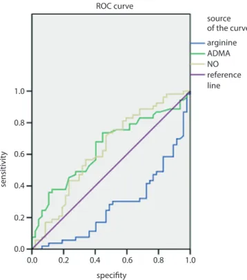
According to the ROC curve analysis (Fig. 2), the NO and ADMA levels were found to be discriminative pa-rameters in the patient group. According to the model, serum NO and ADMA levels may be risk factors for the patient group: the odds ratios (OR) and 95% CI of the risk factors were 1.58 (1.189–2.098) and 1.26 (0.943–1.875), respectively.
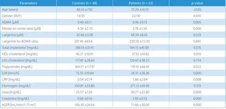
Table 1. Demographic characteristics and biochemistry parameters of patients and controls
Parameters Controls (n = 48) Patients (n = 52) p-value
Age [years] 60.10 ±7.92 75.20 ±10.10 <0.05 Gender (M/F) 13/35 22/30 0.610 ADMA [µM] 0.40 ±0.11 0.46 ±0.13 0.005 Nitrate-to-nitrite ratio [µM] 4.34 ±2.70 2.78 ±1.59 0.000 l-arginine [µM] 67.40 ±5.90 69.30 ±8.65 0.529 l-arginine-to-ADMA ratio 201.45 ±63.4 220.50 ±72.30 0.681 Total cholesterol [mg/dL] 189.10 ±37.41 184.15 ±45.99 0.576 HDL-cholesterol [mg/dL] 43.21 ±10.91 37.32 ±10.82 0.010 LDL-cholesterol [mg/dL] 117.81 ±28.60 120.43 ±38.25 0.714 Triglycerides [mg/dL] 164.57 ±117.97 119.10 ±68.45 0.022 ESR [mm/h] 15.55 ±10.84 28.31 ±20.36 0.000 CRP [mg/dL] 0.54 ±0.14 1.66 ±2.64 0.008 Fibrinogen [mg/dL] 350.81 ±53.80 371.13 ±93.85 0.319 Urea [mg/dL] 25.57 ±7.29 39.77 ±22.80 0.000 Creatinine [mg/dL] 0.66 ±0.16 1.03 ±0.52 0.000 eGFR [mL/min/1.73 m2] 100.30 ±24.04 71.66 ±30.00 0.000
ADMA – asymmetrical dimethylarginine; CRP – C-reactive protein; eGFR – glomerular filtration ratio; ESR – erythrocyte sedimentation rate; HDL – high-density lipoprotein; LDL – low-– high-density lipoprotein; NO – nitric oxide; nitrate-to-nitrite ratio is a measure of NO levels.
Discussion
Risk factors for stroke, such as HT, DM, smoking, hyper-lipidemia, and hyperhomocysteinemia, are associated with endothelial dysfunction. The inhibition of endothelial NO plays an important role in the athero-thrombotic process.14
Asymmetrical dimethylarginine, a post-translationally modified form of arginine,15 is an endogenous inhibitor
of NO synthase and is associated with atherosclerotic diseases.14
In this study, serum ADMA levels were increased and NO levels were decreased in patients who had suffered from an acute ischemic stroke as compared to controls. An increased serum ADMA level was determined to be an independent risk factor for ischemic stroke.
Although studies have examined the relationship be-tween ADMA levels and coronary heart disease (CHD), HT, chronic renal failure, and hypercholesterolemia, stud-ies that have investigated the relationship between ADMA levels and stroke are scarce. Yoo and Lee found that se-rum ADMA levels were significantly higher in 52 patients with stroke than in 36 healthy controls.7 In our study, with
a similar sample size to Yoo and Lee’s study population, ADMA levels were also significantly higher in the patients with acute ischemic stroke than in the controls (p = 0.005).
Prospective clinical studies support the hypothesis that plasma ADMA concentration increases with ischemic
Table 2. ADMA, NO, l-arginine, and l-arginine-to-ADMA ratios in subgroups of stroke according to NIHSS scores
Parameters Control(n = 48) Mild stroke(n = 3) Moderate stroke(n = 40) Severe stroke(n = 9) p-value
ADMA [µM] 0.40 ±0.11 0.45 ±0.06 0.46 ±0.14 0.50 ±0.14 0.329
Nitrate-to-nitrite ratio [µM] 4.34 ±2.70 2.09 ±0.16 2.92 ±1.71 2.39 ±1.18 0.732
l-arginine [µM] 67.40 ±5.90 65.3 ±1.81 69.60 ±6.55 69.20 ±7.19 0.481
l-arginine-to-ADMA ratio 201.45 ±63.40 147.66 ±23.35 241.80 ±74.60 150.50 ±45.00 0.959
ADMA – asymmetrical dimethylarginine; NIHSS – National Institutes of Health Stroke Scale/Score; NO – nitric oxide; nitrate-to-nitrite ratio is a measure of NO levels.
Fig. 1. eGFR–ADMA correlation (left), eGFR–NO correlation (right)
ADMA – asymmetrical dimethylarginine; eGFR – glomerular filtration ratio; NO – nitric oxide.
Fig. 2. ROC curve analyzing the discriminative parameters in patients with acute ischemic stroke
ADMA – asymmetrical dimethylarginine; NO – nitric oxide; ROC – receiver operating characteristic. Diagonal segments are produced by ties.
source of the curve ROC curve 0.0 1.0 0.8 0.6 0.4 0.2 0.0 0.2 0.4 0.6 specifity sensitivit y 0.8 1.0 arginine ADMA NO reference line y = –37.6x + 101.73 R2 = 0.0242 y = –0.1842x + 85.065 R2 = 0.0002 r = 0.223 p = 0.026 r = –0.216 p = 0.031 167.12 117.12 67.12 17.12 167.12 117.12 67.12 17.12 0.1 0.3 0.5 0.7 0.6 5.6 10.6
eGFR–ADMA
eGFR–NO
ADMA [μmol/L] NO [μM] eG FR [m L/ min per 1. 73 m 2] eG FR [m L/ min per 1. 73 m 2]stroke risk factors and in patients with ischemic stroke. Mamatha et al. found significantly higher serum ADMA levels in stroke patients than in controls in a study that included 201 stroke patients and 217 controls (p < 0.001).16
Increased plasma concentrations of ADMA have been reported to be an independent risk factor for ischemic stroke, after correcting for ischemic stroke risk fac-tors (age, alcohol abuse, smoking, HT, DM, low serum HDL-cholesterol and homocysteine).5 In our study, there
was no correlation between ADMA and ischemic stroke risk factors (age, DM, HT, serum cholesterol, and serum triglycerides). In a similar study, Nishiyama et al. found that serum ADMA concentrations were significantly higher in 50 stroke patients and 116 individuals with vas-cular risk factors than in controls with no vasvas-cular risk factors.5 Asymmetrical dimethylarginine concentrations
were found to be associated with vascular risk factors and were suggested as a marker of a future ischemic stroke.16
Elevated ADMA levels have been demonstrated in some clinical conditions and in the presence of conventional car-diovascular risk factors, such as high blood pressure, serum cholesterol levels, serum triglycerides, gestational DM, insulin resistance, and smoking.16–21 Furthermore,
an asso-ciation between ADMA and non-conventional cardiovas-cular risk factors, such as elevated levels of homocysteine, CRP, and the vascular cell adhesion molecule (VCAM) has been demonstrated.22–24
Acutely decreased NO levels result in vasoconstriction, an increased production of free radicals, platelet aggre-gation, and leukocyte adhesion on endothelial surfaces; these processes may in turn aggravate cerebral ischemia.25
This finding suggests a mechanism for the pathogenesis of ischemic stroke.
In our study, we found significantly lower NO levels in the stroke patients compared to the controls (p = 0.000), sug-gesting cerebral ischemia due to acute ischemic stroke. Ce-rebrospinal fluid (CSF) ADMA levels were found to increase in parallel with stroke severity in a study by Brouns et al.25
In our study, serum ADMA levels seemed to be higher in patients with more severe strokes; however, the associa-tion was not statistically significant (p = 0.329).
A recent study has suggested that renal function may have prognostic value for long-term survival in stroke patients and for the occurrence of cardiovascular events after an acute cerebral event.26 In our study, a positive
cor-relation was found between ADMA and creatinine levels (r = 0.224), and a negative correlation was observed between ADMA and eGFR (r = −0.216). Some methylarginines are excreted by the renal route. All symmetrical dimethyl-arginine (SDMA) is excreted by the renal route, where-as ADMA and NG-monomethyl-l-arginine (l-NMMA)
are metabolized extensively. The most important step in ADMA metabolism is its breakdown into citrulline and dimethylamine by dimethylarginine dimethylaminohydro-lase (DDAH). The lack of SDMA measurements is another limitation of this study.
In conclusion, increased serum levels of the NOS inhibi-tor ADMA and decreased levels of NO may be indepen-dent risk factors for ischemic stroke. The small sample size in the stroke subgroups may be another limitation of this study; further studies with more participants should strengthen the findings of the study. Decreased NO lev-els cause vasoconstriction and may be important in the pathogenesis of ischemic stroke. Additional comprehensive studies are needed to validate ADMA and NO as routine risk factors of stroke.
References
1. Kielstein JT, Donnerstag F, Gasper S, et al. ADMA increases arterial stiffness and decreases cerebral blood flow in humans. Stroke. 2006; 37:2024–2029.
2. Landmesser U, Drexler H. The clinical significance of endothelial dys-function. Curr Opin Cardiol. 2005;20:547–551.
3. Leiper J, Vallance P. Biological significance of endogenous methyl-arginines that inhibit nitric oxide synthases. Cardiovasc Res. 1999;43: 542–548.
4. Kielstein JT, Zoccali C. Asymmetric dimethylarginine: A cardiovas-cular risk factor and a uremic toxin coming of age? Am J Kidney Dis. 2005;46:186–202.
5. Nishiyama Y, Ueda M, Otsuka T, et al. Statin treatment decreased serum asymmetric dimethylarginine (ADMA) levels in ischemic stroke patients. J Atheroscler Thromb. 2011;18:131–137.
6. Wanby P, Teerlink T, Brudin L, et al. Asymmetric dimethylarginine (ADMA) as a risk marker for stroke and TIA in a Swedish population.
Atherosclerosis. 2006;185:271–277.
7. Yoo JH, Lee SC. Elevated levels of plasma homocyst(e)ine and asym-metric dimethylarginine in elderly patients with stroke.
Atheroscle-rosis. 2001;158:425–430.
8. Alpoim PN, Sousa LP, Mota AP, Rios DR, Dusse LM. Asymmetric dimethylarginine (ADMA) in cardiovascular and renal disease. Clin
Chim Acta. 2015;440C:36–39.
9. Raptis V, Kapoulas S, Grekas D. Role of asymmetrical dimethylarginine in the progression of renal disease. Nephrology (Carlton). 2013;18:11–21. 10. Baylis C. Nitric oxide synthase derangements and hypertension
in kidney disease. Curr Opin Nephrol Hypertens. 2012;21:1–6. 11. Eiselt J, Rajdl D, Racek J, Vostry M, Rulcova K, Wirth J. Asymmetric
dimethylarginine and progression of chronic kidney disease: A one-year follow-up study. Kidney Blood Press Res. 2014;39:50–57. 12. Brunet P, Gondouin B, Duval-Sabatier A, et al. Does uremia cause
vascular dysfunction? Kidney Blood Press Res. 2011;34:284–290. 13. The National Institute of Neurological Disorders and Stroke rt-PA
Stroke Study Group. Tissue plasminogen activator for acute isch-emic stroke. N Engl J Med. 1995;333:1581–1587.
14. Bonetti PO, Lerman LO, Lerman A. Endothelial dysfunction: A marker of atherosclerotic risk. Arterioscler Thromb Vasc Biol. 2003;23:168–175. 15. Boger RH, Vallance P, Cooke JP. Asymmetric dimethylarginine
(ADMA): A key regulator of nitric oxide synthase. Atheroscler Suppl. 2003;4:1–3.
16. Mamatha SN, Nagaraja D, Philip M, Christopher R. Asymmetric dimethylarginine as a risk marker for early-onset ischemic stroke in Indian population. Clin Chim Acta. 2011;412:139–142.
17. Achan V, Broadhead M, Malaki M, et al. Asymmetric dimethylargi-nine causes hypertension and cardiac dysfunction in humans and is actively metabolized by dimethylarginine dimethylaminohydro-lase. Arterioscler Thromb Vasc Biol. 2003;23:1455–1459.
18. Kielstein JT, Bode-Boger SM, Frolich JC, Ritz E, Haller H, Fliser D. Asymmetric dimethylarginine, blood pressure, and renal perfusion in elderly subjects. Circulation. 2003;107:1891–1895.
19. Lundman P, Eriksson MJ, Stuhlinger M, Cooke JP, Hamsten A, Tornvall P. Mild-to-moderate hypertriglyceridemia in young men is associated with endothelial dysfunction and increased plasma concentrations of asymmetric dimethylarginine. J Am Coll Cardiol. 2001;38:111–116. 20. Mittermayer F, Mayer BX, Meyer A, et al. Circulating concentrations
of asymmetrical dimethyl-l-arginine are increased in women with
21. Stuhlinger MC, Abbasi F, Chu JW, et al. Relationship between insulin resistance and an endogenous nitric oxide synthase inhibitor. JAMA. 2002;287:1420–1426.
22. Boger RH, Bode-Boger SM, Sydow K, Heistad DD, Lentz SR. Plasma con-centration of asymmetric dimethylarginine, an endogenous inhibitor of nitric oxide synthase, is elevated in monkeys with hyperhomocyst(e) inemia or hypercholesterolemia. Arterioscler Thromb Vasc Biol. 2000;20: 1557–1564.
23. Zoccali C, Benedetto FA, Maas R, et al. Asymmetric dimethylarginine, C-reactive protein, and carotid intima-media thickness in end-stage renal disease. J Am Soc Nephrol. 2002;13:490–496.
24. Nanayakkara PW, Teerlink T, Stehouwer CD, et al. Plasma asymmet-ric dimethylarginine (ADMA) concentration is independently asso-ciated with carotid intima-media thickness and plasma soluble vas-cular cell adhesion molecule-1 (sVCAM-1) concentration in patients with mild-to-moderate renal failure. Kidney Int. 2005;68:2230–2236. 25. Brouns R, Marescau B, Possemiers I, Sheorajpanday R, De Deyn PP.
Dimethylarginine levels in cerebrospinal fluid of hyperacute ischemic stroke patients are associated with stroke severity. Neurochem Res. 2009;34:1642–1649.
26. Luneburg N, von Holten RA, Topper RF, Schwedhelm E, Maas R, Boger RH. Symmetric dimethylarginine is a marker of detrimental outcome in the acute phase after ischaemic stroke: Role of renal function. Clin Sci (Lond). 2012;122:105–111.