Antioxidants and Regulation of Antioxidant Enzymes by Cellular
Redox Status
Gökhan SADİ1* Özlem SADİ2
1Department of Biology, Karamanoğlu Mehmetbey University, Karaman, TURKEY
2Department of Educational Science, Karamanoğlu Mehmetbey University, Karaman, TURKEY
*Corresponding Author
e-mail: [email protected] Abstract
It has been well known that free radicals have numerous detrimental effects in both in vitro and in vivo in biological systems because of their unavoidable and tremendous reactivity against biological macromolecules especially proteins, carbohydrates and nucleic acids. They are produced in the biological systems continuously by various mechanisms and eliminated by cellular antioxidant systems. If not eliminated sufficiently, or there is a disturbance in their neutralization, inevitable cellular damage may occur. Activities or the presence of antioxidant enzymes in the cells are under strong regulation with transcriptional, translational and post translational mechanisms as a consequence of changes in cellular redox potential. Redox sensitive metabolic enzymes or proteins; such as protein phosphatases, NF-κB and AP-1, are capable of sensing the oxidant signals by reversible oxidation of their regulatory units which cause them to transduce the signal and adjust the cellular antioxidant metabolism. Redox sensitive proteins execute their functions via kinases, phosphatases, and transcription factors influencing the steady state levels of antioxidant en-zymes. Therefore, cells may sense, transduce, and translate the oxidant signals into appropriate cellular responses depending on the cellular redox state. This rewiev focused on the basic definitions for the free radicals and the tissue defense mechanism and the possible regulation mechanisms of antioxidant enzymes in the situations undergoing reversible changes in cellular redox status.
Key words: Free radicals, Oxidative stress, Antioxidant enzymes, Redox regulation
INTRODUCTION
Free radicals and reactive oxygen species (ROS) play an important role in living systems through their beneficial and detrimental effects. Atoms or molecules having unpaired electrons are called as free radicals and they tend to modify the chemical reactivity of molecules making them more reactive then their unmodified form. They are produced in the cells and tissues by various processes and reactions such as; high energy ionizing radiation, thermal degradation of organic materials and electron transport chain [1]. In healthy cells, production of free radicals is approximately balanced with antioxidant defense systems. If this balance is disturbed or repair or replacement systems to the free radical induced damage fails, the situation having too many radicals in relation to the available antioxidants is raised and called as oxidative stress. In 1991, Sies [2] defined the oxidative stress as a “disturbance in the prooxidant-antioxidant balance in favor of the former, leading to potential damage to the cellular macromolecules”. Increased damage may not only due to increased stress but also due to failure of repair or replacement systems.
As a result; consequences of oxidative stress arises such as cell injury, disruption in cellular homeostasis and accumulation of damaged molecules. Organisms protect themselves by a copious number of antioxidant macromolecules and enzymes.
Low Molecular Weight Radical
Scavengers (Non-enzymatic Antioxidants)
Antioxidant macromolecules are the compounds that are working in the neutralization of the oxidizing effects of the free radicals. Even if they accept the free electrons of the radicals, they do not behave like a radical. A number of compounds can act as non-enzymatic antioxidants such as; vitamin C (ascorbic acid), vitamin E (α-tochopherol), α-lipoic acid (thioctic acid), ubiquinol, polyphenols, flavonoids, resveratrol and reduced glutathione. These molecules have the capability to take free electrons from the radicals without being as reactive as free oxygen radicals.
Vitamin E (α-tocopherol) is a hydrophobic antioxidant mainly present in lipid bilayer and ascorbic acid (vitamin C) that is highly water soluble, present mainly in the cytoplasm.
Together with Vitamin E, Vitamin C cooperates in cellular defense against ROS in both lipid and aqueous phase at the expense of lipoic acid (LA) and dihydrolipoate (DHLA) which serves as a bridge between them. LA is an essential component of the pyruvate dehydrogenase and α-ketoglutarate dehydrogenase multi-enzyme complexes and maintains the concentrations of the other reduced antioxidants; glutathione, thioredoxin, ascorbic acid, vitamin E and ubiquinol [3,4]. Therefore, LA and DHLA are present at the central positions in the antioxidant network [5]. Figure 1 points out to the interaction in between various antioxidants and their relation with lipoic acid network. According to this figure, as vitamin E scavenges a peroxyl radical, produced vitamin E radical may be re-reduced by several other antioxidants such as glutathione, ubiquinol, ascorbate, and DHLA can reduce all these antioxidants at the expense of NADPH. Afterwards, it is regenerated by enzyme lipoamide reductase.
Antioxidant Enzymes
Even though antioxidant molecules are unavoidably essential in protection from oxidation of radicals, there comes through a time when they become not adequate to cope with continuously produced free radicals in the cells. It is known that superoxide radicals and peroxides are continuously produced within the cell by various subcellular reactions and luckily cells evolve specific antioxidant enzymes designed by nature to destroy these compounds.
Using enzymes as an antioxidant provides a cell with various benefits. Firstly, steady-state concentration of radicals and peroxides can be adapted to the cellular requirements. Because, there is a strong regulation mechanism over the antioxidant enzymes in such a way that, they can be induced, inhibited or activated by several mechanism such as transcriptional, translational or post-translational control mechanisms. Therefore, they have imperative functions in the regulation of specific functions and metabolic pathways.
The tissue level of antioxidants critically influences the susceptibility of various tissues to oxidative stress. Figure 2 summarizes the intracellular sources of the oxygen radicals and the downstream antioxidant defense systems developed for the protection against oxidative damage to the tissues. Superoxide
dismutases (SOD) convert superoxide radicals into hydrogen peroxide and Catalases (CAT) remove hydrogen peroxide by converting it into water. Glutathione peroxidases (GPx) get rid of hydrogen peroxide by oxidizing the glutathione (GSH) into its oxidized (GSSH) form. Also, xenobiotic metabolizing phase 2 detoxification enzymes, glutathione S-transferases (GSTs), are functioning in the detoxification reactions of oxidatively modified molecules and peroxides in the cells.
Superoxide Dismutases (E.C:1.15.1.1)
Superoxide dismutases are metalloenzymes, which catalyze the dismutation of superoxide anion (O2-) into H2O2 to protect organisms
against toxic radicals produced during oxidative processes and they have central role in protecting cells and tissues against oxidant stress [6,7]. Four different isozymes of SODs have been characterized in eukaryotes; a copper and zinc containing form (Cu-Zn SOD) localized in the cytosol, a manganese containing form (Mn SOD) in the mitochondria, iron containing form (Fe SOD) in some prokaryotes and plants’ outer mitochondrial membrane, and a copper and zinc containing form in the extracellular matrix (EC SOD) [8]. The active sites of Mn and Fe superoxide dismutases contain the same type of amino acid side chains and exhibit a high degree of sequence and structure similarity, strongly suggesting that these enzymes originate from a common ancestry. The Cu-Zn SODs are inhibited by cyanide (CN-), whereas Fe and Mn containing
SODs are not. Therefore, inhibition by cyanide can be used to distinguish Cu-Zn SOD activities in tissue homogenates. Moreover, prolonged exposure to H2O2 inactivates the Cu-Zn SOD and
Fe SOD but not Mn SOD [1]. Thus, incubating the homogenates with H2O2 inactivates Fe SOD but not Mn SOD allowing the discrimination of these two closely related isoforms.
Catalase (E.C: 1.11.1.6)
Catalase is a tetrameric hemoprotein (porphyrin-containing) with Fe (III) at its active site. A typical catalase activity is the decomposition of the H2O2 into water and oxygen;
in addition to that, they have also peroxidative functions, so as to convert peroxides (ROOH) into alcohol (ROH) and water. Catalase is mainly localized in the peroxisomes [9], but it is also found in the cytosol of human neutrophiles [10]
and in rat-heart mitochondria [11]. Some non-peroxisomal catalase is also found to be present in the livers of some animals such as guinea pigs [12]. Catalase is inhibited by azide, cyanide, peroxynitrite and hypochloric acid [1]. In mammalian cells, NADPH is bound to catalase and it protects the enzyme from inactivation by H2O2 [13].
Enzymes Working in Glutathione Redox Cycle
Three antioxidant enzymes are essential and functioning in the glutathione redox cycle (figure 3). These are Glutathione Peroxidase (GPx), Glutathione S-Transferase (GST) and Glutathione Reductase (GR).
Glutathione Peroxidase (E.C: 1.11.1.9)
Glutathione peroxidases (GPx), water soluble homotetrameric proteins which are present at various locations in the body with different isoforms, catalyze the reduction of peroxides by using reduced glutatihone (GSH) as a reducing power [14,15]. By this process, hydrogen peroxide is reduced to generate water and also organic peroxides are converted into less toxic form which is alcohols. Their active sites contain selenium ions in the form of selenocysteine and they are classified into five isoform according to their expression in different tissue types and location inside of the cells. Cytosolic and mitochondrial forms of this enzyme (GPx1) reduce hydrogen peroxide and fatty acid hydroperoxides using GSH. GPx4, another form, directly reduce the phospholipid hydroperoxides, fatty acid hydroperoxides, and cholesterol hydroperoxides which are produced in oxidized lipoproteins and peroxidized membranes. This form of enzyme is located in the membrane and also cytoplasm [16]. Both GPx1 and GPx4 are present and function as an antioxidant enzyme in most of the tissues. Liver, kidney and erythrocytes predominantly contain GPx1 and renal epithelial cells and testes extremely express GPx4. Two other isoforms, GPx2 and GPx3 are found to be expressed by gastrointestinal tract and kidney, respectively. The last isoform, GPx5 is expressed in epididymis and it is found to be selenium-independent [17].
Glutathione-S-Transferases (E.C:2.5.1.18)
Glutathione S-transferases (GSTs) are (homo or hetero) dimeric, ubiquitous multifunctional
enzymes composed of two polypeptide subunits playing a key role in cellular detoxification [18]. The tripeptide glutathione (GSH) involve in the metabolism of xenobiotics (foreign compounds) many of which metabolized by conjugation with GSH by glutathione S-transferases. In the cells, in addition to selenium dependent neutralization of hydrogen peroxide by by GPx, some isoforms of glutathione S-transferases (GSTs) also reveal selenium independent peroxidase activity on organic hydroperoxidases. There are multiple numbers of different isoforms of GSTs present in cytoplasm and membrane bound organelles in all eukaryotes with different substrate specificity and catalytic functions. Multiple gene families are encoding the various forms of cytosolic GSTs forming homo or heterodimeric enzymes and a range of combinations of different monomers leads to production of different isoforms of GST enzymes. Mamalian GSTs are categorized in at least seven divisions named as alpha, zeta, mu, pi, omega, sigma and theta isoform. The alpha, mu and pi class of enzymes are most abundant in mammals and levels are often increased by the exposure to xenobiotics via antioxidant response elements, which will be discussed in detail later in this manuscript, and they have active roles in drug metabolism. Sigma form is functioning in the prostoglandin synthesis [19]. Theta, pi and tau have been shown to have glutathione peroxides activity to reduce organic hydroperoxides of fatty acids to the corresponding monohydroxy alcahols [20,21]. These GST isoforms are important protectors against lipid peroxidation showing activity toward membrane associated lipid peroxides and metabolizing toxic end products of the lipid peroxidation.
Glutathione Reductase (E.C:1.6.4.2)
Glutathione reductase is an enzyme containg a flavin group (flavoprotein) and catalyzes the reduction of oxidized glutathione (GSSG) to reduced glutathione (GSH) at the expense of NADPH as a reducing power.
GSSG+NADPH+H+→2GSH+NADP+
This enzyme is essential for the GSH redox cycle which maintains adequate levels of reduced cellular GSH. A high GSH/GSSG ratio is important for protection against oxidative stress.
Thiol Specific Antioxidants (Thioredoxins and Peroxiredoxins)
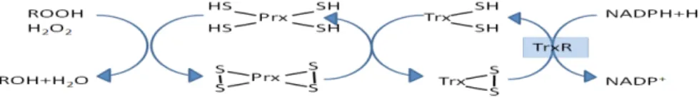
Thioredoxins (Trx) (EC 1.8.4.10) are proteins that act as an antioxidant by facilitating the reduction of other proteins by cysteine thiol-disulfide exchange. Reduced Trx contain two –SH groups that form a disulphide in oxidized thioredoxin. Together with Thioredoxin reductase (TrxR) reducing oxidized thiols, thioredoxin forms an omnipresent oxidation-reduction system having antioxidant and redox regulatory functions. Thioredoxins undergo redox reactions with various proteins with a mechanism described in figure 4.
Peroxiredoxins (Prx) (EC 1.11.1.15) are thiol-dependent antioxidants which are family of peroxidases that reduce H2O2 and organic peroxides at the expense of NADPH together with the thioredoxin systems (Figure 5). They do not have any tightly bound metal ions and have no amino acid sequence similarity with other enzymatic antioxidants such as glutathione peroxidases, catalases, or superoxide dismutases [22]. Peroxiredoxins function slower than the other peroxides in catalyzing H2O2 removal and
inactivated by H2O2 easily [1].
At low H2O2 level, peroxiredoxins are responsible for removal, but as the cells sense extra H2O2, the peroxiredoxins are
partially inhibited to allow gene expression by redox regulation with phosphorylation and dephosphorylation. Peroxiredoxins might be phosphorylated by cyclin dependent kinases that decrease their activity, unlike the CAT and GPx which are phosphorylated by the c-abl and arg kinases which enhance their catalytic activities [23,24].
Redox regulation of gene transcription
Redox regulation can be defined as the modulation of protein and/or enzyme activity by oxidation and reduction potential differences which control the cellular activities in various ways. Intracellular redox balance is tightly controlled and its disturbance leads to modifications in the pattern of gene expressions of several enzymes [25]. Recently, it has been investigated that gene expression of several enzymes and proteins are regulated by antioxidants, oxidants and the factors having an effect on the state of cellular oxidation-reduction (redox) potential [26-28]. It was also shown that redox-sensitive proteins are oxidized by free radicals directly or indirectly [1,29]. Cellular metabolism can be modulated directly by redox
sensitive metabolic enzymes while redox-sensitive signaling proteins exert their functions by means of downstream signaling elements, such as kinases, phosphatases, and transcription factors. Therefore, cells may sense, transduce, and translate the oxidant signals into appropriate cellular responses.
In the cells, there are some proteins having the capability of undergoing reversible oxidative and reductive reactions so called redox-sensitive, and these proteins activate or slow down the downstream signaling pathways depending on cellular redox status. Thioredoxins and peroxiredoxins are such proteins having highly conserved cysteine (sulfhydryl) groups in their active and regulatory points and these sites are prone to reversible oxidation and reduction reactions. Therefore they are important components of redox signaling pathways. Several genes, which are involved in tissue antioxidant defense, have DNA binding sequences on which several transcription factors such as NF-κB and AP-1 can bind and modulate the transcription of their products serving as a key point in the control of gene expression of several proteins [30]. Therefore, critical steps in the signal transduction cascade are sensitive to changes in oxidant/antioxidant balance and both endogenous and exogenous antioxidants such as glutathione, thioredoxin, lipoate and dihydrolipoate may be the efficient regulators of redox-sensitive signal transduction and gene expression.
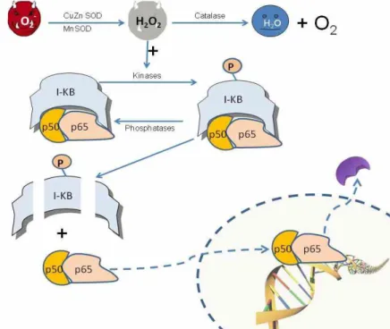
A dimeric transcription factor NF-κB is composed of p50 and p65 subunits and mainly resides in the cytoplasm where it is associated with a transcriptional repressor IκB. It is involved in the regulation of transcription of several genes thought to include antioxidant enzymes, responsible for immune and inflammatory responses. It has been demonstrated that a wide range of stimuli such as cytokines, radiation and oxidative stress (e.g. exposure to H2O2)
may activate this transcription factor causing it to enter nucleus and initiate transcriptional responses [31]. These activating factors leads IκB phosphorylation and its dissociation from NF-κB causes it to be localized in nucleus [32]. Figure 6 summarizes the oxidant stress induced NF-κB activation mechanism.
Recent studies have been demonstrated that NF-κB activation may be suppressed by antioxidant administration. Application of
antioxidant α-tocopherol reduces the NF-κB activity and translocation to the nucleus [31]. Moreover, H2O2, UV, and ionizing radiation
stimulate the IκB degradation and activate NF-κB translocation [31-33]. It was also found that overexpression of catalase suppress the NF-κB activation while overexpression of superoxide dismutase increases the NF-κB activation in TNF cells [34]. These observations lead to understand the role of reactive oxygen intermediates, especially H2O2, involved in the
activation of NF-κB and thereby antioxidant enzymes’ gene expressions. Although, the effects of antioxidants on activation or reduction of NF-κB is not known precisely, it has been proposed that a redox sensitive antioxidant, thioredoxin peroxidase may regulate the phosphorylation of inhibitory protein, IκB, relieving the suppression over the NF-κB [35].
Another redox sensitive mediator of gene expression is a protein which links extracellular signals to the intracellular signaling pathways, and activates the transcription of certain genes responsible for growth, differentiation and stress [36,37]. This transcription factor is named as activator protein-1 (AP-1) and it is composed of homodimeric (Jun/Jun) or heterodimeric (Jun/Fos) complexes. In the cells, two proteins; Fos and Jun are expressed at basal levels, on the other hand under the presence of a wide range of signals; such as changes in cellular redox status and UV radiation, they are induced quickly and momentarily by forming AP-1 complex to initiate the transcription machinery of several oxidative stress response genes [26,32]. Post-transcriptional and transcriptional events regulate the activation mechanism of AP-1. Former perform its effects on preexisting AP-1 protein and the letter provides an enhancement in the amount of AP-1 binding proteins increasing its transcription. What’s more; Schenk and coworkers [38] found that reversible modification of DNA binding domain of Fos, which contains cysteine residues, may have power over the DNA binding ability of this transcription factor.
There is a basic distinction in the mechanism of oxidative signal transduction (redox signaling) caused by AP-1 and NF-κB complexes in that NF-κB passes a signal from cytoplasm to nucleus by bridging these two compartments [39,40], whereas, AP-1 (i.e. Fos and Jun family proteins) are stacked to the nucleus to where
several other protein kinases, such as MAPK, are thought to pass messages from cytoplasm to nucleus [41,42].
Ref-1 is a hydrolytic endonuclease catalyzing the repair of oxidative lesions (primarily abasic sites) in DNA and stimulating the DNA-binding ability of AP-1 proteins (i.e. Fos and Jun) by a mechanism depending on redox status of the cell. Studies demonstrated that, redox-sensitive cysteine residues of the transcription factor AP-1 are reduced by Ref-1 protein and thus it assists AP-1’s DNA-binding and transcriptional activities [43]. Both DNA repairing activities and stimulating the DNA-binding ability of certain transcription factors, demonstrate the importance of Ref-1 protein in cellular response to oxidative stress [44].
Not only transcription factors, AP-1 and NF-κB, but also protein tyrosine phosphatases (PTPs) have cysteine residues at active domains, which are susceptible to oxidation by reactive oxygen species and altered reversibly in a redox dependent manner [45-48]. Therefore, in a situation of changed redox status, expression and activities of antioxidant enzymes are thought to be regulated with these reversible and mandatory modifications at post-translational level.
Kinases and phosphatases in redox signal transduction
Serine, threonine or tyrosine residues on proteins can be phosphorylated or dephosphorylated by protein kinases (PK) or protein phosphatases by adding a phosphate group to abovementioned residues covalently and reversibly. Phosphoregulation is defined as regulation of proteins by phosphorylation and it determines the degree of activity of a particular protein. The most common and the operative phosphoregulation are carried out by tyrosine phosphorylation which is controlled by the differing activities of protein tyrosine kinases (PTKs) and protein tyrosine phosphatases (PTPs). Recent studies indicated that, there is a strong relationship in between redox signaling and steady state level of protein phosphorylation because; reactive oxygen species (especially H2O2) may alter the cellular phosphorylation
status influencing activities and expressions of antioxidant enzymes [47-49]. H2O2 has been found to modulate the intracellular protein phosphorylation level by changing the degree of phosphorylation status by modifying the
cysteine residues of several PTPs reversibly. Furthermore, various cellular and intracallular reductants and antioxidants restore the oxidized cysteine residues into their reduced form and refurbish the biological activities of oxidized redox sensitive proteins.
Protein tyrosine phosphatases (PTPs) are one of the important redox regulated proteins in the cells due to thiols groups in active sites and this makes them vulnerable to reversible inactivation by oxidation of the conserved catalytic site cysteine groups [50] which is an important regulatory mechanism in cellular signal transduction cascades [51]. Figure 7 demonstrates the possible effects of oxidized cysteine on PTPs regulating the overall phosphorylation pattern throughout the cell.
Antioxidant Response Elements
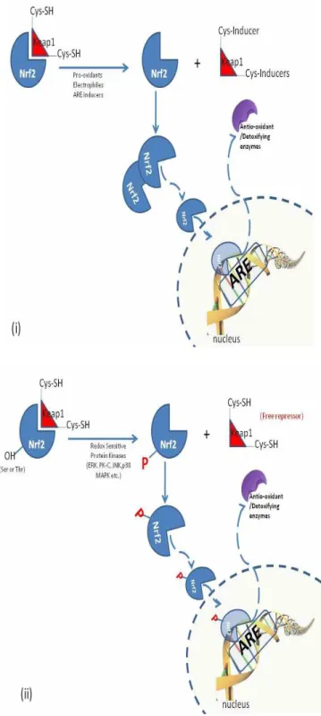
In cells, there are various genes and their products, which are continuously expressed and coordinately induced as the amounts of the reactive oxygen species, antioxidants and xenobiotics fluctuate within a time period. This group of genes on DNA structure is named as antioxidant response elements (AREs) defined as a cis-acting enhancer element which is upstream of many phase II detoxification and antioxidant enzymes [52,53]. Antioxidant response elements (ARE) are DNA sequences to which transcription factors bind when xenobiotics are present. Binding of transcription factors allows increased transcription of genes encoding xenobiotic metabolizing enzymes responsible for the metabolic detoxification of drugs and carcinogens as well as protecting the cells against redox cycling and oxidative stress [54]. Phase II detoxification enzymes such as glutathione S-transferase (GST) are proved to contain ARE in their promoter regions [55]. Moreover, superoxide dismutase (SOD), glutathione peroxidase (GPx), catalase (CAT) genes are considered to be regulated with such kind of antioxidant response elements [28] which are subjected to fine transcriptional control by a transcription factor called Nuclear factor-erythroid-2-related factor 2 (Nrf2). Nrf2 is essential for the coordinated induction of those genes and related proteins. It is the central protein being in the charge of the ARE that activate the constitutive gene transcription and oxidative stress response genes. Under normal circumstances, Nrf2 is kept hold on in the cytosol
associating with a cytoskeletal protein Keap1 which is rich in cysteine residues (figure 8). Upon the oxidation and covalent modifications of critical cysteine residues of Keap1 protein with oxidative elements, Nrf2 is released to be free to accumulate in nucleus and hence induce genes containing ARE elements [28].
Another activation mechanism is the release of Nrf2 from its repressor by the phosphorylation of Nrf2 with kinases. Cytosolic Nrf2 is phosphorylated with MAPK in response to PK-C activation by cellular stress and translocates into the nucleus. In the nucleus, Nrf2 activate genes through AREs by interacting with other transcription factors [28].
In the cells, oxidation and reduction potential difference (redox balance) is firmly controlled and its disturbances may lead to modifications in the pattern of gene expressions and catalytic activities as well as protein amount of antioxidant enzymes [56]. Furthermore, application of some antioxidants such as lipoic acid or ascorbic acid has been found to be beneficial in restoring the changes in the expression and activities of these enzymes. Recent studies demonstrated that tyrosine phosphorylation at specific tyrosine residues of antioxidant enzymes such as catalase (CAT) and glutathione peroxidase (GPx) may regulate its activity and its proteolytic degradation [19,24]. When ROS levels are low in cells, enzymatic CAT activity is stimulated by phosphorylation and as it is very high, activity is diminished by targeting the phosphorylated CAT for degradation [57]. Moreover, c-Abl and Arg which are the non-receptor tyrosine kinases are found to be activated in the presence of oxidative stress [24] and they modulate the phosphorylation levels of peroxide detoxifying enzymes; CAT and GPx by forming an active association having kinase activity. This activated complex phosphorylates the GPx and CAT at specific tyrosine residues, augmenting the activity and providing a protection to cells against oxidative stress. Additionally, changes in activities of antioxidant enzymes are linked to redox sensitive protein tyrosine phosphatases regulating the enzyme activities by phosphorylation dephosphoylation cascades. In addition to these post-translational mechanisms controlling the activities of these two antioxidant enzyme, the gene expressions of these enzymes were also found to be regulated in a situation where oxidative stress present [58]
by a transcriptional mechanism as in case of AP-1, Nrf-2 and NF-κB discussed earlier in this manuscript.
The actual regulation mechanisms of the antioxidant enzymes could not be solved so far and we have tried to explain this in part or whole by reversing the changes in redox sensitive transcriptional factors such as NF-κB, AP-1, Nrf2 and phosphatases which sense the oxidative stress and modify the expressions and activities of antioxidant enzymes. Since, DNA binding sites and active regulatory cysteine groups of these transcription factors are located in the promoter region of antioxidant enzymes, and any changes in intracellular redox state modulate the mRNA and protein expressions of antioxidant enzymes providing a protection to cell or causing a cell to undergo apoptotic pathways by reversible modification of these redox sensitive proteins. Knirsch and Clerch [59] found that a cytoplasmic protein can bind to the 3’untranslated region of Mn SOD mRNA and this binding was regulated by the phosphorylation of Mn SOD binding protein affecting the Mn SOD protein expression. Therefore, expression and so activities of Mn SOD are also under the control by phosphorylation/dephosphorylation cascade systems.
In order to solve the actual regulation mechanism over the antioxidant enzymes, the researchers must focus on to determine how transcriptional factors are modulated whose effects were seen at antioxidant enzymes’ gene transcription. Any change observed in the levels of expression of antioxidant enzymes may provide new insights for analyzing cellular mechanism for protection of cells against oxidative damage solving the molecular nature of the regulation of the antioxidant enzymes in a state of redox misbalance. From all these present knowledge of related literature, we conclude that, little have been known about the molecular regulation mechanisms and it is clear that oxidative stress coordinately regulates the activities of the antioxidant enzymes at molecular level in a very intricate mechanism that should be elucidated.
REFERENCES
[1] Halliwell B, Gutteridge J (2007) Free Radicals in Biology and Medicine (4th edition) Oxford University Press, USA
[2] Sies H (1991) Oxidative Stress: Introduction. In Oxidative stress: Oxidants and Antioxidants (H. Sies, Ed.). Academic Press, London
[3] Oberley LW (1988) Free-Radicals and Diabetes. Free. Radic. Biol. Med. 5(2): 113-124
[4] Packer L, Witt EH, Tritschler HJ (1995) Alpha-Lipoic Acid As A Biological Antioxidant. Free Radic. Biol. Med. 19(2): 227-250
[5] Packer L, Kraemer K, Rimbach G (2001) Molecular aspects of lipoc acid in the preventation of diabetic complications. Nutrition 17: 888-895
[6] Fridovich I, Freeman B (1986) Antioxidant defenses in the lung. Annu Rev Physiol. 48; 693-702
[7] Tsan MF (1997) Superoxide dismutase and pulmonary oxygen toxicity. Proc. Soc. Exp. Biol. Med. 214; 107-113
[8] Marklund SL (1982) Human copper-containing superoxide dismutase of high molecular weight. Proc. Natl. Acad. Sci. 79: 7634-7638
[9] Davies P, Drath DB, Engel EE, Huber GL (1979) The localization of catalase in the pulmonary alveolar macrophage. Lab. Invest. 40: 221-226
[10] Ballinger CA, Mendishandagama SMLC, Kalmar JR, Arnold RR, Kinkade JM (1994) Changes in the localization of catalase during differentiation of neutrophilic granulocytes. Blood 83: 2654-2668 [11] Radi R, Turrens JF, Chang LY Bush KM,
Crapo JD, Freeman BA (1991) Detection of catalase in rat heart mitochondria. J. Biol. Chem. 266: 22028-22034
[12] Bulitta C, Ganea C, Fahimi HD, Volkl A (1996) Cytoplasmic and peroxisomal catalase of the guinea pig liver: evidence for two distinct proteins. Biochim Biophy Acta 1293(1):55-62
[13] Kirkman H, Gaetani G (1984) Catalase: A Tetrameric Enzyme with Four Tightly Bound Molecules of NADPH, Proc. Natl. Acad. Sci. 81: 4243
[14] Flohe L (1989) In Glutathione: Chemical, Biochemical and Medical Aspects (Dolphin, D., Poulson, R., and Avramovic, O., eds) Part A, pp. 643–731, John Wiley & Sons Inc., New York
KD, Roveri A, Schomburg D, Flohe L (1995) Diversity of glutathione peroxidases, Methods Enzymol. 252: 38-53
[16] Chance B, Sies H, Boveris A (1979) Hydroperoxide metabolism in mamalian organs. Physiol Rev 59: 527-605
[17] Imai H, Narashima K, Arai M, Chiba N, Nakagawa Y (1998) Suppression of leukotriene formation in RBL-2H3 cells that overexpressed phospholipid hydroperoxide glutathione peroxidase. J. Biol. Chem. 273: 1990-1997.
[18] Dixon DP, Lapthorn A, Edwards R (2002) Plant glutathione transferases, Genome Biology 2002, 3(3):reviews3004.1–3004.10
[19] Jowsey IR, Thomson AM, Flanagan JU, Murdock PR, Moore GBT, Meyer DJ, Murphy GJ, Smith SA, Hayes JD (2001) Mammalian class sigma glutathione S-transferases: catalytic properties and tissue-specific expression of human and rat GSH-dependent prostaglandin D2 synthases. Biochem J. 359: 507-516 [20] Roxas VP, Smith RK, Allen ER, Allen
RD (1997) Overexpression of glutathione S-transferase/glutathione peroxidase enhances the growth of transgenic tobacco seedlings during stress. Nat. Biotechnol. 15: 988-991
[21] Cummins I, Cole DJ, Edwards R (1999) A role for glutathione transferases functioning as glutathione peroxidases in resistance to multiple herbicides in black-grass. Plant J. 18: 285-292
[22] Aalen RB (1999) Peroxiredoxin antioxidants in seed physiology. Seed Sci. Res. 9, 285–295
[23] Cao C, Leng Y, Kufe D (2003) Catalase activity is regulated by c-Abl and Arg in the oxidative stress response. J. Biol. Chem. 278(32): 29667-29675
[24] Cao C, Leng YM, Huang W, Liu X, Kufe D (2003) Glutathione peroxidase 1 is regulated by the c-Abl and Arg tyrosine kinases J. Biol. Chem. 278(41):39609-614 [25] Schoonbroodt S, Piette J (2000) Oxidative
stress interference with the nuclear factor-kappa B activation pathways. Biochemical Pharmacology 60(8): 1075-1083 [26] Sen CK, Packer L (1996) Antioxidant and
redox regulation of gene transcription.
FEBS Journal 10(7): 709-720
[27] Watai Y, Kobayashi A, Nagase H, Mizukami M, McEvoy J, Singer JD, Itoh K, Yamamoto M (2007) Subcellular localization and cytoplasmic complex status of endogenous Keap1. Genes to Cells 12: 1163-1178
[28] Surh YJ, Kundu JK, Na HK (2008) Nrf2 as a Master Redox Switch in Turning on the Cellular Signaling Involved in the Induction of Cytoprotective Genes by Some Chemopreventive Phytochemicals. Planta Med. 74: 1526-1539
[29] Storz G, Imlay JA (1999) Oxidative stress. Current Opinion In Microbiology 2(2): 188-194
[30] Holbrook NJ, Fornace AJ (1991) Response to adversity: molecular control of gene activation following genotoxic stress. New Biologist 3: 825–833.
[31] Gius D, Botero A, Shah S, Curry HA (1999) Intracellular oxidation:reduction status in the regulation of transcription factors NF-KB and AP-1. Toxicology Letters 106: 93-106
[32] Vranova E, Inze D, Van Breusegem F (2002) Signal transduction during oxidative stress. J of Exp. Bot. 53(372): 1227-1236
[33] Muller J, Rupec RA, Baeuerle PA (1997) Study of the gene regulation by NF-κB and AP-1 in response to reactive oxygen intermediates. Methods 11: 301–312 [34] Schmidt KN, Traencker EB, Meier B,
Baeuerle PA (1995) Induction of oxidative stress by okadaic acid is required for activation of NF-κB. J. Biol. Chem. 270: 27136–27142
[35] Jin DY, Chae HZ, Rhee SG, Jeang KT (1997) Regulatory Role for a Novel Human Thioredoxin Peroxidase in NF-κB Activation J. Biol. Chem. 272: 30952-30961
[36] Angel P, Karin M (1991) The role of Jun, Fos and the AP-1 complex in cell-proliferation and transformation. Biochim Biophys Acta. 1072(2-3):129–157 [37] Herrlich P, Ponta H, Rahmsdorf HJ (1992)
DNA damage-induced gene expression: signal transduction and relation to growth factor signaling. Rev. Physiol. Biochem. Pharmacol. 119:187–223
W, Schulzeosthoff K (1994) Distinct effects of thioredoxin and antioxidants on the activation of transcription factors NF-kB and AP-1 Proc. Nati. Acad. Sci. USA 91: 1672-1676
[39] Baeuerle PA, Baltimore D (1996) NF-kappa B: Ten years after. Cell 87(1): 13-20 [40] Xanthoudakis S, Curran T (1996) Redox regulation of AP-1: a link between transcription factor signaling and DNA repair. Adv. Exp. Med. Biol. 387: 69-75 [41] Karin M, Smeal T (1992) Control
Of Transcription Factors By Signal Transduction Pathways-The Beginning of The End. Trends in Biochemical Sciences 17(10): 418-422
[42] Xanthoudakis S, Curran T (1994) Analysis of C-Fos And C-Jun Redox-Dependent Dna-Binding Activity. Methods in Enzymology 234: 163-174
[43] Xanthoudakis S, Curran T (1992) Identification and characterization of Ref-1, a nuclear protein that facilitates AP-1 DNA-binding activity. EMBO J. 11: 653-664
[44] Xanthoudakis S, Smeyne RJ, Wallace JD, Curran T (1996) The redox/DNA repair protein, Ref-1, is essential for early embryonic development in mice. Proc. Natl. Acad. Sci. U S A. 93(17): 8919-8923. [45] Xanthoudakis S, Miao GG, Wang F et al.,
(1992) Redox activation of fos-jun DNA binding activity is mediated by a DNA repair enzyme. EMBO J. 11: 3323–3335. [46] Walker LJ, Craig RB, Harris AL, Hickson
ID (1994) A role for the human DNA repair enzyme HAP1 in cellular protection against DNA damaging agents and hypoxic stress. Nucleic Acids Res. 22: 4884-4889 [47] Groen A, Lemeer S, Wijk TVD,
Overvoorde J, Heck AJR, Ostman A, Barford D, Slijper M, den Hertog J (2005) Differential Oxidation of Protein-tyrosine Phosphatases J. Biol. Chem., 280: 10298-10304
[48] Rhee SG, Kang SW, Jeong W, Chang TS, Yang KS, Woo HA (2005) Intracellular messenger function of hydrogen peroxide and its regulation by peroxiredoxins. Curr. Op. in Cell Biol. 17(2): 183-189 [49] Chiarugi P, Cirri P (2003) Redox
regulation of protein tyrosine phosphatases
during receptor tyrosine kinase signal transduction. Trends In Biochemical Sciences 28(9): 509-514
[50] Jackson M and Denu JM (2001) Molecular reactions of protein phosphatases--Insights from structure and chemistry, Chemical Reviews, 101, 2313-2Hertog JD, Groen A, Wijk TVD (2004) Redox regulation of protein-tyrosine phosphatases. Arch Biochem. Biophys. 434(1): 11–15 [51] Rushmore TH, Morton MR, Pickett
CB (1991) The antioxidant responsive element. Activation by oxidative stress and identification of the DNA consensus sequence required for functional activity. J. Biol. Chem. 266: 11632-11639
[52] Radjendirane V, Joseph P, Jaiswal AK (1997) Oxidative Stress and Signal Transduction (In: Cadenas, E. and Forman, H.J., Editors) Chapman and Hall, New York
[53] Matés JM (2000) Effects of antioxidant enzymes in the molecular control of reactive oxygen species. Toxicology 153: 83-104
[54] Motohashi H, Yamamoto M (2004) Nrf2-Keap1 defines a physiologically important stress response mechanism. Trends in Molecular Medicine. 10(11): 549-557 [55] Sadi G, Guray T (2009) Gene expressions
of Mn-SOD and GPx-1 in streptozotocin induced diabetes: effect of antioxidants. Mol. Cell. Biochem. 327: 127-134 [56] Cao C, Leng Y, Liu X, Yi YP, Li P,
Kufe D (2003) Catalase is regulated by ubiquitination and proteosomal degradation. Role of the c-Abl and Arg tyrosine kinases. Biochemistry 42(35): 10348-10353
[57] Sadi G, Yilmaz O, Guray T (2008) Effect of vitamin C and lipoic acid on streptozotocin-induced diabetes gene expression: mRNA and protein expressions of Cu-ZnSOD and catalase. Mol. Cell. Biochem. 309(1-2): 109-116
[58] Knirsch L, Clerch LB (2001) Tyrosine phosphorylation regulates manganese superoxide dismutase (MnSOD) RNA-binding protein activity and MnSOD protein expression. Biochem 40(26):7890-95.
Figure 1. Presentation of antioxidant network indicating the relations between vitamin E, ubiquinol,
vitamin C, glutathione, and lipoic acid redox cycles [5].
Figure 2. Metabolic pathways of reactive oxygen radical generation in cells and antioxidant systems
Figure 3. Glutathione redox cycle in which antioxidant enzymes Glutathione
Peroxidase (GPx), Glutathione S-transferase (GSTs) and Glutatihone Reductase (GR) is functioning.
Figure 4. Roles of Trx and TrxR system in the NADPH dependent
reduction of oxidized proteins and several other substrates.
Figure 5. Thioredoxin dependent reduction of organic hydroperoxides and hydrogen peroxide with
Figure 6. Proposed mechanism for oxidative activation of NF-κB. Increase in
superoxide anion and so H2O2 activates kinases or inhibit phosphatases bringing
about augmentation of steady state phosphorylation level of inhibitory protein IκB which is then degraded and leaving the subunits of NF-κB (p50/p65) [35].
Figure 7. Fate of protein tyrosine phosphatases (PTPs) under the presence of
increased H2O2 which inactivates them catalytically by oxidation of thiol groups
at active sites. Inhibition of PTPs leads to sustained phosphorylated proteins in the downstream signaling pathways.
Figure 8. Mechanisms of the induction of antioxidant response element with oxidative stress. Nrf2
can be activated by at least two mechanisms; (i) stabilization of Nrf2 via Keap1 cysteine thiol modification and (ii) phosphorylation of Nrf2 by upstream kinases [28].
![Figure 1. Presentation of antioxidant network indicating the relations between vitamin E, ubiquinol, vitamin C, glutathione, and lipoic acid redox cycles [5].](https://thumb-eu.123doks.com/thumbv2/9libnet/4558340.83148/10.892.209.706.263.537/figure-presentation-antioxidant-indicating-relations-ubiquinol-vitamin-glutathione.webp)