28
Makale Kodu/Article code: 412 Makale Gönderilme tarihi: 07.10.2010 Kabul Tarihi: 31.12.2010
ABSTRACT
Purpose: The aim of this study was to investigate the
radiopacity of provisional crown and bridge materials (PCMs) in comparison with human and bovine dental hard tissues.
Material and Methods: A total of 56 samples, measuring 6
mm in diameter and 1 mm in thickness, with eight samples of each material were prepared from PCMs; Dentalon Plus L (DL), Dentalon Plus M (DM), Dentalon Plus D (DD), EDE Temdent (TD), Imident (ID), Keen (KE) and Voco Structur 2 SC (ST). The optical densities of each material, along with one tooth section (human molar tooth), bovine dentin and enamel samples, and an aluminium step wedge, were measured from radiographical images using a transmission densitometer. The data were analyzed by nonparametric 1-way ANOVA (Kruskal-Wallis) and Student-Newman-Keuls multiple range tests for post hoc comparison (α=0.05).
Results: The mean equivalent aluminium thickness values
(mean ± standard deviation) of the materials were for KE (0.671 ± 0.053), DL (0.674 ± 0.046), TD (0.684 ± 0.091), DD (0.702 ± 0.091), DM (0.703 ± 0.063), ID (0.722 ± 0.032), ST (1.079 ± 0.114), human dentin (1.225 ± 0.0762), bovine dentin (1.282 ± 0.045), bovine enamel (1.797 ± 0.124). Among PCMs, Voco Structur 2 SC had statistically significantly higher radiopacity values (p<0.05). The radiopacity values of the other materials did not reveal statistically significant difference (p>0.05).
Conclusion: The radiopacity values of all PCMs were lower
than the human and bovine dentin and enamel. The radiopacity values of ST were significantly higher than other PCMs.
Key words: provisional restorative materials, provisional
crown, radiopacity, radiodensity, step wedge
ÖZET
Amaç: Çalışmanın amacı geçici kron ve köprü (GKK)
materyallerinin radyopasitelerinin insan ve sığır diş sert dokuları ile karşılaştırarak araştırılmasıdır.
Gereç ve yöntem: GKK materyallerinin [Dentalon Plus L
(DL), Dentalon Plus M (DM), Dentalon Plus D (DD), EDE Temdent (TD), Imident (ID), Keen (KE) and Voco Structur 2 SC (ST)] her birinden 8 adet olmak üzere çapları 6 ve kalınlıkları 1 mm olan toplam 56 örnek hazırlandı. Her bir materyalin optik dansitesi ayrıca bir dişin kesitinin (insan molar dişi), sığır dentin ve minesinin ve bir aluminyum “step wedge’in” optik dansitesi röntgen görüntülerinden bir transmisyonlu dansitometre kullanılarak hesaplandı. Veriler parametrik olmayan tek yönlü varyans analizi (Kruskal-Wallis) ve Student-Newman-Keuls çoklu karşılaştırma testleri ile analiz edildi (α=0.05).
Bulgular: Materyallerin ortalama eşdeğer aluminyum kalınlık
değerleri (ortalama ± standart sapma); KE (0.671 ± 0.053), DL (0.674 ± 0.046), TD (0.684 ± 0.091), DD (0.702 ± 0.091), DM (0.703 ± 0.063), ID (0.722 ± 0.032), ST (1.079 ± 0.114), insan dentini (1.225 ± 0.0762), sığır dentini (1.282 ± 0.045), sığır minesi (1.797 ± 0.124) belirlendi. GKK materyalleri arasında Voco Structur 2 SC istatistiksel olarak anlamlı şekilde yüksek radyopasite değerine sahip bulundu (p<0.05). Diğer materyallerin radyopasite değerleri istatistiksel olarak anlamlı bir farklılık göstermedi (p>0.05).
Sonuç: Test edilen GKK materyallerinin hepsinin radyopasite
değerleri insan ve sığır dentini ve minesinden düşük bulundu. ST’nin radyopasite değerleri diğer GKK materyallerinden anlamlı derece yüksek bulundu.
Anahtar kelimeler: geçici restorasyon materyalleri, geçici
kron, radyopasite, radyodansite, step wedge
COMPARATIVE RADIOMETRIC EVALUATION OF SOME PROVISIONAL CROWN AND BRIDGE MATERIALS
BAZI GEÇİCİ KRON VE KÖPRÜ MATERYALLERİNİN KARŞILAŞTIRMALI RADYOMETRİK DEĞERLENDİRİLMESİ
Yrd. Doç. Dr. Gürel PEKKAN*
29 INTRODUCTION
Within the last decade, we have been moved quickly to esthetic dentistry. Provisional crown and bridge materials (PCMs) are needed in many stages in esthetic dentistry. Successful fixed dental prosthetic work relies heavily upon the precise use of PCMs.1 Two major groups of PCMs are currently available: methacrylate resins (liquid/powder, hand-mixed) and composite resin based materials (paste/paste, mainly auto-mixed).2
As with permanent restorations, the marginal integrity of PCMs and their relationship between teeth and periodontal tissues are extremely important.3 Furthermore, adequate radiopacity of PCMs lead an appropriate radiographic assessment of a restoration when needed. The necessary corrections of the PCMs for the formation of the periodontal tissues and teeth will guide the future of the permanent restoration.1,3
On the other hand, in case of insufficient radiopacity of the intra-oral restorations, it may be very difficult to detect the localization of them if they are aspirated or impacted in the tissue due to the trauma or iatrogenic reasons.4-6 This may cause the patients expose to advanced imaging techniques such as computerized tomography.7-9
According to author’s knowledge, there is no study about the radiopacity of the PCMs in the literature. There are only few studies about the radiopacity of some acylic resin base materials that are in similar chemical composition with PCMs.10-12
The aim of this study was to investigate the radiopacity PCMs in comparison with human and bovine dental hard tissues. The null hypotheses were that: (1) Radiopacity levels of PCMs are not different from that of human and bovine dentin; (2) Radiopacity levels of PCMs are not different from each other.
MATERIALS AND METHODS
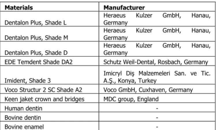
In this study, the radiopacity level of 5 different brand PCMs (Dentalon Plus with 3 colors) were tested. The materials, as well as the manufacturers used in this study, are listed in Table 1. Human molar dentin (HD), bovine dentin (BD), bovine enamel (BE) and an aluminium (Al) step wedge were used as references. For fabricating the specimens teflon moulds were used. Eight specimens, measuring 6 mm in diameter
and 1 mm in thickness, were prepared from PCMs; Dentalon Plus L (DL), Dentalon Plus M (DM), Dentalon Plus D (DD), EDE Temdent (TD), Imident (ID), Keen (KE) and Voco Structur 2 SC (ST) according to manufacturer’s instructions. In total, 56 (n=8) specimens were obtained.
Table 1. Materials used in the study
Materials Manufacturer
Dentalon Plus, Shade L Heraeus Kulzer GmbH, Hanau, Germany
Dentalon Plus, Shade M Heraeus Kulzer GmbH, Hanau, Germany
Dentalon Plus, Shade D Heraeus Kulzer GmbH, Hanau, Germany
EDE Temdent Shade DA2 Schutz Weil-Dental, Rosbach, Germany
Imident, Shade 3 Imicryl Diş Malzemeleri San. ve Tic. A.Ş., Konya, Turkey Voco Structur 2 SC Shade A2 Voco GmbH, Cuxhaven, Germany Keen jaket crown and bridges MDC group, England
Human dentin -
Bovine dentin -
Bovine enamel -
All specimens were ground through 400-grit silicon carbide paper (SiC) (Struers, Willich, Germany) to create a flat surface and measured with a digital caliper (Youfound Precision Co., Ltd, Zhejiang, China) after fabrication to verify the critical tolerance of 1.0 ± 0.01 mm. All specimens were ultrasonically cleaned in distilled water for 5 minutes using an ultrasonic bath (Eurosonic 4D; Euronda S.p.A., Vicenza, Italy). Eight bovine enamel and dentin disk specimens were prepared from bovine lower incisor teeth (6 x 1 mm). Bovine dentin and enamel specimens were obtained by longitudinal sectioning of the buccal side of teeth after separating the roots. Longitudinal sections of human permanent molar teeth were also prepared to the same thickness using a micro-slicing device (Accutom, Struers Co, Copenhagen, Denmark) (n=8 for each). A step wedge (Alu-Keil; PEHA Med. Geräte GmbH, Sulzbach, Germany) were prepared. The step wedge’s maximum thickness was 8 mm; each step had a thickness of 1mm, length of 5 mm, and width of 14 mm (Fig. 1.). All specimens were soaked in distilled water using a water bath NB9 (Nüve Temel Laboratuar Cihazları, Ankara, Turkey) at 37°C for 24 hours. One specimen of each material, bovine dentin and enamel,
30 a molar tooth section and the Al step wedge were positioned side by side on occlusal D speed radiographic film (Kodak Ultra-speed; Eastman Kodak Company, Rochester, NY). A special holder like a trivet was fabricated from wood. It had stabilization pin perpendicular to its legs to tighten the cone. It was mounted to ensure a fixed focus/film distance. The films were exposed for 0.38 seconds with a dental X-ray system (Trophy; Vincennes, France) at 70 kV and 8 mA; the object-to-film distance was 30 cm. All films were processed immediately in a standard automatic processor (Velopex Extra-X; Medivance, Harlesden, UK) using fresh developer and fixer (Velopex Ready Mixed Developer and Fixer; Hexagon International (GB) Ltd, UK).
Fig. 1. A graph of the Al step wedge used in this study
The optical densities of the radiographic images were measured by the author with a transmission densitometer (Pehamed Denso-Dent Densitometer; PEHA Med. Geräte GmbH) (mean of at least 3 readings per specimen) with an aperture size of 3 mm (DIN 6868/55).Following the method of El-Mowafy and Benmergui,13 the optical density data for the Al steps were entered into a computer, and the best possible exponential fit was used for curves of Al optical density. A graph was plotted to illustrate the relationship between step wedge thickness and optical density values (ODVs) with the following equations: [y=-0.5095Ln(x) + 2.1314] (Fig. 2). From that graph, ODVs of the specimens were used to determine the equivalent Al thickness (eq Al) values. One-way analysis of variance (ANOVA) (Kruskal-Wallis) and Student-Newman-Keuls (SNK) multiple range tests were conducted to statistically analyze the ODVs and
eq Al values of the materials. SPSS 13.0 for Windows (SPSS Inc., Chicago, IL) statistical software was used for the analyses.
y = -0,5095Ln(x) + 2,1314 R2 = 0,9836 0 0,5 1 1,5 2 2,5 0 2 4 6 8 10
Aluminium Ste p We dge Thickness (mm)
Op ti ca l D e n s it y V a lu es
Aluminium Step Wedge
Log. (Aluminium Step Wedge)
Fig. 2. Optical density calibration curves for Al step wedge
RESULTS
The mean ODVs and eq Al values (mean ± standard deviation) of the materials are listed in Table 2. Among PCMs, Voco Structur 2 SC had statistically significantly different eq Al values and ODVs (p<0.05). The radiopacity values of the different colors of the Dentalon Plus and other materials did not reveal statistically significant difference (p>0.05). The radiopacity values of all materials were statistically lower than human and bovine dentin and enamel (p<0.05).
Table 2. Mean and standard deviation (SD) of optical density and equivalent aluminium thickness values of materials with the results of statistical categorization
Material code
Optical density mean values ± SD
Equivalent Al Step wedge mean values ± SD Statistical category* KE 2.336 ± 0.041 0.671 ± 0.053 A DL 2.334 ± 0.034 0.674 ± 0.046 A TD 2.329 ± 0.069 0.684 ± 0.091 A DD 2.315 ± 0.060 0.702 ± 0.091 A DM 2.312 ± 0.047 0.703 ± 0.063 A ID 2.297 ± 0.022 0.722 ± 0.032 A ST 2.095 ± 0.055 1.079 ± 0.114 B HD 2.029 ± 0.032 1.225 ± 0.076 C BD 2.005 ± 0.018 1.282 ± 0.045 C BE 1.834 ± 0.037 1.797 ± 0.124 D
Step
St
Step heigth: 1 mm Step width: 14 mm Step length: 5 mm 40 mm 8 mm31 DISCUSSION
The results of this study revealed that all PCMs used in this study had lower radiopacity values than bovine enamel. All PCMs had also statistically significantly lower radiopacity values than human and bovine dentin. Therefore, the first null hypothesis that the radiopacity levels of PCMs are not different from that of human and bovine dentin was rejected. According to some studies, when bovine teeth are compared to human teeth, they have similar morphological and histological characteristics.14,15 In accordance with these studies, the radiopacity values of human and bovine dentin did not show statistically significant difference.
The radiopacity values of PCMs except ST had statistically similar radiopacity values. Nevertheless, the second null hypothesis that the radiopacity levels of PCMs are not different from each other was also rejected. All PCMs, except ST were methacrylate resins (liquid/powder, hand-mixed). ST was self polymerizing composite resin based material (paste/paste, auto-mixed).16 Composite resin based materials are preferred versus methacrylate resins due to more favorable mechanical properties.1-3,17 If a high mechanical strength is indispensable directly after fabrication, a dual polymerizing provisional material is recommended.2,18 According to results of this study, the further advantage of composite resin based PCMs may be their higher radiopacity compared to methacrylate resins.
Most of current prosthetic acrylic resins are radiolucent and cannot be imaged using standard radiographic techniques. If accidentally impacted or ingested, delays in localizing or removing the foreign body may be life-threatening. CT and ultrasonography has substantial advantages over conventional radiography in the detection of radiolucent polymeric materials.8,9 When the studies are evaluated in the literature, the desirability of using radiopaque plastics in dentistry is seen.4-6 There are continuous research into the production of a radiopaque material adding radiopaque glasses, salts or metals powders in radiolucent polymer. In one study, triphenylbismuth at a final concentration of 30% wt/wt added to polymethyl methacrylate injection-molded resins to provide a high level of detection.9 However, current
technology cannot produce clinically acceptable levels of radiopacity without degrading physical and esthetic properties of the acrylic resin.12
In the inorganic phase of composite resins, quartz, lithium, aluminum, glass and particles such as silica are not radiopaque.19 For this reason, barium, strontium, yttrium and high atomic number elements are added to composite resins for their radiopacity. However, the radiopaque particles increase the thermal expansion, hydrolyze the silane bonding agent, and cause opacity. Therefore very limited amount of inorganic phase is added.19
It was reported that the variability in radiopacity measurements of the same restorative materials among different studies depends on a number of factors, including speed of the X-ray film, exposure time, voltage used and the age of the developing and fixing solutions.13,20 Furthermore, source-film distance, intensifying screens and grids are also factors that affect measurements of radiopacity. In the present study, X-ray films were processed with fresh developing and fixing solutions using occlusal D speed radiographs with 0.38-second exposure times and 70 kV with a source-film distance of 30 cm. ISO standards require that the minimum radiopacity of restorative materials be equal to or greater than that of an equivalent thickness of Al.21 Although the radiopacity of dentin and enamel specimens varies, pure Al provides a constant reference value. According to Watts and McCabe22, in the radiopacity studies, the thickness of the specimens that have Al equivalent thickness radiopacity values lower than 1 mm Al, have to be 2 mm or thicker. The specimens’ thickness of 1 mm is one limitation of this study. Thicker specimens may affect the results. Another limitation may the limited number of the materials used in this study. Further studies are needed to be conducted using other types of composite resin based PCMs.
CONCLUSIONS
The radiopacity of composite resin based material ST revealed significantly higher radiopacity values than the other PCMs that were methacrylate resin based. The radiopacity values of all PCMs were lower than the human and bovine dentin and enamel. The radiopacity values of all PCMs tested, except ST,
32 seem to be insufficient according to the minimum radiopacity requirements for the restorative materials.
REFERENCES
1. Gough M. A review of temporary crowns and bridges. Dent Update 1994;21: 203-7.
2. Balkenhol M, Mautner MC, Ferger P, Wöstmann B. Mechanical properties of provisional crown and bridge materials: chemical-curing versus dual-curing systems. J Dent 2008;36: 15-20.
3. Gratton DG, Aquilino SA. Interim restorations. Dent Clin North Am 2004;48: 487-97.
4. Price C. A method of determining the radiopacity of dental materials and foreign bodies. Oral Surg Oral Med Oral Pathol 1986;62: 710-718.
5. Firth AL, Moor J, Goodyear PWA, Strachan DR. Dentures may be radiolucent. Emerg Med J 2003;20: 562-563.
6. The desirability of using radiopaque plastics in dentistry: a status report. Council on Dental Materials, Instruments, and Equipment. J Am Dent Assoc 1981;102: 347-9.
7. Kavanagh PV, Mason AC, Müler NL. Thoracic foreign bodies in adults. Clinical Radiology 1999;54: 553-60.
8. Aras MH, Miloglu O, Barutcugil C, Kantarci M, Ozcan E, Harorli A. Comparison of the sensitivity for detecting foreign bodies among conventional plain radiography, computed tomography and ultrasonography. Dentomaxillofac Radiol 2010;39: 72-8.
9. Newton JP, Abel RW, Lloyd CH, Yemm R. The use of computed tomography in the detection of radiolucent denture base material in the chest. J Oral Rehabil 1987;14: 193-202.
10. Lang LA, Mattie PA, Rawis HR. The effect of triphenylbismuth on the radiopacity and performance properties of compression- and injection-molded denture resins. J Prosthodont 2000;9: 23-9.
11. Mattie PA, Rawls HR, Cabasso I. Development of a radiopaque, autopolymerizing dental acrylic resin. J Prosthodont 1994;3: 213-8.
12. Bloodworth KE, Render PJ. Dental acrylic resin radiopacity: literature review and survey of practitioners' attitudes. J Prosthet Dent 1992;67: 121-3.
13. El-Mowafy OM, Benmergui C. Radiopacity of resin-based inlay luting cements. Oper Dent 1994;19: 11-5.
14. Fonseca RB, Haiter-Neto F, Fernandes-Neto AJ, Barbosa GAS, Soares CJ. Radiodensity of enamel and dentin of human, bovine and swine teeth. Arch Oral Biol 2004;49: 919-22.
15. Soares CJ, Mitsui FHO, Neto FH, Marchi GM, Martins LRM. Radiodensity evaluation of seven root post systems. Am J Dent 2005;18: 57-60.
16. Diliūnas S, Varinauskas V, Šimelis M. Investigation of Temporary Bridges Prostheses. Materials Science (MEDŽIAGOTYRA) 2006;12(3):226-9. 17. Bağış B, Basmacı FÇ, Ustaömer S, Özen B. Sabit
geçici restorasyonlar. Atatürk Üniv Diş Hek Fak Derg 2006;16:42-9.
18. Aydın C, Yılmaz H, Korkmaz T, Atlı Y, Zan T. Değişik kron-köprü veneer materyallerinin sertliklerinin incelenmesi. Cumhuriyet Üniv Diş Hek Fak Derg 1998;1:93-6.
19. O’Brien W.J. Dental Materials and Their Selection, 3rd Ed., Quintessence Publishing Co, Inc., Chicago, 2002, p. 210-221.
20. El-Mowafy OM, Brown JW, McComb D. Radiopacity of direct ceramic inlay restoratives. J Dent 1991;19: 366-8.
21. International Standards Organisation. ISO 4049. Dentistry – polymer-based filling, restorative and luting materials, 3rd Ed. 2000.
22. Watts DC, McCabe JF. Aluminium radiopacity standards for dentistry: an international survey. J Dent 1999;27: 73-8.
Yazışma Adresi:
Yard. Doç. Dr. Gürel Pekkan
Dumlupınar Üniversitesi, Diş Hekimliği Birimi, Merkez Kampüs, Tavşanlı Yolu 10. Km., Kütahya.
Tel: +90-274-2652031 (2172) Faks: +90-274-2652277 E-mail: [email protected]