1
SHORT-TERM MUTAGENICITY and CARCINOGENICITY TESTS
Nermin Gozukirmizi, Elif Karlık
Istinye University, Faculty of Art and Science, Department of Molecular Biology and Genetics, Istanbul/Turkey
Abstract
The millions of natural and/or synthetic substances with the unknown biological effects found in our environment, their mutagenic and carcinogenic potentials are still unknown. For this reason, it is very important to evaluate the mutagenic and carcinogenic potentials of natural and/or synthetic substances that are placed in our daily lives. Considering the cost and duration of the tests, researchers have developed tests are easy to use and inexpensive in a short time. The most common short-term test systems are bacterial mutagenicity tests. Various tests for the detection of mutagenic and carcinogenic activities of chemicals in bacteria, insects, plants and all mammalian cells for DNA damage, chromosome alterations, and cell cycle are often used at the same time. Recently retrotransposon based systems came into consideration. Use of various molecular markers in an effort to establish high-throughput screening methods that could revolutionize future toxicology studies.
2 Introduction
Toxicants can be classified as carcinogens causing cancer, mutagens causing DNA mutations and teratogens causing birth defects, allergens and neurotoxins. These toxicants lead to several problems such as cancer, birth defects, overactivation of the immune system, assoulting of the nervous system, interfere with the endocrine. Therefore, mutagenicity is a subset of genotoxicity, resulting in events that alter the DNA and/or chromosomal number or structure that are irreversible and, capable of being passed to subsequent cell generations if they are not lethal to the cell in which they occur, or, if they occur in germ cells, to the offspring. These mutations can be in in a single base pairs; partial, single or multiple genes; or chromosomes. Chromosomes breaks can be resulted in the stable (transmissible) deletion, duplication or rearrangement of chromosome segments. In addition, choromosome number changes, including gain or loss of chromosomes (i.e. aneuploidy) cause that the cell have not an exact multiple of the haploid number. Morover, mitotic rekombinastion can be resulted DNA changes. However, test chemicals in mutagenicity tests can cause positive results that do not directly effect and alter DNA. For examples, topoisomerase inhibitors can cause aneuploidy or metabolic inhibition of nucleotide synthesis can be resulted with gene mutations (COM, 2011).
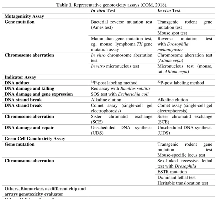
Genotoxicity is a broader term, including mutagenicity, DNA damages which may also be reversed by DNA repair or other cellular processes, thus which may or may not result in permanent alterations in the structure or information content in a surviving cell or its progeny. Therefore, genotoxicity tests mostly evaluate induced damage to DNA (but not direct evidence of mutation) via effects such as unscheduled DNA synthesis (UDS), DNA strand breaks (e.g. comet assay) and DNA adduct formation, i.e. primary DNA damage tests (COM, 2011). Genotoxicity test systems were summarize at Table 1.
3
Table 1. Representative genotoxicity assays (COM, 2018).
In vitro Test In vivo Test
Mutagenicity Assay
Gene mutation Bacterial reverse mutation test
(Ames test)
Transgenic rodent gene mutation test
Mouse spot test Mammalian gene mutation test,
eg, mouse lymphoma TK gene mutation assay
Reverse mutation test with Drosophila
melanogaster
Chromosome aberration In vitro chromosome aberration test
Chromosome aberration test (Allium cepa)
In vitro micronucleus test Micronucleus test (mouse, rat, Allium cepa)
Indicator Assay
DNA adduct 32P-post labeling method 32P-post labeling method
DNA damage and killing Rec assay with Bacillus subtilis DNA damage and gene expression SOS test with Escherichia coli
DNA strand break Alkaline elution Alkaline elution
DNA strand break Comet assay (single-cell gel
electrophoresis)
Comet assay (single-cell gel electrophoresis)
Chromosome aberration Sister chromatid exchange
(SCE)
Sister chromatid exchange (SCE)
DNA damage and repair Unscheduled DNA synthesis
(UDS)
Unscheduled DNA synthesis (UDS)
Germ Cell Genotoxicity Assay
Gene mutation Transgenic rodent gene
mutation test
Mouse-specific locus test
Chromosome aberration Sex-linked recessive lethal
test with Drosophila ESTR mutation Dominant lethal test Heritable translocation test Others, Biomarkers as different chip and
arrays genotoxicity evaluator Others Cell transformation assay
Prokaryotic systems
The Ames test (bacterial mutagenesis test):
The Ames test is one of the most widely used methods to assess genotoxicity. In 1973, Ames et al. developed the test as a fast and sensitive way to evaluate the ability of a compound to induce mutations in DNA. Up to date, the Ames test has been utilized to determine mutagenicity of various compounds that the test have been validated by using 300 chemicals which were known as carcinoges. Also, its afficacy have been improved by a number of modifications. The test includes plating His– Salmonella typhimurium onto media containing trace amounts of histidine and adding chemicals to be tested for mutagenicity, resulting colonies that if the compound is
4
capable of reversing the mutation in S. typhimurium. His- mutation is consequently converted to His+ (Ames et al., 1973;Flückiger-Isler et al., 2004).
Eukaryotic systems
The yeast DEL assay and yeast mitotic gene-conversion assay:
The yeast deletion (DEL) assay is similar to the Ames test wherein the eukaryote yeast is employed as the test organism. The test scores the DNA deletions in the yeast Saccharomyces cerevisiae is capable of to determine a wide range of carcinogens. The test is a simple and fast technique to measure the frequency of reversion of a disrupted his3 gene due to interruption by short repeats that the test detects mutagens by its ability to revert the strain back to His+, either by plating on media lacking histidine or in a microtiter plate format using a colorimetric readout. It has been demonstrated that the DEL assay has a high level of sensitivity and specificity toward carcinogens, many of which are poorly determined by bacterial mutagenicity and other short-term genotoxicity assays (Brennan and Schiestl, 2004; Musgrove and Camps, 2012).
The comet assay/single-cell gel technique:
The comet assay or single-cell gel (SCG) test allows the visualization of DNA damage and studying with DNA repair in eukaryotic cells. The assay can detect single- and double-strand breaks, alkali labile sites, basic sites, oxidative damage, and cross-linking of DNA with DNA, protein, or drug. In this microgel electrophoresis method, cells with increased DNA damage exhibit increased migration of chromosomal DNA from the nucleus toward the anode, which resembles the shape of a comet (Speit and Hartmann, 1999). Intact DNA which are not disrupted are too large and remain in the cavity, however the small fragments migrate in a given period. Bu using the comet assay, novel chemicals can be tested for genotoxicity, environmental contamination, human biomonitoring and molecular epidemiology, and fundamental research in DNA damage and repair. Moreover, the sensitivity and specificity of the assay can be increased by using bacterial repair endonucleases that recognize specific kinds of damage in the DNA and
5
convert lesions to DNA breaks which is resulted with enhanced he amount of DNA in the comet tail (Collins, 2004).
The micronucleus tests:
The in vitro micronucleus can determine genotoxic damage novel chemicals in interphase cells. The micronucleus or small nucleus can be defined as a third nucleus containing a portion of acentric or entire chromosome. This formation could not be carried to the opposite poles during anaphase of mitosis or meiosis resulted in a daughter cell that lacks either a part or a complete chromosome. The micronucleus test can be used as an alternative to the other chromosome aberration tests, because the test allows to examine cells at interphase, micronucleus can be scored faster and is more amenable to automation (Doherty, 2012).
Animal models
The most used animal model is the mice due to their close resemblance to human. Especially, transgenic mouse models, including Muta™Mouse, LacZ Plasmid Mouse, and the Big Blue®
mouseassay were developed to evaluate genotoxic effects of novel chemicals (Musgrove and Camps, 2012). Mutant frequency corrected for clonal expansion defines the mutation frequency. Thus, clonal expansion can be calculated by correcting for mutations that repeat one or more times in a given tissue from a given mouse, by counting a given mutation only once per animal per tissue. However, mouse models have mostly time-,labor-, and resource-intensive nature in comparison to other alternative tests (Eastmond et a., 2009).
The zebrafish, Danio rerio, represents a vertebrate species serves as a model for development and embryogenesis which also have been used to study genotoxicity. Zebrafish and mammals exhibit a high degree of similarity that have been demonstrated to share 70% sequence homology with humans and possess about 84% of the same genes as humans that are associated with disease (Howe et al., 2013). Using zebrafish model system for genotoxicity have several advantages which
6
are ease of breeding, low-cost maintenance, smallness, permeable and transparent larvae. Moreover, fertilization and development occurs externally which would allow the use of non-invasive imaging technology. Zebrafish lack some of the mammalian organs, lung, prostate, skin, and mammary gland. Therefore, zebrafish are not expected to replace the classical mammalian test systems. However, zebrafish can be used as a first step in vertebrate modeling of disease and drug discovery (Kari et al., 2007).
The plant-based biotests
The use of plant-based biotests in the identification of mutagen/carcinogens is a process that began in the early 1980s and continues to the present day; cytogenetic aberrations and gene mutations that lead to monitoring and control of environmental chemicals have been reported to be very useful systems (Constantin and Owens, 1982; Grant, 1994). In order to determine the mutagenic/carcinogenic potentials of chemicals, they met in Geneva in 1985, together with the International Labor Organization (ILO), the United Nations Environment Program and the WHO Environmental Health Criteria (The Environmental Health Criteria). The International Program on Chemical Safety (IPCS) (Ashby et al., 1988). In this study, Arabidopsis thaliana chlorophyll and embryo tests (Redei, 1982) and Tradescantia clone 4430 stamen hair test (Vant’t Hof and Schairer, 1982) are used for gene mutation tests. Tradescantia clone 4430 micronucleus test was used in cytogenetic tests and it was used for chromosome aberration test in Vicia faba (Kihlman and Andersson, 1984). In addition, other plant species commonly used to evaluate the mutagenic/carcinogenic effects of a chemical are Allium cepa, Crepis capillaris, Glycine max,
Hordeum vulgare, Lycopersicum esculentum, Nicotiana tabaccum, Pisum sativum and Zea mays
(Grant, 1994). Large plants have large chromosome size makes them very useful for cytogenetic analysis (Fiskesjö, 1985).
7
Plant-based tests are commonly used to detect mutagen/carcinogen effects of chemical compounds and to determine genotoxicity, often in situ monitoring of environmental contaminants that do not require any preliminary concentration or a very long, difficult and expensive preparation step (Grant, 1994; 1999). The use of plant-based mutagen/carcinogen test systems has some advantages. Especially high build plants; they are basically good indicators of the cytotoxic (damaging effects on cell structure or function), cytogenetic (effects on chromosomes) and mutagenic (effects of genetic change) of the chemicals to be tested. For this reason, high-structured plants have specific advantages in experimental studies as alternative test systems that will be used primarily in determining the use of the chemical to be tested or the damage they may cause to the environment (Ma et al., 1995). However, the life cycle of many plants is longer than that of bacteria, yeast and Drosophila, and the presence of basic pharmacokinetic and biochemical differences between plants and animals imposes limitations on the use of plant systems. Grant et al. (1981) published comparative results of some chemical substances in terms of genetic abnormalities in both plant systems and animal systems. The Allium test is frequently used to determine the mutagenic/carcinogenic effect of the test chemical and is easy, inexpensive and particularly correlates well with mammalian test systems (Fiskesjo, 1985).
The advantages of using high-structure plants in biotests in testing and monitoring the substance to be tested can be listed as follows (Grant, 1994); Plants of eukaryotic organisms have chromosome structure similar to humans. They exhibit mitosis and meiosis cell division. They mutate. It is easy to apply. It is cheaper than other test systems and is easy to culture. The life cycles of some plants such as Arabidopsis are completed in a short time. Tests can be performed in a wide range of environmental conditions, pH and temperature ranges. Plants can be regenerated from a single haploid plant or regenerated from a diploid plant. Plant genotoxicity tests can be used to evaluate a wide range of chemicals, from a single chemical to complex mixtures. The
8
reliability has been proven for long years. Their utility in mutagenesis studies has also been proven. The availability of genotoxicity results for many chemicals has allowed comparisons between different tests. The results of the studies showed that mammalian cytogenetic tests and plant biotest showed a positive correlation. Plant biotests can be combined with microbial tests to identify mutagenic metabolites (promutagens). The sensitivity of plant biotests has been shown to be high in determining the carcinogenicity of the substance to be tested.
To date, seven high-structure plants (A. cepa, A. thaliana, Glycine max, Hordeum vulgare,
Tradescantia paludosa, Vicia faba and Zea mays) have been widely used in many studies to
determine the mutagenic/carcinogenic or genotoxic effects of the material/chemical tested. However, most studies have focused on structural changes in chromosomes or chromatids, called chromosomal aberrations, i.e. fractures, deletions, voids, fragment changes, inversions, ring structures, and other disorders such as adhesion, clustering wear. In addition, disorders such as spindle thread inactivation, c-mitosis, or non-dissociation, particularly in mitotic and meiotic divisions, have also been implicated in chromosome distribution during anaphase resulting in polyploid or aneuploid cells. In addition, events occurring during recombination such as somatic crossing ovary and sister chromatid exchange, inefficiency and embryonic death, pollen grains, point mutations expressed in chlorophyll or offspring (embryo) are also studied (Choy, 2001; Uhl et al., 2003; Ma et al., 2005).
Among these stress conditions, tissue culture conditions are also accepted and among the studies conducted on the examination of retrotransposon activity changes under tissue culture conditions, studies on retrotransposon activity are remarkable and it has been shown that retrotransposon activity under stress changes and moves in the genome (Bonchev et al., 2010; Bayram; et al., 2012; Hamat-Mecbur et al., 2012; Yılmaz and Gozukirmizi, 2013; Yılmaz et al., 2014). In 2010,
9
Bonchev et al. investigated the transcriptional activity changes of BARE1 and WIS2-1A retrotransposons in the hexaploid wheat and rye mutants obtained from ethyl methanesulfonate (EMS) using SSAP, IRAP and REMAP marker techniques. The results of the study showed that EMS caused retrotransposon-based insertion polymorphism, but also increased transcriptional activity of retrotransposon in mutant plants. Bayram et al., (2012) examined the activity movements of Nikita retrotransposon in barley calluses under tissue culture conditions using IRAP method. In this study, the presence of polymorphic bands in two different 90 days callus originating from different embryos was shown by using 30-, 60- and 90-days barley calluses originating from a single embryo under tissue culture conditions. Hamat-Mecbur et al. (2012) in tissue culture conditions germinated barley plant for 10 and 20 days for two different concentrations (250 and 500 μg/ml) by applying epirubicin antibiotic BAGY2, BARE1 and Sukkula investigated using the polymorphism of the IRAP marker method. According to the results, it was shown that epirubicin application at different concentrations had no effect on the activity of BAGY2 and Sukkula retrotransposon, whereas epirubicin at 500 μg/ml caused polymorphic bands in BARE1. Finally, Yılmaz et al. (2014) examined the insertion polymorphism in barley callus and shoots due to activity movements of BAGY2 retrotransposon, which may occur depending on the culture period. According to the findings, the rate of polymorphism between calluses of 45 and 90 days and shoots obtained from them was shown as 0-21%.
Biomarkers as genotoxicity evaluators
The application of assessing the genotoxicity of various molecules can be diverse, including prokaryotic systems, eukaryotic systems, and whole-animal systems. However, test systems is still need the improvement to analyse the dose responsiveness of multiple compounds at a single time point, or multiple time points in real time, resulting in the availability of quantitative data in short intervals of time. Therefore, some strategies have been developed that some of them are miniaturized three-dimensional cell-culture arrays or the data toxicology-assay chip which allow
10
to screen of various compounds to an all new level, allowing the assessment of toxicity of the compound, as well as its metabolites. The strategy encapsulates human cells in three-dimensional hydrogel matrices such as collagen or alginate on a glass slide in specific spatial arrangements which allow to screen multiple compounds simultaneously along with the appropriate controls (Lee et al., 2008). However, this strategy has only been used to evaluate the cytotoxicity of drug candidates, has not yet been adapted for genotoxic studies (Fernandes et al., 2009). Recently, Agilent technologies has developed a microarray-based method which detects and analyses DNA-damage and -repair mechanisms across the genome. The alteration of acetylation levels in histone proteins is utilized to evaluate for genotoxicity (Powell et al., 2015).
Another genotoxicity tests, is named as Cell Ciphr, was developed by Cellumen Inc. in 2015. The technology combines several aspects of genotoxicity assessment such as tissue-specific cells, multiplexed functional biomarkers, and a compound reference library, into a single cell-based assay. Also, this technology only allows to assess genotoxicity for potential drug candidates by using more than ten biomarkers at a single stretch (Zanella et al., 2010). Thermo Fisher Scientific has also introduced another cell-based technique, CellSensor® cell lines coupled with GeneBLAzer® technology to analyse several pathways (PubChem, 2020). The most common cell line used in the analysis is the CellSensor® p53RE-bla HCT-116, which includes a β-lactamase gene under the control of a p53 response element, a major element that is involved in DNA repair (Ranganatha et al., 2016). SA Biosciences offers the Oligo GEArray® allows to screen approximately 113 genes which are involved in DNA damage signalling pathway. Moreover, real-time polymerase chain reaction (PCR)- based format of this assay were also improved to screens approximately 84 genes (SA Biosciences, 2015).
11
GADD45A promoter in human cell lines is induced upon exposure to clastogens, aneugens, and
mutagens that this gene expression is overexpressed under genotoxic conditions (Knight et al., 2009). Gentronix developed the GreenScreen HC Assay which utilize the human derived p53-competent TK6 cell line with a patented green fluorescent protein (GFP) reporter system that utilizes the regulation of the GADD45A gene. The GFP reporter complex recognize mutagens, aneugens, clastogens, and topoisomerase and polymerase inhibitors which increase the expression of GFP based on overexpression of the GADD45A gene. Additionally, this strategy is highly specific and sensitive, also providing in vitro negative results for non-genotoxins (Walmsley and Tate, 2012). A variant of this assay, BlueScreen HC assay, includes a Gaussia luciferase-reporter system instead of the GFP-reporter system (Simpson et al., 2013). This assay is reported to have reduced interference from auto fluorescent compounds and easier to analyse compared to GreenScreen HC Assay (Ranganatha et al., 2016).
Conclusion
Prokaryotic and eukaryotic models for the assessment of genotoxicity have established to be extremely useful and complement each other. Prokaryotic systems are cheaper and have the potential to be incorporated into high-throughput format, however their feasibility is limited with the detection of point mutations and frame shifts. Besides, eukaryotic systems demonstrate more sensitivity toward DNA damage with the challenging of being time-consuming and labour-intensive. The major challenge in the development of genotoxicity systems is that the system should be high-throughput, rapid, non-labour-intensive, and cost-effective, yet accurately mimic the human environment.
Acknowledgement
I also would like to thank Istanbul University Molecular Biology Genetics Department, Istinye University Research Fund supporting us during our short time mutagenesis research.
12 References
Ames BN, Durston WE, Yamasaki E, et al. Carcinogens are mutagens: a simple test system combining liver homogenates for activation and bacteria for detection. Proc Natl Acad Sci U S A. 1973; 70(8):2281–2285.
Ashby J, De Serres FJ, Shelby MD, et al. “Evaluation of short-term tests for carcinogens”. Report on the International Programme on Chemical Safety’s Collaborative Study on in vivo assays. Vols.I/II, Cambridge: Cambridge University Press. 1988.
Bayram E, Yilmaz S, Hamad-Mecbur H, et al. 2012. Nikita retrotransposon movements in barley (Hordeum vulgare L.) callus culture. Plantomics. 2012; 5(3):211-215.
Bonchev G, Georgiev S, Pearce S. Retrotransposons and ethyl methanesulfonate-induced diversity in hexaploid wheat and Triticale. Cent Eur J Biol. 2010; 5(6):765-776.
Brennan RJ, Schiestl RH. Detecting carcinogens with the yeast DEL assay. Genetic Recombination. Methods in Molecular Biology. 2004; 262:111–124.
Choy WN. Genetic toxicology and cancer risk assessment. New York: Marcel Dekker. 2001. Collins AR. The comet assay for DNA damage and repair: Principles, applications, and limitations.
Mol. Biotechnol. 2004; 26:249–261.
COM. Guidance On A Strategy For Genotoxicity Testing Of Chemical Substances. 2011. COM. Guidance On A Strategy For Genotoxicity Testing Of Chemical Substances. 2018.
Constantine MJ, Owens ET. Introduction and perspectives of plant genetic and cytogenetic assays, A report of the US Environmental Protection Agency Genotox Program. Mut Res. 1982; 99:1-12.
Doherty AT. The in vitro micronucleus assay. Methods Mol Biol. 2012; 817:121–141.
Eastmond DA, Hartwig A, Anderson D, et al. Mutagenicity testing for chemical risk assessment: update of the WHO/IPCS Harmonized Scheme. Mutagenesis. 2009; 24:341–349.
13
Fernandes TG, Diogo MM, Clark DS, et al. High-throughput cellular microarray platforms: applications in drug discovery, toxicology and stem cell research. Trends Biotechnol. 2009; 27(6):342–349.
FiskesjöG.The Allium test as a standard in environmental monitoring Hereditas.1985;102:99112. Flückiger-Isler S, Baumeister M, Braun K, et al. Assessment of the performance of the Ames II
assay: a collaborative study with 19 coded compounds. Mutat Res. 2004; 558:181-197. Grant VF. Plant Speciation. New York: Columbia University Press. 1981.
Grant WF. The present status of higher plant bioassays for the detection of environmental mutagens. Mut Res. 1994; 310:175-185.
Grant WF. Higher plant assays for the detection of chromosomal aberrations and gene mutationsda brief historical background on their use for screening and monitoring environmental chemicals. Mutat Res. 1999; 426:107e112.
Hamad- Mecbur H, Yilmaz S, Temel A, et al. Effects of epirubicin on barley seedlings. Toxicol Ind Health. 2012; 30(1):52-59.
Howe K, Clark MD, Torroja CF, et al. The zebrafish reference genome sequence and its relationship to the human genome. Nature. 2013; 496(7446):498–503.
Kari G, Rodeck U, Dicker AP. Zebrafish: an emerging model system for human disease and drug discovery. Clin Pharmacol Ther. 2007; 82:70‐ 80.
Kihlman BA, Andersson HC. Root tips of Vicia faba for the study of the induction of chromosomal aberrations and sister chromatid exchanges. Handbook of Mutagenicity Test Procedures. Eds: Kilbey BJ, Legator M, Nichols W, Ramel C. Amsterdam, New York, Oxford: Elsevier. 1984.
Knight AW, Birrell L, Walmsley RM. Development and validation of a higher throughput screening approach to genotoxicity testing using the GADD45a-GFP GreenScreen HC assay. J Biomol Screen. 2009; 14(1):16–30.
14
Lee MY, Kumar RA, Sukumaran SM, et al. Three-dimensional cellular microarray for high-throughput toxicology assays. Proc. Nat. Acad. Sci. USA. 2008; 105:59–63.
Ma TH, Xu ZD, Xu C, et al. 1995. The improved Allium/Vicia root tip micronucleus assay for clastogenicity of environmental pollutants. Mut Res. 1995; 334:185-195.
Ma TH, Cabrera GL, Owens E. Genotoxic agents detected by plant bioassays. Rev Environ Health. 2005; 20:1–13.
Musgrove C, Camps M. Models for detection of genotoxicity in vivo: present and future. In: Mishra DR, editor. Mutagenesis. Rijeka, Croatia: InTech; 2012; 31–50.
National Center for Biotechnology Information. PubChem Database. Source=NCGC, AID=743292, https://pubchem.ncbi.nlm.nih.gov/bioassay/743292 (accessed on Jan. 9, 2020).
Powell JR, Bennett MR, Evans KE, et al. 3D-DIP-Chip: a microarraybased method to measure genomic DNA damage. Sci Rep. 2015; 5:7975.
Ranganatha R, Chakravarthy S, Sukumaran S. High-throughput approaches for genotoxicity testing in drug development: recent advances. 2016; 6:1-12.
Redei GP. Mutagen assay with Arabidopsis: A report of the US Environmental Protection Agency Gene-Tox Program. Mut Res. 1982; 99:243-255.
SABiosciences. Oligo GEArray® human DNA damage signaling pathway microarray. Available from:http://saweb2.sabiosciences.com/gene_array_product/HTML/OHS-029.html.
(accessed on Nov. 4, 2015).
Simpson K, Bevan N, Hastwell P, et al. The BlueScreen-384 assay as an indicator of genotoxic hazard potential in early-stage drug discovery. J Biomol Screen. 2013; 18(4):441–452. Speit G, Hartmann A. The comet assay (single-cell gel test). A sensitive genotoxicity test for the
15
Uhl M, Plewa M, Majer BJ, et al. Basic principles of genetic toxicology with an emphasis on plant bioassays”. Bioassays in plant cells for improvement of ecosystem and human health. Editörler: Maluszynska, J., Plewa, M. Katowice: Wydawnictwo Uniwersytetu Śląskiego. 2003.
Van’t Hof J, Schairer LA. Tradescantia assay system for gaseous mutagens report of the US Environmental Protection Agency GeneTox Program. Mut Res. 1982; 99:303-315.
Walmsley RM, Tate M. The GADD45a-GFP GreenScreen HC assay. Methods Mol Biol. 2012; 817:231–250.
Yılmaz S, Gozukirmizi N. Variation of Retrotransposon Movement in Callus Culture and Regenerated Shoots of Barley. Biotechnol Biotechnol Equip. 2013; 27:4227-4230.
Yilmaz S, Marakli S, Gozukirmizi N. BAGY2 retrotransposon analyses in barley calli cultures and regenerated plantlets. Biochem Genet. 2014; 52:233-244.
Zanella F, Lorens JB, Link W. High content screening: seeing is believing. Trends Biotechnol. 2010; 28(5), 237–245.