© Turkish Society of Radiology 2010
P
rogressive massive fibrosis (PMF) of the lung is a type of late stage pneumoconiosis and pathologically consists of fibrotic lesions more than 1 cm in greatest diameter (1). The chest radiographic and computed tomography (CT) findings of PMF have been reported (2, 3). In our geographic region, PMF is mostly seen as a complication of coal workers’ pneumoconiosis (CWP). Evaluation and follow-up of PMF lesions are generally performed by using CT scans in CWP patients. However, imaging has to provide detailed information about the ana-tomical extension of the PMF. Since CT imaging requires ionizing radia-tion and applicaradia-tion of CT contrast agents is limited in patients with al-lergies to ionized contrast media or in patients with renal insufficiency, alternative imaging methods for diagnosis and follow-up are always of interest. If the fast magnetic resonance imaging (MRI) of PMF lesions of the lungs is proven to be diagnostically significant, this modality can be used instead of CT imaging. The rapid development of MRI techniques during the last years has resulted in excellent soft tissue imaging capa-bilities. MRI of the lung is difficult and hampered by three factors: first (and very important) is signal loss due to physiological motion (respira-tion and cardiac pulsa(respira-tion); second is low proton density in lung results in a low signal-to-noise-ratio (SNR); and third is the unique combination of air and soft tissue resulting in significant susceptibility to artifact. De-spite these difficulties, fast MRI techniques for evaluating lung patholo-gy have been developed and addressed in a number of articles describing preliminary results (4–6). However, correct evaluation of PMF lesions by fast MRI techniques has not yet been well described. Volumetric interpo-lated breath-hold examination (VIBE) and half-Fourier single-shot turbo spin echo (HASTE) MR sequences are very fast imaging techniques, and these techniques can also be used for fast pulmonary imaging.In the present study, we hypothesized that fast pulmonary MRI of the PMF lesions by using VIBE and HASTE fast MRI protocols would enable detection of lesions and might have a role in the management of PMF, especially in long-term follow-up. Thus, the purpose of our study was to determine the feasibility of fast MRI in the management of PMF in comparison with CT imaging.
Materials and methods Study design
Twenty-three patients were enrolled in this prospective study be-tween January 2007 and July 2008. Three patients were unable to un-dergo MRI because of claustrophobia and had to be excluded from the study. Thus, data sets of 20 patients were analyzed for this study. All patients were male, with an age range of 52–82 years (mean, 72 years). The patients had worked as coal miners for 15–25 years (mean, 15 years) working underground. They had pulmonary mass lesions
rang-C H E S T I M A G I N G
O R I G I N A L A R T I C L EFast MRI evaluation of pulmonary progressive massive fibrosis with
VIBE and HASTE sequences: comparison with CT
Koray Hekimoğlu, Tanzer Sancak, Meltem Tor, Halit Beşir, Bora Kalaycıoğlu, Sadi Gündoğdu
From the Department of Radiology (K.H. korayhekim@yahoo.
com.tr), Başkent University School of Medicine, Ankara, Turkey;
the Department of Radiology (T.S., S.G.), Ufuk University School of Medicine, Ankara, Turkey; the Departments of Pulmonary Diseases (M.M.) and Radiology (H.B., B.K.), Karaelmas University School of Medicine, Zonguldak, Turkey.
Received 22 September 2008; revision requested 26 March 2009; revision received 31 May 2009; accepted 11 June 2009.
Published online 16 December 2009 DOI 10.4261/1305-3825.DIR.2313-08.2
PURPOSE
The aim of this prospective study was to evaluate the diagnos-tic utility of volumetric interpolated breath-hold examination (VIBE) and half-Fourier-acquisition single-shot turbo spin-echo (HASTE) fast magnetic resonance imaging (MRI) sequences in the evaluation of pulmonary progressive massive fibrosis (PMF) in comparison with computed tomography (CT) imag-ing. If fast MRI is proven to be diagnostically significant, this modality can be used for diagnosis and follow-up studies of PMF patients.
MATERIALS AND METHODS
Twenty-two PMF lesions from 20 coal workers were evalu-ated. After CT imaging, patients underwent pre-contrast VIBE, contrast-enhanced VIBE, and HASTE MRI studies for detection and evaluation of the PMF lesions. Measurements of the three groups were evaluated with intra-class coefficients. Correla-tion levels between sizes, image quality, and artifact were evaluated with linear Pearson correlation analysis.
RESULTS
There was almost perfect agreement among radiologists for lesion detection with kappa analysis. There was significant agreement between three MRI study groups and gold stand-ard CT images. We found the best agreement values with con-trast-enhanced VIBE images for lesion detection and image quality in comparison with CT imaging. Presence of artifact was also lowest with this protocol.
CONCLUSION
With fast MRI sequences in pulmonary imaging, image quality has significantly improved being very close to that of CT stud-ies. In this study, contrast-enhanced VIBE protocol provided the best depiction of PMF lesions. This protocol may be an alternative choice for CT, avoiding the use of iodinated con-trast material and minimizing exposure to ionizing radiation for follow-up studies.
Key words: • pulmonary fibrosis • magnetic resonance imaging • computed tomography • comparative study
MRI was performed with two 1.5 T MRI scanners (Intera Master Gyros-can, Philips Medical Systems, Best, The Netherlands; and Magnetom Sympho-ny, Siemens Medical Solutions, Erlan-gen, Germany) with a maximum gra-dient strength of 30 mT/m and a slew rate of 150 mT/m per ms. A standard phased-array body coil was used for signal reception. ECG triggering with an active fiberoptic ECG system was used for reduction of cardiac motion artifact in HASTE sequence imaging. Seventeen patients were scanned with the Philips scanner, and the remaining three with the Siemens scanner. Fast T1-weighted MRI sequences
T1-weighted VIBE sequence was chosen for fast T1-weighted MRI. VIBE sequence parameters were adapted to Philips scanner for the ultrafast 3D gradient echo (T1-TFE) sequence. Im-aging parameters for VIBE sequence were as follows: TR/TE, 5.12/2.51 ms; flip angle, 10°; partition thickness, 5 mm without interslice gaps; matrix size, 256 × 116 with three-dimesional (3D) breath-hold imaging technique. The 3D VIBE sequence is a 3D-gradi-ent echo (GRE) sequence (volumetric interpolated breath-hold examina-tions) and has been presented as a fast MR sequence for liver and pulmonary imaging. It is similar to the 3D radiof-requency-spoiled GRE sequence used to perform 3D MR angiography. How-ever, this sequence differs from MR angiography sequences by symmetri-cal k-space acquisition in the phase encoding and the partition encoding directions (ky and kz, respectively), which decreases truncation artifact and improves image quality. On the other side, ultrafast spoiled gradient echo (T1-TFE) sequence was used on Intera-Philips scanner for fast T1-weighted MRI with these parameters: TR/TE, 4.67/1.67 ms; flip angle, 20°; matrix size, 256 × 192. Fat saturation tech-niques were not employed. The field of view (FOV) ranged from 375 mm to 450 mm for the acquisition of coro-nal and axial MRI images for the two MRI scanners. Breath-hold protocol was applied for better image contrast. Acquisition time was 20 s. Pre- and post-contrast images were obtained in all patients. We used a power injector (Medrad Vistron MR injection system, Medrad Inc.) to inject 0.1 mmol/kg of gadopentate dimeglubine (Magnevist,
Schering AG) at a rate of 2 mL/s in all patients. Contrast-enhanced MR imag-es were obtained 60–70 s after comple-tion of the intravenous injeccomple-tion. Fast T2-weighted MRI sequences
T2-weighted HASTE was used for fast T2-weighted MRI. The HASTE sequence is described as a useful breath-hold fast T2 imaging process in lung parenchy-ma. In HASTE sequence, the data were acquired during a train of 180° refo-cusing pulses. The central portion of k-space was acquired immediately after the radiofrequency (RF) pulse, and im-age reconstruction was performed with a half-Fourier method by using k-space symmetry. ECG triggered, breath-hold, black blood (SPIR) prepared HASTE im-ages were obtained in the axial and coronal orientation with the following parameters: TR/TE, 2000 (2 R-R inter-vals)/53 ms; flip angle, 160°; effective slice thickness, 5 mm without interslice gaps; matrix size, 256 × 256; NSA, 3; phase-encoding direction, anteropos-terior. In HASTE scanning, 35 slices covering the entire lung area were col-lected in two interleaved concatena-tions of 20 s for each set. No contrast agent was given. For patients evaluated with the Philips Intera scanner, this sequence was adapted as being single-shot turbo spin echo (TSE) sequence with the following parameters: TR/TE, 2000/80 ms; effective slice thickness, 5 mm without interslice gaps; matrix size, 256 × 192; turbo factor, 17 se-lected. FOV range was 375–400 mm for these sequences. The T2 TSE sequence was combined with spectrally selective attenuated inversion recovery (SPAIR) for fat saturation.
Image analysis
The analysis of the imaging data was performed in a four-step manner after completion of data acquisition for all patients. It was based on reviewing hard and soft copies, which were avail-able on workstations for the two MRIs. In step 1 of the analysis, the gold standard reference images were defined by two experienced radiologists in con-sensus by reviewing and interpreting the CT scans. They remained blinded to the MRI findings of these patients. All PMF lesions previously confirmed by biopsy were counted for the study. The number, location, and three di-mensional sizes of the detected lesions (transverse, sagittal, and craniocaudal ing from 25 mm to 50 mm in
diam-eter (mean, 35 ± 3 mm) on CT images. Transthoracic biopsy was applied to all lesions by interventional radiol-ogy department, and PMF diagnosis was histopathologically confirmed. Follow-up of the PMF lesions in these patients were performed with CT. We selected lesions that showed no signif-icant change on CT for at least 2 years in order to exclude the possibility of a coexistent lung cancer.
One week after CT study, all pa-tients underwent a pre-contrast VIBE, HASTE, and contrast-enhanced VIBE MRI for the evaluation of the PMF le-sions. Both MRI and CT examinations were well tolerated with no adverse reactions. None of the data sets were excluded for serious respiratory or mo-tion artifact, and no examinamo-tion had to be repeated because of poor image quality. The mean in-room time was 10 ± 5 min for MRI examination and 8 ± 2 min for CT examination.
Images were divided into four groups for each patient (Group I, CT images; Group II, pre-contrast VIBE images; Group III, contrast-enhanced VIBE im-ages; and Group IV, HASTE images). Upon completion of imaging in every patient, all imaging groups were inter-preted by five experienced radiologists. Measurement of lesions on each imag-ing sequence was performed for further statistical analysis.
This study was approved by the in-stitutional ethics committee of Kara-elmas University School of Medicine. Informed consent was obtained from all patients.
Imaging techniques
CT scans were performed on spiral CT (Philips Secura 2000, Philips Medi-cal Systems, Best, The Netherlands) us-ing the followus-ing parameters: 120 kV; 150 mA; pitch factor, 1,5; slice width, 7 mm; effective slice thickness and re-construction, 5 mm; in-plane matrix size, 512 × 512. A contrast agent (100 mL iopromide [300 mgI/mL, Ultravist, Schering AG, Berlin, Germany]) was used with CT injector system (Medrad Vistron CT injection system, Medrad Inc., Warrendale, Pennsylvania, USA) via vena brachialis with 3 mL/s flow rate. After a delay of 30 s, the image set covering the entire lung was col-lected over 12–15 s. This protocol is our standard for routinely performed thoracic CT.
diameters) were recorded (Group I). To avoid miscounting, the observers marked each detected lesion on hard-copy film.
In step 2 of the analysis, each MRI group was analyzed independently by two experienced radiologists who were unaware of the results of the CT exami-nations. Each radiologist recorded the sizes, number, and location of PMF le-sions on each MR image.
In step 3, the corresponding CT and MRI data sets were reviewed again si-multaneously for one-to-one compari-son of the size and location of the de-tected lesions in all groups of images for each patient. Three radiologists in-dependently evaluated the general im-age quality (A) and presence of artifact (B) of detected pulmonary lesions. In the grading of image quality (A), radi-ologists used a semiquantitative grad-ing system as follows: 1 = poor (images which are non-diagnostic), 2 = fair (not optimal quality but sufficient to permit a diagnosis to be established), 3 = good (optimal image quality in resolution, sharpness, and clarity), and 4 = excellent (best image quality). All MRI images (Groups II–IV) were evalu-ated with regard to artifact (B) caused by breathing or cardiac pulsation sepa-rately. Total artifact scores were also calculated. In all patients, each lobe of the lung and PMF lesions were scored for the presence of artifact (score 0, no artifact; score 1, minor artifact; score 2, moderate artifact; score 3, severe arti-fact with insufficient imaging quality for diagnosis).
In step 4 of the image analysis, PMF lesions were evaluated for signal and post-contrast enhancement patterns of the lesions by two radiologists in consensus by reviewing the MRI im-ages. The pre-contrast signal intensity (SI) was supposed to reflect the status of massive fibrosis, and the signal pat-tern including post-contrast enhance-ment was supposed to reflect the sec-ondary change or vascular nature of PMF. Quantitative analysis was not performed in this study because the region of interest (ROI) measurements showed considerable variability of SI within the same lesion in the pilot study. In this analysis, the relative in-tensities of PMF lesions were compared with skeletal muscle on the same MRI images and categorized as hypointense, isointense, or hyperintense.
Contrast-enhanced VIBE images (Group III) were classified into three patterns: (a) no enhancement, (b) rim enhancement, and (c) diffuse enhance-ment. Contrast enhancement of the PMF lesions was defined visually as increase of SI after intravenous admin-istration of contrast media at the same contrast window and level setting. On the other side; the signal pattern of le-sions on pre-contrast VIBE and HASTE images (Groups II, IV) were classified into four types: (a) homogenous isoin-tense SI, (b) homogenously low SI, (c) high SI only at the rim, and (d) high SI areas and high SI rim.
Statistical analysis
All results were statistically described using commercial software (SPSS 11, SPSS Inc., Chicago, lllinois, USA; Excel 2003, Microsoft, Redmond, Washing-ton, USA). Type I error was accepted as 0.05, All reported P values were type-3 Wald significance levels and were declared to indicate a statistical significance if <0.05. Measurement of the results in three groups (II, III, IV) on three sizes were evaluated with in-tra-class coefficient (ICC). Correlation levels between these results and gold standard CT results (Group I) were evaluated with linear Pearson correla-tion analysis. The significance of differ-ences between groups were determined with randomized block design for ar-tifacts. In addition, group differences in image quality and spatial resolution were calculated using Friedman test. Simple kappa coefficients were used to assess interobserver agreement for lesion detection (0.00–0.20 indicated slight agreement; 0.21–0.40 fair agree-ment; 0.41–0.60 moderate agreeagree-ment; 0.61–0.80 substantial agreement; and 0.81–1.00 almost perfect agreement). Results
Diagnoses based on imaging findings Twenty-two PMF lesions were identi-fied and evaluated in 20 patients with CT and MRI. Based on the one-to-one correlation between CT and MR im-ages, the findings in all imaging tech-niques correlated well. All MRI images were of acceptable diagnostic quality. However, the image quality of lung pa-renchyma and PMF lesions was better detected with contrast-enhanced VIBE images than pre-contrast VIBE and HASTE images (Figs. 1–3). MRI inter-pretations did not show false-negative
or false-positive findings. None of the lesions detected on CT were missed on MRI images.
Evaluation of measurements on imaging findings
Linear correlation of the results between Group I (gold standard CT images) and the other groups (II-IV) which were pre-contrast VIBE, post-contrast VIBE, and HASTE sequence images respectively, were evaluated for three dimensions of the lesions. Correlation (r) values for x dimen-sion between Group I and the other groups (II–IV) were 0.972, 0.989, and 0.829, respectively. With respect to y dimension, r values were 0.987, 0.996, and 0.862, respectively. Finally, r val-ues for z dimension between Group I and the other groups were calculated as 0.997, 0.999, and 0.959, respective-ly. For all correlations P values were <0.01. Agreement values (ICC) for each dimension between three study groups and gold standard Group I are shown in Table 1. We found signifi-cant agreement between three study groups (II–IV) and gold standard Group I. Best agreement with gold standard CT imaging values for r and ICC were obtained with Group III (post-contrast VIBE sequence studies).
Interobserver agreement
There was almost perfect agreement among radiologists regarding report quality for both the CT lesion detec-tion group (kappa score, 0.85) and for the MRI lesion detection group (kappa score, 0.83).
Image quality
In all study groups, post-contrast VIBE (Group III) images had the best diagnostic quality. For Group III, aver-age quality score (± standard deviation, SD) was 3.65 ± 0.49 with a median val-ue of 4 (excellent). Pre-contrast VIBE (Group II) images had the second best diagnostic quality; statistical values for this group were 3.2 ± 0.41 with a medi-an value of 3 (good). HASTE (Group IV) images did not have a high diagnostic quality. Degradation was observed on these images because of respiratory or cardiac motion artifact and short scan times. The average quality score for Group IV was 2.25 ± 0.44, with a me-dian value of 2 (poor). All groups had statistically significant differences with each other (P < 0.01).
Artifacts
With regard to comprehensive scor-ing of breathscor-ing and cardiac pulsation artifacts, post-contrast VIBE scans pro-vided the lowest artifact scores com-pared with the pre-contrast VIBE and HASTE sequences in all parts of the lung. The average total artifact score ranged from 0.1 to 1 for pre-contrast VIBE (Group II) images, from 0.1 to 1 for post-contrast VIBE (Group III) im-ages, and from 0.8 to 1.8 for HASTE (Group IV) images.
In all MRI sequences, respiratory artifact was more prominent than cardiac or vessel pulsation artifact. This may be related to our breath-hold choice while determining MRI sequences. Pulsation and cardiac arti-fact ranged from 0 to 0.8 for pre-con-trast VIBE (Group II), from 0 to 0.8 for post-contrast VIBE (Group III) im-ages, and from 0.8 to 1.8 for HASTE (Group IV) images. Respiratory arti-fact ranged from 0.1 to 1.2 in Group II for pre-contrast VIBE images from 0.1 to 0.8 in Group III for post-con-trast VIBE sequence, and from 0.8 to
1.8 for Group IV (HASTE) images. We did not find significant differences in artifact between Group II and III im-ages. However, we found a significant difference between group IV and oth-er groups (II–III) (P < 0.05). Avoth-erage artifact scores in different pulmonary locations are shown in Table 2.
Signal intensity
On pre-contrast VIBE images (Group II), homogenous isointense SI compared with skeletal muscle (n = 16, 72%) was the most frequent SI, followed by homogenous low SI (n = 2, 9%), and high SI rim only (n = 2, 9%). The PMF lesions did not show Table 1. Intra-class coefficients (ICC) between gold standard (Group I) and the other groups for “x”, “y”, and “z” dimensions (single measures ICC)
Group II Group III Group IV
“x” dimension Group I 0.967 0.989 0.774 P values <0.0001 <0.0001 <0.0001 “y” dimension Group I 0.982 0.996 0.784 P values <0.0001 <0.0001 <0.0001 “z” dimension Group I 0.990 0.999 0.873 P values <0.0001 <0.0001 <0.0001 Group I, CT images; Group II, pre-contrast VIBE images; Group III, contrast-enhanced VIBE images; Group IV, HASTE images.
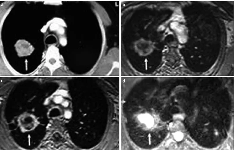
Figure 1. a–d. Axial plane CT (a), pre-contrast VIBE (b), contrast-enhanced VIBE (c), and HASTE MR (d) images of a 47-year-old coal worker who presented with huge progressive massive fibrosis lesion on the right side (white arrows).
b
a
d
c
both high SI areas and high SI rim in this group. On HASTE images (Group IV), the PMF lesions showed mostly homogenous low SI (n = 20, 91%), and the other lesions showed iso-intense SI (n = 2, 9%). PMF lesions did not show high SI rim at the rim only or in the lesion and at the rim
in Group IV. In evaluation enhance-ment patterns, Group III showed rim enhancement in 10 PMF lesions (45%) and diffuse enhancement in the other 12 lesions (55%). There were no non-enhancing PMF lesions in this group (Table 3).
Discussion
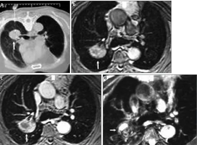
In this study, we combined two fast MRI sequences for evaluating PMF lesions. T1-weighted VIBE sequence was performed pre- and post-contrast enhancement. The gold standard modality for evaluating pulmonary lesions is spiral or multidetector CT, Figure 2. a–d. CT-guided biopsy (a), pre-contrast VIBE (b), contrast-enhanced VIBE (c), and HASTE MR (d) images of a 55-year-old coal worker who had large progressive massive fibrosis lesion on the right lower lobe (white arrows). The lesion showed diffuse enhancement pattern on contrast-enhanced image (c) with excellent image quality and minor artifact scores.
b
a
d
c
Table 2. Average artifact score as found in the study groups in different pulmonary localizations
Pulsation-cardiac artifacts Breathing artifacts Total artifacts
Group II Group III Group IV Group II Group III Group IV Group II Group III Group IV
Right upper lobe 0 0 1 0.1 0.1 1 0.1 0.1 1
Right middle lobe 0 0 1.4 0.2 0.2 1.6 0.2 0.2 1.5
Right lower lobe 0.2 0.2 1.8 0.5 0.4 1.8 0.4 0.3 1.8
Left upper lobe 0 0 1.5 0.1 0.1 1.5 0.1 0.1 1.5
Left lower lobe 0.2 0.2 1.8 0.5 0.4 1.8 0.4 0.3 1.8
Hilar zone 0.6 0.6 1.4 0.3 0.2 1.5 0.5 0.4 1.5
Mediastinum 0.8 0.8 0.8 1.2 0.8 0.8 1 1 0.8
PMF lesions 0 0 1 0.1 0.1 1 0.1 0.1 1
Group II, pre-contrast VIBE images; Group III, contrast-enhanced VIBE images; Group IV, HASTE images.
although several studies have pre-sented MRI assessment of pulmonary lesions as a similarly efficient modal-ity (5–9).
PMF is defined as a lesion of fibrosis and pigment deposition larger than 1
cm in diameter and is sometimes des-ignated as “complicated” pneumoco-niosis. These lesions commonly occur in coal workers who have had exposure to large amounts of heavy dust. Radio-logically, PMF starts near the periphery
of the lung and may closely resemble pulmonary carcinoma (2, 6, 10).
According to the results of this study, results of fast MRI in PMF patients may be identical to results of CT, which is accepted as gold standard in pulmo-nary pathologies. Compared with the pre-contrast VIBE and HASTE images, the contrast-enhanced VIBE images provided superior visualization of PMF lesions in pulmonary parenchyma. These results show that post-contrast VIBE sequence imaging has the best agreement with gold standard CT im-aging.
VIBE sequence is a 3D gradient-echo MRI technique tailored toward mini-mizing acquisition time and partial volume effects and maximizing image contrast, permitting imaging of tis-sue, with the first studies focusing on the liver (11). VIBE sequence has been proven to provide high spatial resolu-tion, good visualization of lung anato-my, and low rates of artifact in healthy volunteers. These image qualities have been confirmed in recent studies using 3D-GRE sequences in a wide spectrum Table 3. Qualitative analysis of images for signal and enhancement patterns (number/total
number of lesions and percentage)
Group II Group III Group IV Signal pattern
Homogenous iso SI 16/22 72% 2/22 9%
Homogenous low SI 2/22 9% 20/22 9%
High SI rim 2/22 9% -
-High SI rim and high SI areas - - -
-Enhancement pattern
No enhancement -
-Rim enhancement 10/22 45%
Diffuse enhancement 12/22 55%
Group II, pre-contrast VIBE images; Group III, contrast-enhanced VIBE images; Group IV, HASTE images. SI, signal intensity.
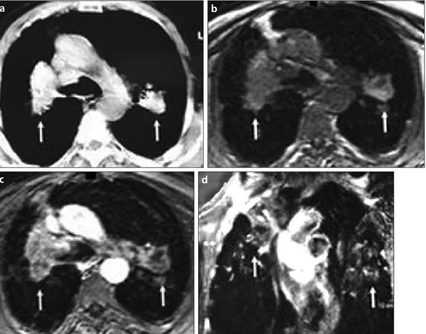
Figure 3. a–d. Axial plane CT (a), pre-contrast VIBE (b), and contrast-enhanced VIBE MR (c) images of a 52-year-old coal worker with a 20-year underground working history who had progressive massive fibrosis lesions on each lung (white arrows). The lesion revealed rim enhancement pattern on coronal post-contrast MR image (d, white arrows).
b
a
d
c
of malignant and benign pulmonary diseases (4, 12). This technique in-volves two independent directions— perpendicular (phase-encoding) and parallel (partition-encoding) to the plane of excitation. Asymmetric sam-pling performed in each of the phase-encoding directions improves spatial resolution. The main advantages of the VIBE sequence are its ability to rapidly acquire a volumetric data set during a single breath-hold, which enables ac-quisition of contiguous thin-slice im-ages with no interslice gap. Another important point of this sequence is the relative lack of phase artifact secondary to cardiac motion. This is achieved by the very short acquisition time for the central 10% of k-space, because this area of k-space contributes the most to image contrast but is most suscep-tible to phase artifact. The very short TR of VIBE sequence (4.67 ms) allows rapid imaging of lungs during one breath hold time. However, a short TE of only 1.67 ms minimizes susceptibil-ity effects due to short T2* relaxation time of pulmonary parenchyma and increases the SNR, resulting in success-ful visualization of pulmonary paren-chyma (13). The reduction in image artifact is another factor that improves image quality in this technique.
In this sequence, fat saturation tech-niques were not employed; it could have increased TR, which would have increased acquisition time and imaging artifact. Fat suppression was thought to improve visualization of the medi-astinum, chest wall, and axilla for this sequence and was critical for the detec-tion of the PMF lesions, which were seen as a focus of high signal intensity on the darkened background of sup-pressed fat.
HASTE sequence is a half-Fourier single-shot of TSE fast imaging MRI technique. This sequence has been re-ported to be a useful breath-hold T2-weighted sequence for imaging of the lung parenchyma (14). Because of its rapid data acquisition, HASTE imag-ing is relatively insensitive to motion artifact and may be particularly valu-able in patients who are unvalu-able to hold their breath for a long acquisition peri-od. HASTE images are characterized by high signal intensity in water-rich tis-sues. Thus, pulmonary lesions and ves-sels appear bright, whereas surround-ing air-filled parenchyma display low signal intensity (15, 16). Distinct
blur-ring artifact was seen on virtually all HASTE images, and the VIBE sequence had scores indicating a “minor” level, but the differences in artifact level were not significant. However, this limita-tion of image quality of HASTE images entailed lower sensitivities in lesion detection compared to VIBE sequence. Small PMF lesions could hardly be dif-ferentiated from vessels because of blurring (16).
Similar to CT, VIBE and HASTE techniques allow continuous data ac-quisition during a single breath-hold. In this study, the smallest lesion was 25 mm in diameter. No lesions in the three study groups were missed; thus, sensitivity for lesion detection was 100% in all groups. VIBE sequence, es-pecially post-contrast studies, revealed values close to those of CT images (17). Evaluation of lesion size with HASTE sequence, however, did not show high accuracy compared to gold standard CT images. Although CT has been re-garded as a standard reference tech-nique for detection of pulmonary nod-ules, it is associated with a relatively high level of ionizing radiation—from 10 to 100 times as much as chest radi-ography (18). Fast MRI techniques may be particularly useful in patients who would otherwise be exposed to a sub-stantial cumulative dose of radiation from undergoing repeated chest CT, such as our study patients in whom periodic chest CT follow-ups for PMF lesions had been requested for the pur-poses of ruling out lung cancer and/or of occupational compensation.
There were several limitations in this study. First, our study population was small. However, our intent was to show the feasibility of using the VIBE and HASTE sequences for fast pulmo-nary MRI. So the results were statisti-cally significant for our hypothesis. Second, in post-contrast imaging we used single delay time and we did not choose dynamic MRI study because quantitative analysis of ROI measure-ments showed considerable variability within the same lesion. Thus, we did not use prolonged dynamic study of PMF lesions for SI evaluation.
In conclusion, using fast MRI se-quences such as VIBE and HASTE have potential clinical utility for the MRI evaluation of PMF lesions in pneumo-coniosis. Of these techniques, post-contrast VIBE modality significantly reduces artifact and better depicts the
PMF lesions than pre-contrast VIBE and HASTE modalities. Although CT is a much more widely available modity, fast pulmonary MRI could be an al-ternative modality, particularly if min-imizing exposure to ionizing radiation or avoiding the use of iodinated con-trast material is of concern. Additional studies are warranted to establish the clinical value of fast pulmonary MRI in patients with various pulmonary dis-eases.
References
1. Gibbs AR. Occupational lung disease. In: Halsleton PS, ed. Spencer’s pathology of the lung. 5th ed. New York: McGraw Hill, 1996; 461–504.
2. Williams JL, Moller GA. Solitary mass in the lungs of coal miners. Am J Roentgenol Radium Ther Nucl Med 1973; 117:765– 770.
3. Soutar CA, Collins HP. Classification of progressive massive fibrosis of coalminers by type of radiographic appearance. Br J Indust Med 1984; 41:334–339.
4. Biederer J, Both M, Graessner J, et al. Lung morphology: fast MR imaging assessment with a volumetric interpolated breath-hold technique: initial experience with patients. Radiology 2003; 226:242–249.
5. Both M, Schultze J, Reuter M, et al. Fast T1-and T2-weighted pulmonary MR-imaging in patients with bronchial carcinoma. Eur J Radiol 2005; 53:478–488.
6. Jung JI, Park SH, Lee JM, Hahn ST, Kim KA. MR characteristics of progressive massive fibrosis. J Thorac Imaging 2000; 15:144– 150.
7. Matsumoto S, Mori H, Miyake H, et al. MRI signal characteristics of progressive mas-sive fibrosis in slicosis. Clin Radiol 1998; 53:510–514.
8. Kauczor HU, Kreitner KF. MRI of the pul-monary parenchyma. Eur Radiol 1999; 9:1755–1764.
9. Bruegel M, Gaa J, Woertler K, Waldt S, Hillerer C, Rummeny EJ. MRI of the lung: value of different turbo spin-echo, single-shot turbo spin-echo, and 3D gradient-echo pulse sequences for the detection of pulmonary metastases. J Magn Reson Imaging 2007; 25:73–81.
10. Matsumoto S, Miyake H, Oga M, Takaki H, Mori H. Diagnosis of lung cancer in a patient with pneumoconiosis and progres-sive masprogres-sive fibrosis using MRI. Eur Radiol 1998; 8:615–617.
11. Rofsky NM, Lee VS, Laub G, et al. Abdominal MR imaging with a volumet-ric interpolated breath-hold examination. Radiology 1999; 212:876–884.
12. Bader TR, Semelka RC, Pedro MS, Armao DM, Brown MA, Molina PL. Magnetic reso-nance imaging of pulmonary parenchymal disease using a modified breath-hold 3D gradient-echo technique: initial observa-tions. J Magn Reson Imaging 2002; 15:31– 38.
13. Biederer J, Reuter M, Both M, et al. Analysis of artifacts and detail resolution of lung MRI with breath-hold T1 weighted gradi-ent-echo and T2 weighted fast spin-echo sequences with respiratory triggering. Eur Radiol 2002; 12:378–384.
14. Hatabu H, Gaa J, Tadamura E, et al. MR imaging of pulmonary parenchyma with a half-Fourier single-shot turbo spin-echo (HASTE) sequence. Eur J Radiol 1999; 29:152–159.
15. Vogt FM, Herborn CU, Hunold P, et al. HASTE MRI versus chest radiography in the detection of pulmonary nodules: com-parison with MDCT. AJR Am J Roentgenol 2004; 183:71–78.
16. Schroeder T, Ruehm SG, Debatin JF, Ladd ME, Barkhausen J, Goehde SC. Detection of pulmonary nodules using a 2D HASTE MR sequence: comparison with MDCT. AJR Am J Roentgenol 2005; 185:979–984.
17. Karabulut N, Martin DR, Yang M, Tallaksen RJ. MR Imaging of the chest using a con-trast-enhanced breath-hold modified three-dimensional gradient-echo tech-nique: comparison with two-dimensional gradient-echo technique and multidetector CT. AJR Am J Roentgenol 2002; 179:1225– 1233.
18. Diederich S, Wormanns D, Lenzen H, Semik M, Thomas M, Peters PE. Screening for asymptomatic early bronchogenic car-cinoma with low dose CT of the chest. Cancer 2000; 89(11 Suppl):2483–2484.