DEVELOPMENT OF BIOACTIVE PEPTIDE NANOFIBERS FOR INTERVERTEBRAL DISC REGENERATION
A THESIS SUBMITTED TO
GRADUATE SCHOOL OF ENGINEERING AND SCIENCE OF BILKENT UNIVERSITY
IN PARTIAL FULFILLMENT OF THE REQUIREMENTS FOR THE DEGREE OF
MASTER OF SCIENCE IN NEUROSCIENCE By ÖZGE UYSAL August, 2017
ii
DEVELOPMENT OF BIOACTIVE PEPTIDE NANOFIBERS FOR INTERVERTEBRAL DISC REGENERATION
By Özge Uysal, August, 2017
We certify that we have read this thesis and that in our opinion it is fully adequate, in scope and in quality, as a thesis for the degree of Master of Science.
Ayşe Begüm Tekinay (Advisor)
Çağlar Elbüken
Yusuf Şükrü Çağlar
Approved for the Graduate School of Engineering and Science:
Ezhan Karaşan
iii ABSTRACT
DEVELOPMENT OF BIOACTIVE PEPTIDE NANOFIBERS FOR INTERVERTEBRAL DISC REGENERATION
Özge UYSAL
M.Sc. in Neuroscience Advisor: Ayşe Begüm Tekinay
August, 2017
Lower back pain (LBP) and neck problems are the most common orthopedic diseases worldwide, and the main reason behind LBP is intervertebral disc degeneration (IVDD). Therefore, specialized therapeutic applications to induce intervertebral disc (IVD) regeneration is a necessity. Here, we report the use of a collagen-mimetic bioactive peptide nanofiber scaffold (Col-PA/E-PA) for the improvement of disc regeneration by recapitulating the structure and function of the natural extracellular matrix (ECM) of intervertebral connective tissue. Following two weeks of degeneration, the bioactive nanofiber scaffold was topically applied and the IVD regeneration process was observed through histochemical analyses. The collagen-mimetic bioactive peptide nanofiber system was found to significantly promote glycosaminoglycan and collagen deposition at the site of injury compared to control
iv
nanofiber system and saline groups. In addition, the bioactive scaffold was consistently associated with lower values in degeneration scoring analyses, confirming the functional recovery of the tissue. Overall, the collagen-mimetic peptide nanofiber scaffold was able to prevent the progression of IVD degeneration and provide further functional recovery to the tissue.
Keywords: Peptide nanofiber, lower back pain, intervertebral disc degeneration, needle, regeneration, collagen
v ÖZET
İNTERVERTEBRAL DİSK REJENERASYONU İÇİN BİYOAKTİF PEPTİT NANOFİBERLERİN GELİŞTİRİLMESİ
Özge UYSAL Nörobilim, Yüksek Lisans Tez Danışmanı: Ayşe Begüm Tekinay
Ağustos, 2017
Bel ağrısı ve boyun sorunları dünyadaki en yaygın ortopedik hastalıklardandır ve bel ağrısının ana nedeni intervertebral disk dejenerasyonu (IVDD)'dur. Bu nedenle, intervertebral disk (IVD) rejenerasyonunu tetikleyebilecek terapötik uygulamaların geliştirilmesi bir zorunluluktur. Bu tez kapsamında, intervertebral bağ dokusunun doğal ekstraselüler matris yapısını ve fonksiyonunu yenileyebilecek disk rejenerasyonunun iyileştirilmesi için kullanılabilecek kolajeni taklit edebilen biyoaktif peptit nanofiber iskele (Col-PA / E-PA) geliştirilmesi ve kullanımı anlatılmaktadır. İki haftalık dejenerasyonun ardından, biyoaktif nanofiber iskelet topik olarak uygulanmıştır ve histokimyasal analizler yoluyla IVD yenilenme süreci izlenmiştir. Kolajen-mimetik biyoaktif peptit nanofiber sisteminin, kontrol nanofiber sistemi ve salin gruplarına kıyasla, yaralanma yerinde glikozaminoglikan ve kolajen depozisyonunu önemli ölçüde teşvik ettiği bulunmuştur. Buna ek olarak, biyoaktif iskeletin dejenerasyon puanlama analizlerinde sürekli olarak düşük değerlerle ilişkili
vi
olması, dokunun fonksiyonel iyileşmesini teyit etmektedir. Sonuç olarak, kolajen-mimetik peptit nanofiber iskele, IVD dejenerasyonunun ilerlemesini önlemiş ve dokuya daha fazla fonksiyonel iyileşme sağlayarak IVD dokusunun iyileştirilmesinde etkili bir tedavi yöntemi olarak kullanılabileceği gösterilmiştir.
.
Anahtar sözcükler: Peptit nanofiber, bel ağrısı, intervertebral disk dejenerasyonu, rejenerasyon, kolajen
vii
ACKNOWLEDGEMENTS
First, I would like to express my gratitude to my advisor Prof. Ayşe Begüm Tekinay. I am grateful for the opportunity to have a part in her laboratory, additionally I am grateful for her extreme patience and guidance throughout my master education. I appreciate her vast knowledge in many areas. I also would like to thank Prof. Mustafa Özgür Güler for his support during my master education.
I would like to thank TÜBİTAK (The Scientific and Research Council of Turkey) ARDEB 114S913 and for the financial support.
In addition, before the biggest thank to my family in the final, here I would like to thank my family for their financial support.
First and for most, I would like to thank Dr. Elif ARSLAN for guiding me throughout my Master’s degree education. Literally we shared every thing. Before, we shared many experiments and now we shared the day for our thesis presentations and also the jury members. She is one of the cleverest and the most knowledable PhD student that I have ever met. I learnt almost everthing from her during my Master education and I know she will be an excellent PI in the future. I also would like to thank Dr. Gülcihan Gülseren for her support in the experiments and her friendship. I also would like to thank Çağla EREN ÇİMENCİ for her support and teach me the first things that I have to learn at the beginning of my Master’s education and her amaizing friendship. She always smiles and helps to everyone about whatever they want. And of course I also would like to thank Alper Devrim ÖZKAN for his amazing knowledge about
viii
everything and for his amazing stories about the weird things that I wouldn’t know anything without him.
I would like to thank our former lab members: Merve ŞEN for her weird but very funny kind of friendship; Fatih YERGÖZ for his countless technical helps and his friendship; Burak DEMİRCAN for his fruitful conversations and I wish a good luck on his very healthy diets. I also would like to thank Melike SEVER for her support about the experiments and Ahmet Emin TOPAL for his “Özge Uysal merhaba!” song. I would like to thank Canelif YILMAZ for her unexpected atars (I know Mustafa also wrote the same thing but I couldn’t find any word better than that.); Zeynep ORHAN for her supports and being from my hometown and now hopefully she will be my neighbor. I also would like to thank Gökhan GÜNAY for his amazing friendship and his continuing “I am bored” words and he always knows which Friends scene that I am talking about (of course this is also true for İdil; but her thank you part will come later.) I also would like to thank Oğuz TUNCAY for his enjoyable and admirable personality. I also would like to thank Göksemin ŞENGÜL for her weird (but again good weird) friendship. She is always there for me whenever I want, and sometimes even I don’t want. Göksemin, Oğuz and I were always together from the beginning of our MBG adventure. They will always be my friends.
Moreover, I would like to thank my special Breakfast Club friends: Mustafa BETER, Elif ARSLAN, Begüm DİKEÇOĞLU and Gülistan TANSIK. We shared lots of foods but also we shared lots of memories. They supported me so much and specifically I would like to thank Mustafa for his continuing amazing support and friendship. I believe our friendship will always continue; even if we will be in different countries and even in different continents.
ix
Moreover, I would like to thank Nurcan HAŞTAR, Nuray GÜNDÜZ and İdil UYAN. We shared lots of things but especially in the last few weeks, I think we know each other much better. All the good things and the bad things made our friendship stronger. Especially I would like to thank Nurcan, she is the most affectionate and reliable person that I have ever met. She is always very helpful to everyone and I always complain about my problems and she never gets tired about that or at least she haven’t told me that.
Most importantly, I would like to thank my family: my father Necati, my mom Saadet for their constant and unconditional love and support throughout my life. They taught me lots of things but most importantly they made me who I am. I would like to thank my dearest sister Bilge for being my little mom since I was born.
My special thanks to Ömer ÖZDEMİR to being my best friend and my love; he is literally everything to me. He is a really special person and I admire his personality so much. He is the most positive person that I have ever met. He always supported me and I know he will always. I believe we will experience much more things and together there isn’t any thing that we cannot achieve.
Finally, without my family and Ömer, I couldn’t achieve any of these things; therefore I would like to dedicate this thesis to them.
x CONTENTS
ABBREVIATIONS ... xix
1. Introduction ... 1
1.1. Lower Back Pain and Socio-Economic Impact ... 1
1.2. Anatomy of IVD ... 2
1.3. Extracellular Matrix (ECM) of Intervertebral Disc ... 4
1.4. IVD Degeneration and Aging ... 6
1.4.1 Changes in the ECM of Degenerated IVD ... 8
1.4.2 Role of Inflammatory Cytokines and Growth Factors in the Degeneration Process ... 10
1.4.3 Changes in Vascularization and Innervation in Degeneration Process…… ... 10
1.5. Treatment Options for IVD Degeneration ... 13
1.6. Peptide Amphiphiles for Regenerative Medicine Applications ... 16
2. Objectives ... 19
xi
Materials ... 20
Synthesis and Purification of Peptide Amphiphiles ... 20
Scanning Electron Microscopy (SEM) ... 21
Transmission Electron Microscopy (TEM) ... 21
Circular Dichroism (CD) ... 21
Rheological Analyses of Materials ... 22
Cell Culture and Viability Analyses of MSCs ... 22
Analysis of GAG Deposition by MSCs ... 23
Gene Expression Profile Analysis... 23
IVD Cell Isolation from Rats ... 24
Enzyme-Linked ImmunoSorbent Assay (ELISA) for Nerve Growth Factor (NGF) ... 24
Collagen-Mimetic Peptide Nanofiber Treatment of an In Vivo Rabbit Intervertebral Disc Degeneration Model... 25
xii
Histological Scoring Method ... 29
Statistical Analysis ... 31
4. Results ... 32
4.1 Synthesis and Characteization of Peptide Nanofiber Systems... 32
4.2 Peptide Nanofiber System Provides a Biocompatible Environment for rMSCs ... 40
4.3 Collagen-mimetic Peptide Nanofibers Induce Chondrogenic Differentiation of MSCs In Vitro ... 41
4.4 Evaluation of NGF Release from the IVD Cells Treated with Collagen-mimetic Peptide Nanofibers ... 46
4.5 Morphology Tracking of Isolated IVD Cells Cultured on Peptide Nanofiber Systems ... 48
4.6 Collagen-mimetic Peptide Nanofiber Scaffold Induces Regeneration of the IVD Tissue Defect in the Rabbit Needle Puncture Model ... 50
5. Discussion ... 63
xiii
TABLE OF FIGURES
Figure 1 Anatomical structure of intervertebral disc. Reused from Raj et al. 2008 with the permission of John Wiley and Sons Publisher [11]. ... 2
Figure 2 Illustration showing morphologies and locations of the IVD cells. Adapted from Sakai et al. (2015) with the permission of Nature Publishing Group [15]. ... 3
Figure 3 Schematic representation of the factors that influence the balance between nutrient supply and demand. a. The balance between demand and supply in the normal disc. b. Imbalance between demand and supply in degenerating disc, demands exceed supply. c. Establishment of new balance in degenerated disc. Reused from Huang et al. (2014) with the permission of Nature Publishing Group [28]. ... 7
Figure 4 Schematic representation of nutrient pathways; a. in the normal intervertebral disc; b. in the degenerated intervertebral disc. Adapted from Huang et al. (2014) with the permission of Nature Publishing Group [28]. ... 11
Figure 5 Schematic representation of the processes observed in healthy disc vs. degenerated disc. Reused from McCann et al. (2016) with the permission from PMC [26]. ... 13
Figure 6 Representative image of nanofiber formation through self-assembly of peptide amphiphile molecules. Adapted from Hartgerink et al. (2001) with the permission from The American Association for the Advancement of Science [73]. 17
xiv
Figure 7 Chemical structures of peptide amphiphile molecules that were used in this study. ... 34
Figure 8 Liquid chromatogram and mass spectra of Col-PA. [M+H]+ (calculated) = 922.00, [M+H]+ (observed) = 922.7732 ... 35
Figure 9 Liquid chromatogram and mass spectra of E-PA. [M-H]- (calculated) = 654.82, [M-H]- (observed) = 654.4181 ... 35
Figure 10 Liquid chromatogram and mass spectra of K-PA. [M+H]+ (calculated) = 654.88, [M+H]+ (observed) = 654.5002 ... 35
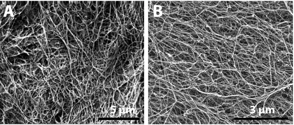
Figure 11 Structural characterization of peptide nanofibers by using SEM A. Representative SEM image of Col-PA/E-PA structure; B. Representative SEM image of K-PA/E-PA structure. ... 37
Figure 12 Structural characterization of peptide nanofibers by using TEM; A. Representative TEM image of Col-PA/E-PA structure; B. Representative TEM image of K-PA/E-PA structure. ... 38
Figure 13 CD spectra of Col-PA peptide amphiphile molecules, Col-PA/E-PA and K-PA/E-PA peptide nanofibers ... 39
Figure 14 Rheological analysis of Col-PA/E-PA and K-PA/E-PA peptide nanofibers ... 39
xv
Figure 15 Viability analyses for rMSCs that were cultured on Col-PA/E-PA, K-PA/E-PA and TCP. Representative fluorescence images of Live/Dead assay on day 1 and quantification of Alamar Blue assay. ... 41
Figure 16 Glycosaminoglycan deposition analysis by Safranin-O staining of rMSCs cultured on peptide nanofiber systems in chondrogenic media (scale bars= 100 µm) ... 43
Figure 17 Chondrogenic differentiation analysis by qRT-PCR. Expression level of Sox9 was quantified on day 7 and day 14 (**p<0.01, ***p<0.001 by one-way ANOVA with Tukey post-test, mean ± s.e.m) ... 45
Figure 18 Measurement of secreted NGF concentration of isolated IVD cells that were cultured on peptide nanofiber systems by ELISA. ... 47
Figure 19 Morphology tracking of isolated IVD cells that were cultured on Col-PA/E-PA, K-PA/E-PA and TCP under light microscope. ... 49
Figure 20 Representative images of in vivo IVD Tissue Defect in the Rabbit Needle Puncture Model ... 50
Figure 21 Glycosaminoglycan deposition analyses of Delayed Degeneration Groups; Representative images of Safranin-O/Fast Green/ Hematoxylin staining and Alcian Blue staining (scale bars = 1 mm) ... 52
xvi
Figure 22 Glycosaminoglycan deposition analyses of Immediate Degeneration Groups; Representative images of Safranin-O/Fast Green/ Hematoxylin staining and Alcian Blue staining (scale bars = 1 mm) ... 53
Figure 23 Representative images for hematoxylin&eosin staining of delayed degeneration groups ... 54
Figure 24 Representative images for hematoxylin&eosin of immediate degeneration groups ... 55
Figure 25 Analyses of degenerative changes in the NP and AF in Delayed Degeneration Groups; A. Schematic representation of the scoring method; B. Grading score for delayed degeneration in the NP, as analyzed by hematoxylin and eosin staining; C. Grading score of delayed degeneration in the AF, as analyzed by Safranin-O staining (5 and 0 are maximum and minimum degeneration scores, respectively) (**p<0.01 by Student’s t-test, mean ± s.e.m.) ... 56
Figure 26 Analyses of degenerative changes in the NP and AF in Immediate Degeneration Groups; A. Grading score for immediate degeneration in the NP, as analyzed by hematoxylin and eosin staining; B. Grading score of immediate degeneration in the AF, as analyzed by Safranin-O (5 and 0 are maximum and minimum degeneration scores, respectively) (*p<0.05 by Student’s t-test, mean ± s.e.m.) ... 58
Figure 27 Collagen deposition analysis of Delayed Degeneration by Immunohistochemistry against collagen II antibody (scale bars = 1 mm). ... 59
xvii
Figure 28 Collagen deposition analyses of Delayed Degeneration; Representative images of picro serius red staining and Masson’s trichrome staining under light microscope (scale bars = 1 mm). ... 60
Figure 29 Collagen deposition analyses of Immediate Degeneration; Representative images of picro serius red staining and Masson’s trichrome staining under light microscope (scale bars = 1 mm). ... 61
xviii
LIST OF TABLES
Table 1 Types of collagen and glycoprotein in the ECM of IVD tissue…………... 5
Table 2 Main molecules involves in IVD degeneration. Reused from Baptista et al. (2015) with the permission of Coluna/Columna [36]……… 9
Table 3 Experimental Plan & Animal Groups……….. 27
xix
ABBREVIATIONS
ANOVA Analysis of variance
AF Annulus Fibrosus
CD Circular dichroism
DCM Dichloromethane
DIEA N,N-diisopropylethylamine
DMEM Dulbecco's modified Eagle's medium
DMF N,N-Dimethylformamide
ECM Extracellular matrix
ELISA Enzyme-linked ImmunoSorbent Assay
FBS Fetal bovine serum
Fmoc 9-Fluorenylmethoxycarbonyl
GAG Glycosamionglycan
GAPDH Glyceraldehyde 3-phosphate dehydrogenase HBTU N,N,N′,N′-Tetramethyl-O-(1H-benzotriazole-1-yl)
uronium hexafluorophosphate
HPLC High pressure liquid chromatography
IVD Intervertebral disc
IVDD Intervertebral disc degeneration
LC-MS Liquid chromatography-mass spectroscopy
xx
NP Nucleus Pulposus
MSC Mesechymal Stem Cell
PA Peptide amphiphile
PBS Phosphate buffered saline
PG Proteoglycan
qRT-PCR Quantitative real-time polymerase chain reaction
SD Standard deviation
SEM Scanning electron microscopy s.e.m. Standard error of the mean TCP Tissue culture plate
TEM Transmission electron microscopy TFA Trifluoroacetic acid
TIS Triisopropyl silane
1
Development of Bioactive Peptide Nanofibers for Intervertebral Disc Regeneration
This work is partially described in the following publication:
Ozge Uysala,b, Elif Arslana,c, Gulcihan Gulserena,c, Mustafa Cemil Kılıncd, Ihsan Dogand, Yusuf Sukru Caglard, *, Mustafa O. Gulere, *, Ayse B. Tekinaya, b, c, * (Manuscript in preparation)
1. Introduction
1.1. Lower Back Pain and Socio-Economic Impact
Many people experience lower back pain (LBP) in a particular period of their lives [1]. Especially today, LBP and the neck problems are the most common orthopedic disorders throughout the world because of the need to spend long hours sitting at the desk in the modern life working conditions. Most people experience this disease when they become older due to the aging [2-4]. In general, LBP and neck problems affect about 632 million people worldwide [5]. The cost of treatment of these diseases are around $86 billion in United States, around £12 billion in United Kingdom and €16.5– 50 billion in Germany; which is higher than many other diseases [6-10].
Intervertebral disc (IVD) is found between two vertebrae and degeneration of IVD is the main reason behind the LBP. IVDs play important mechanical role for the body; since they are constantly exposed to mechanical loading of the whole body. Due to
2
their cartilage-like structure, they provide flexibility and protection to the spinal column.
1.2. Anatomy of IVD
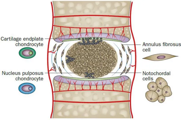
As a tissue, IVD comprises of three main structures: Nucleus Pulposus (NP), Annulus Fibrosus (AF) and cartilaginous endplates (Figure 1).
Figure 1 Anatomical structure of intervertebral disc. Reused from Raj et al. 2008 with the permission of John Wiley and Sons Publisher [11].
NP occupies the inner part of the tissue and it comprises randomly organized collagen fibers (especially collagen type-II) and radially arranged elastin fibers which are embedded in hydrated aggrecan-rich gelatinous structure. Chondrocyte-like cells are
3
interspersed in this gelatinous aggrecan-rich structure and sometimes they are embedded into a capsule within this matrix [12, 13].
NP is surrounded by AF which consists of highly oriented collagen type-I forming lamellae structures [14]. However, the inner region of AF also has a proteoglycan rich microenvironment which is responsible for the resistance to compression. There are up to 25 concentric lamellae structures within this region. The morphology of the cells which are found in the outer region of AF resembles more to fibroblasts. They are more elongated and aligned parallel to the collagen lamellae. However, the cells found in the inner region of AF have more rounded morphologies, as shown in Figure 2 [11, 15].
Figure 2 Illustration showing morphologies and locations of the IVD cells. Adapted from Sakai et al. (2015) with the permission of Nature Publishing Group [15].
4
The third structure of the IVD is called cartilage endplates which lie between vertebral body and disc. They are composed of hyaline cartilage and osseous components with less than 1 mm thickness. It contains collagen fibers in parallel with the vertebral bodies [16, 17].
1.3. Extracellular Matrix (ECM) of Intervertebral Disc
Both NP and AF cells are derived from mesenchymal stem cells (MSCs), however they have different morphologies and produce different ECM components. In addition, the compositon of ECM changes with age and degeneration [18].
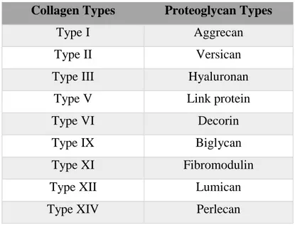
Water content is very high in the nucleus pulposus depending on the proteoglycan and collagen content of NP, because proteoglycans provide swelling pressure to increase the water holding capacity of the tissue and collagen is required to resist this swelling pressure. As it is shown in Table 1, there are several collagen and proteoglycan types in the IVD tissue. Among these collagen molecules, types I, II, III, V, IX, XI, XII and XIV contribute to the formation of fibrillar and fibril-associated structures; whereas collagen types VI and X contribute to the formation of pericellular structures in the IVD.
5
Table 1 Types of collagens and glycoproteins in the ECM of IVD tissue
Collagen Types Proteoglycan Types
Type I Aggrecan
Type II Versican
Type III Hyaluronan
Type V Link protein
Type VI Decorin
Type IX Biglycan
Type XI Fibromodulin
Type XII Lumican
Type XIV Perlecan
The collagens that are found in the IVD have different properties compared to the other tissues. Especially type II collagen has more hydroxylated proline and lysine compared to the other tissues suggesting an increase in the resistance for collagenase digestion [19]. However, during the degeneration process, collagen molecules lose this resistance and are digested by collagenase. At the later stages of the degeneration, this digestion eventually results in weakening of the mechanical strength of the tissue.
In addition to the various collagen types in the IVD tissue, there are also a variety of proteoglycan molecules as shown in Table 1. Aggrecan is the one that is found as the most abundant in the tissue and in mature IVD structure, there are both chondroitin sulfate and keratan sulfate chains that are bound on aggrecan molecule [20, 21]. With age, chondroitin sulfate chain length decreases; whereas length of keratan sulfate chains increases. This change in the composition of glycosaminoglycan chains may be the result of the disrupted oxygen supply into the inner parts of the tissue because oxidation is required for the chondroitin sulfate synthesis, but there is no need for the
6
oxidation for the keratan sulfate synthesis. Another type of proteoglycan found in IVD is versican which has the ability to bind to hyaluronan. It is found throughout the disc, but it is more abundant in the nucleus [22]. In the IVD, there are also various leucine-rich proteoglycans, such as decorin and biglycan, which can interact with the collagen fibers. In addition, fibromodulin and lumican, which are keratan sulfate proteoglycans, can also interact with the collagen fibers [23].
In addition to collagen and proteoglycan content of the IVD, the matrix proteins are also found in other connective tissues. One of the most abundant matrix proteins is fibronectin which has a role in the cell-matrix interactions. The other matrix protein is elastin which provides resistance for stretches [24]. Elastin is found in the elastin fibers that lies parallel to the collagen fibers.
1.4. IVD Degeneration and Aging
During aging, ECM of disc changes. This could be caused by senescence of MSCs, loss of notochondral cells, decrease in vascularization, calcification of cartilage endplates, and decrease in water content [25]. In a healthy disc, anabolism and catabolism rates are balanced but with aging and degeneration, degradation of ECM molecules predominates resulting in the loss of biochemical structure and biomechanical function of the tissue [26]. Because of its avascular structure, maintaining proper nutrient and oxygen supply and elimination of metabolite products can be challenging, especially for the inner parts of the disc. The only parts that blood vessels can reach through the vertebral bodies to the disc are cartilage endplates. From there, nutrients and oxygen are carried by diffusion to the inner parts in which concentration of nutrients and oxygen are at their lowest [27]. However, to keep the cells alive and active, a threshold shoud be exceeded. Therefore, if there are some
7
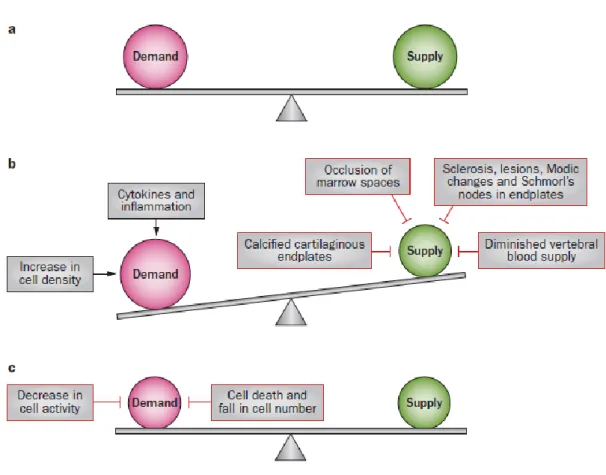
factors which negatively influence the balance between cellular demand and the supply; degeneration in the disc is observed as depicted in Figure 3.
Figure 3 Schematic representation of the factors that influence the balance between nutrient supply and demand. a. The balance between demand and supply in the normal disc. b. Imbalance between demand and supply in degenerating disc, demands exceed supply. c. Establishment of new balance in degenerated disc. Reused from Huang et al. (2014) with the permission of Nature Publishing Group [28].
When nutrient and oxygen supply is disrupted in the disc, as in the case of degeneration, it causes a reduction in progenitor and stem cell production, a decrease in matrix molecules and also an increase in cell death [29-31].
8
1.4.1 Changes in the ECM of Degenerated IVD
It is known that ECM components of the degenerated disc decrease due to their degradation. In addition to the modified synthesis of collagen type I and II, aggrecan synthesis decreases in the NP. This reduction in collagen and aggrecan concentrations can extend to the AF of the tissue, leading to reduction in IVD space [32]. Because of these biochemical changes, biomechanical imbalance between NP, AF and endplates may occur and this may lead to an increase in load onto the spine during motion and cause pain [33]. Synthesis of collagen and proteoglycans also change in the degenerative state. In the first stages of degeneration, synthesis of collagen increases, probably as a repair mechanism; whereas production of aggrecan and glycosaminoglycans decreases. All these changes in ECM composition cause decrease in IVD hydration [2, 34].
This remodeling of ECM components are controlled by the enzymes called matrix metalloproteinases (MMPs) and a disintegrin and metalloproteinase with thrombospondin (ADAMTs). These enzymes can break down every type of proteins in the ECM and their action can be inhibited by endogenous inhibitors called tissue inhibitor matrix metalloproteinase (TIMPs). The level of enzymes and their inhibitors change according to the healthy or diseased state of the tissue as shown in Table 2 [35].
9
10
1.4.2 Role of Inflammatory Cytokines and Growth Factors in the Degeneration Process
IVD cells can synthesize several proinflammatory cytokines such as 1β, 6, IL-8, IL-10 and TNF-α as briefly shown in Table 2. These cytokines promote ECM degradation, change in cell phenotype in the degenerated IVD, apoptosis or senescence of the cells and chemokine production [37, 38]. Also this chemokine production causes the accumulation of immune cells into the degenerated area and further proinflammatory response is generated. TNF-α and IL-1 are the most studied cytokines for the IVD degeneration.
In the previous studies, it has been reported that TNF-α has a role in the disc herniation and nerve ingrowth [39, 40] . Expression levels of both TNF-α and IL-1 is observed to elevate in the degenerated IVD compared to the normal discs. The study performed by Le Maitre et al. showed that mRNA level of IL-1 receptor type I (IL-1R1) was increased in the degenerated disc; whereas there was no increase in the expression level of TNF receptor 1 (TNF-R1) suggesting that the role of IL-1 in the IVD degeneration might be more prominent than TNF-α [41].
1.4.3 Changes in Vascularization and Innervation in Degeneration Process Atherosclerosis of the blood vessels that supply nutrients and oxygen to the disc causes disc degeneration [42, 43]. In addition, this situation is also observed in other diseases which affect the blood circulation [44]. Blood supply can also be blocked in the cartilage endplates because of the increase in calcification of these structures [45, 46]. Besides calcification, sclerosis, lesions and Modic changes in the endplates also cause the disruption of blood supply through the endplates shown in Figure 4 [28, 47].
11
Figure 4 Schematic representation of nutrient pathways; a. in the normal intervertebral disc; b. in the degenerated intervertebral disc. Adapted from Huang et al. (2014) with the permission of Nature Publishing Group [28].
Besides vascularization, innervation of the disc also changes during the degeneration process (Figure 5). Normal IVDs are the largest aneuronal tissues in the body but still they may contain some nerves in the outer collagen lamellae [11]. However, cartilage endplates are mostly aneuronal tissues as in the case of other cartilage tissues. The innervation process regulated by growth factors and this may be interrupted in the disease condition which may cause inappropriate neurite extensions into some aneuronal tissues. Normally, extracellular components of healthy IVD tissue inhibit
12
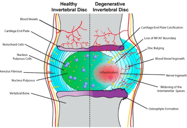
the ingrowth of nerves [48]. However, in the degenerated discs, nerves can penetrate into AF or sometimes even further into NP regions which may lead enhanced pain in the lower back [49-51]. In the study performed by Stefanakis et al., it has been demonstrated that lower swelling pressure of the degenerated disc makes it favorable for both vascularization and innervation through the inner parts of the tissue, whereas swelling pressure of the healthy disc prevents them [52]. Furthermore, neurogenic factors are generated by IVD cells and the immune cells that accumulate into the IVD during degeneration. Due to the increase in the concentration of neurogenic factors, pain-associated cation channels are generated in the dorsal root ganglion and it is likely that activation of these channels promote dicogenic pain in the degenerated IVD [53].
13
Figure 5 Schematic representation of the processes observed in healthy disc vs. degenerated disc. Reused from McCann et al. (2016) with the permission from PMC [26].
1.5. Treatment Options for IVD Degeneration
The treatment options for the IVD degeneration are mostly invasive surgical treatments such as total disc excition, arthrodesis, intradiscal steroids, chemonucleolysis, intradiscal decompression and annuloplasty [54]. Currently the least invasive surgery option is lumbar discectomy. In this operation, prolapsed disc region can be removed by using a camera-attached endoscope [11].
These surgical options mostly focus on IVD fusions to eliminate the pain but they do not regenerate the tissue function. Therefore, regenerative medicine techniques should be more preferred treatment options for the IVD degeneration. For this purpose, the delivery of therapeutic genes to produce growth factors or direct injection of growth
14
factors have great potential as treatment strategies [55, 56]. However, in the late-stage of the disease, IVD cells may not respond to the growth factor injection therapy because they may enter apoptosis or senescence [57]. In previous studies, specific progenitor cells and stem cells in the IVD have been identified. Some of these cells are able to renew themselves and differentiate into specific IVD cells [58-60]. Activating these progenitor cells and stem cells could be a very effective treatment strategy to induce regeneration of the tissue. However, in this technique there are also some limitations because with aging and in the case of degeneration, cells can be exhausted [61]. Therefore, lately, cell transplantation techniques have been also intensified. For this purpose, MSCs could be a very promising choice; since they can differentiate into specific IVD cells and also they can induce endogenous IVD cell migration to the degenerated region [15]. In cell transplantation techniques, cell leakage from the injected site can be observed and this leakage may cause osteophyte formation in the tissue [62]. Thus a scaffold for both cell and growth factor delivery is a necessity for more effective and promising treatments. Since NP is a highly hydrated tissue, injectable hydrogel systems could be more suitable to maintain cellular viability and functionality and to provide mechanical support during the transplantation process [63].
Besides cell transplantation applications, biomaterial applications for the IVD regeneration have been attempted to generate biocompatible and non-immunogenic biomaterials as an IVD substitute. For this purpose, injectable crosslinked hydrogel-based systems have been widely investigated. In the study performed by Miles et al. an injectable peptide:GAG hydrogel system which is capable of rapid in situ self-assembly was used for IVD regeneration. Peptide:GAG composition was injected into
15
the extracted NP from a bovine tail to test its mechanical properties and they showed that this peptide system, which has a β-sheet forming unit and hydrogen bonding capacity, can be combined with chondroitin sulphate (GAG) chain and this combination could restore biomechanical properties of IVD ex vivo. In addition, it could prevent GAG leakage from the injection site of the tissue [64]. However, this system needs to be tested in in vivo conditions, since there will be more mechanical loading onto the tissue and this may cause different problems during regeneration process.
Since one of the most important components of the natural ECM of the IVD is collagen, there are several studies that have been trying to increase and restore the collagen composition in the degenerated IVD tissue. For this purpose, Sato et al. [65] have shown that NP cells grown on atelocollagen scaffolds can be implanted to stimulate recovery in a rabbit IVD degeneration model. However, atelocollagen fibers are only soluble under acidic conditions, which is not consistent with the normal physiological environment of the cells. In another study, Alini et al. [66] used a collagen-I/hyaluronan scaffold to induce proliferation and glycosaminoglycan (GAG) synthesis in NP cells, but also noted that most of the synthesized GAGs were released to the culture medium rather than being retained in the scaffold.
Limitations associated with collagen-based scaffolds can be circumvented through the use of peptide nanofibers, which are highly hydrated and resemble the natural ECM of tissues. Peptide nanofiber scaffolds are one of the most common materials in tissue engineering and can provide a biocompatible, non-immunogenic and aqueous environment for the cells of the NP, which has a very high water content in its healthy state [11, 67]. These scaffolds are formed under physiological conditions through
16
noncovalent interactions between primary amino acid sequences of individual peptide molecules [68]. In addition, peptide amphiphiles can be functionalized with the addition of bioactive sequences derived from the natural ECM components of a specific tissue. Owing to this composition, biomimetic peptide nanofibers can trigger specific biological activities such as proliferation, differentiation and migration of the cells [69-71].
1.6. Peptide Amphiphiles for Regenerative Medicine Applications
Self-assembling peptide nanofiber system is one of the major classes of biomaterials for regenerative medicine applications. Their complex architecture and ECM mimicking capability are very crucial for tissue regeneration processes [72]. Besides its ECM mimicking properties, they are biodegradable, have oxygen permeability and have high water storage capacity with their hydrophilic nature. They can be functionalized with a specific bioactive sequence of a natural protein such as IKVAV for generating laminin-mimetic peptide amphiphiles or RGD for mimicking many ECM proteins that have a role in the adhesion of the cells like fibronectin. By using specific bioactive sequences, distinct cell surface receptors can be induced and cellular responses such as adhesion, differentiation, migration and spreading can be achieved [68].
17
Figure 6 Representative image of nanofiber formation through self-assembly of peptide amphiphile molecules. Adapted from Hartgerink et al. (2001) with the permission from The American Association for the Advancement of Science [73].
Peptide amphiphiles can be used for various biomedical applications. The most common applications are bone regeneration and biomineralization [74, 75], cartilage regeneration [76], neural tissue regeneration [77] and angiogenesis [78].
It has been shown that glycosaminoglycan mimetic peptide nanofibers (GAG-PA) have the ability to induce bone regeneration. These nanofibers provide biocompatible environments for the rMSCs and promote their osteogenic differentiation in vitro and are able to enhance bone regeneration in vivo in the tibial bone defect model. With Alkaline phosphatase assay and Alizarin Red staining, the ability of osteogenic differentiation, induction and biomineralization was investigated. Enhanced ostogenic differentiation activity of the cells cultured on GAG-PA scaffold was also investigated
18
in in vivo rabbit tibial bone defect model. Histological analyses and Micro-CT analysis showed that GAG-PA treatment supported the bone regeneration by enhancing the biomineralization of the natural ECM [74].
As biomimetic biomaterials, peptide amphiphile molecules have very versatile properties. Overall, they offer very promising improvements in various biomedical applications. However, use of these biomaterials in clinics is very limited since there are many issues which need to be covered in more detail. Solving the mechanisms behind specific cellular changes is very challenging and more defined systems should be improved to understand these mechanisms.
19 2. Objectives
The major cause of disability is LBP and it affects many people globally. LBP is frequently associated with IVD degeneration which may be caused by obesity, lifestyle, lack of physical activity and genetic predisposition. In addition to that, the aging global population is at greater risk from IVD degeneration. During IVD degeneration, change in ECM components, increase in cell death and decrease in stem cell production, calcification of endplates and decrease in water content occur in the tissue. However, the treatment of LBP is symptomatic and mostly entails the reduction of pain. In contrast, inducing the differentiation of MSCs or progenitor cells to NP and AF cells and supporting the ECM production by these cells must be the main strategy for long-term recovery of the tissue. Therefore, the objective of this thesis was to design and synthesize bioactive peptide amphiphile molecules that can induce regeneration of IVD tissue. For this purpose, collagen mimetic peptide molecules were synthesized and tested in an in vivo IVD degeneration model.
20 3. Experimental Section
Materials
9-Fluorenylmethoxycarbonyl (Fmoc) and tert-butoxycarbonyl (Boc) protected amino acids, lauric acid, 4-(20, 40-dimethoxymethyl- aminomethyl)-phenoxyacetamido-norleucyl MBHA resin (Rink amide MBHA resin), Fmoc-Asp(OtBu)-Wang resin, 2-(1H-benzotriazol-1-yl)-1,1,3,3-tetramethyluronium hexafluorophosphate (HBTU) and diisopropylethylamine (DIEA) were purchased from NovaBiochem and ABCR. All other chemicals and materials were obtained from Invitrogen, Fisher, Merck, Alfa Aesar and Sigma-Aldrich.
Synthesis and Purification of Peptide Amphiphiles
Peptide synthesis and characterization was carried out with the standard solid state synthesis procedure. Collagen peptide was constructed on MBHA Rink amide resin at 0.25 mmol scale. Amino acid couplings were done with 2 equivalents of Fmoc-protected amino acid, 1.95 equivalents of HBTU, and 3 equivalents of DIEA for 2 h. Fmoc removal was performed with 20% piperidine/dimethylformamide (DMF) solution for 20 min. Cleavage of the peptides from the resin was carried out with a mixture of TFA: TIS: H2O in the ratio of 95:2.5:2.5 for 3 h. Excess TFA was removed by rotary evaporation. The remaining viscous peptide solution was triturated with ice-cold ether and the resulting white product was freeze-dried.
21 Scanning Electron Microscopy (SEM)
Coverslip surfaces were coated with 10 mM Col-PA, E-PA and K-PA peptide solutions and incubated for 15 min for gel formation. Critical point drying procedure was applied for water removal. FEI Quanta 200 FEG environmental scanning electron microscope was used for the acquisition of SEM images. Coverslips were coated with 3 nm Au−Pd prior to imaging. Image acquisition was performed under 10−15 kV voltage.
Transmission Electron Microscopy (TEM)
A 1% (w/v) Col-PA peptide solution was prepared and mixed with 1% (w/v) E-PA solution and 1% (w/v) K-PA peptide solution was prepared and mixed with again, 1% (w/v) E-PA. The peptide gels were diluted with water and solution was dropped to a carbon-covered copper grid. 2% (w/v) uranyl acetate solution was applied to the grid to visualize the organic peptide fibers. The carbon grid was air-dried following staining. A FEI Tecnai G2 F30 transmission electron microscope was used to image the samples.
Circular Dichroism (CD)
Peptide solutions were dissolved in water. Each peptide sample was prepared by mixing with E-PA and was incubated for 15 min to observe nanofiber formation. The samples were diluted with water to a final concentration of 1 x 10⁻⁴ M. Used parameters were DIT: 4 s; band width: 1.00 nm; data pitch: 0.2 nm; start mode: 1 mm; scanning mode: continuous.
22 Rheological Analyses of Materials
Oscillatory rheology measurement was performed with Anton Paar Physica RM301 for determining the viscoelastic properties of the samples. 25 mm parallel plate configuration was used for the rheometer operating at 25 °C. 125 µL of each sample (Col-PA/E-PA and K-PA/E-PA) was used, and 250 µL of total volume with a final peptide concentration of 1 wt % was carefully loaded on the center of the lower plate and incubated for 15 min before measuring. After equilibration, the upper plate was lowered to a gap distance of 0.5 mm. Storage moduli (G’) and loss moduli (G’’) values were screened from 100 rad/s to 0.1 rad/s of angular frequency, with a 0.5% shear strain.
Cell Culture and Viability Analyses of MSCs
In vitro cell culture experiments were performed by using rMSCs (rat Mesenchymal Stem Cells, Invitrogen) at passage number 8. Cells were cultured with DMEM supplemented with 10% fetal bovine serum (FBS), 1% GlutaMAX and 1% penicillin-streptomycin (Maintenance Media) and incubated in the humidified atmosphere with %5 CO2 at 37 °C. For the differentiation analysis, cells were cultured with chondrogenic differentiation media (Gibco).
For the in vitro experiments, peptide amphiphile molecules were dissolved in ddH2O at a concentration of 1 mM, sonicated for 30 min, and sterilized under UV for 1 h. Then, well plates (96-well plates or 48-well plates) were coated with 1 mM Col-PA and 1 mM E-PA at 1:1 ratio for the Col-PA/E-PA group and 1 mM K-PA and 1 mM E-PA at 1:1 ratio for the K-PA/E-PA group. These peptide-coated plates were dried at room temperature overnight for hydrogel formation. On the next day, rMSCs were
23
seeded at a density of 3000 cells/cm2. Tissue culture plate (TCP) was used as a control group.
Cell viability was analyzed by Alamar Blue and Live/Dead assays on day 3 by quantifying both the absorbance and fluorescence of the reagent using spectrophotometry for the Alamar Blue assay, and imaging the cells by fluorescent microscopy for the Live/Dead assay.
Analysis of GAG Deposition by MSCs
Glycosaminoglycan deposition was assessed by Safranin-O Staining at the end of day 7 and day 14. Briefly, the culture medium was removed and cells were washed with PBS prior to fixation with 4% paraformaldehyde for 15 min at room temperature. After fixation, cells were incubated with 1% BSA in PBS for 1 h. Cells were then stained with 0.1% Safranin-O in 0.1% acetic acid for 5 min at room temperature, cells were washed twice with PBS to remove unspecific binding of the Safranin-O dye. The samples were subsequently observed by light microscope.
Gene Expression Profile Analysis
The expression profile of Sox9 gene was assessed by quantitative real time PCR (qRT-PCR) as a measure of chondrogenic differentiation. RNA from the cells that were cultured on the peptide nanofibers and tissue culture plate were isolated with TRIzol reagent (Invitrogen) according to the manufacturer’s instructions. Purity and yield of the samples were determined with Nanodrop 2000 (Thermo Scientific). A one-step qRT-PCR kit (SuperScript III Platinum SYBR Green) was used to perform both cDNA and mRNA amplification according to the manufacturer’s instructions. In brief, the reaction was performed as follows: 55 °C for 5 min, 95 °C for 5 min, 40 cycles of 95
24
°C for 15 s, 61.5 °C for 30 s and 40 °C for 1 min. After the reaction, melting curve analysis was performed to confirm the product specificity. Expression level of Sox9 was analyzed at days 7 and 14. Gene expression data were normalized to the expression levels of GAPDH in each run as an internal control gene and analyzed by using comparative Ct method.
IVD Cell Isolation from Rats
All experimental procedures involving animals were approved and performed according to the Animal Ethics Committee of Diskapi Yildirim Beyazit Research and Training Hospital. IVD cells were isolated according to the protocol adapted from Lee et al. [79]. Briefly, IVD tissues were dissected from two rats and placed into 1X PBS. Here, pieces of IVD tissues were finely diced with a blade and then incubated with 0.05% collagenase in DMEM+Ham’s F-12 medium supplemented with 15% FBS for the ECM digestion process overnight at 37 °C. After overnight incubation, pipetting was done for further digestion of ECM molecules. Collagenase containing residual IVD tissues were retrieved, and cell solution was filtered by using a cell strainer to remove tissue debris. After filtering, cell solution was centrifuged at 2500 rpm for 5 min. Then, cells were seeded into culture medium at a density of 2.5x10⁴ cells/cm2.
Enzyme-Linked ImmunoSorbent Assay (ELISA) for Nerve Growth Factor (NGF)
To assess the concentration of released NGF from the IVD cells, a well plate was coated with the coating antibody of NGF which was diluted with coating buffer, and then was incubated at +4 °C overnight. After the overnight incubation, wells were washed with washing buffer and blocked with 1X blocking buffer or 0.5% BSA
25
solution for 2 h at room temperature. Then, media that were collected from the isolated IVD cells were added into the wells and kept overnight incubation at +4 °C. After the overnight incubation, excess washing steps were applied. Biotin conjugated detection antibody against NGF was added into the wells and incubated for 2 h at room temperature. Wells were washed and then, streptavidin conjugated HRP solution was added into the wells for 30-45 min of incubation at room temperature. To measure the absorbance, TMB chromogen solution was used and incubated for 15 min. This reaction was stopped with the stop solution and absorbance was measured by using Microplate Reader at 450 nm and 650 nm.
Collagen-Mimetic Peptide Nanofiber Treatment of an In Vivo Rabbit Intervertebral Disc Degeneration Model
Animal studies were performed in the animal facility of Ankara University Faculty of Medicine Ibni Sina Hospital. Protocols for animal care and handling were approved by local and governmental Ethics Committees. 18 healthy male, 2.5-3 kg New Zealand rabbits were used in this study (n=3). To avoid any effects due to hormonal fluctuations of female rabbits, only male rabbits were used in the study. Rabbits were housed in standard cages at a temperature- and light-controlled environment (24 °C and 12/12 hours light/dark cycles). Throughout the experimental period, they were allowed access to food and water ad libitum.
The annulus needle puncture technique was performed through the posterolateral retroperitoneum to create a rabbit animal model of IVD degeneration. For this purpose, following anesthesia with intramuscular injection of ketamine (20 mg/kg) and xylazine (2 mg/kg); animals were positioned in their lateral decubitus with a 20° angle. A
26
longitudinal skin incision was performed to the paraspinal muscles from rib cage to pelvic rim. To reach the intervertebral disc, retroperitoneal adipose tissue and muscle tissue were incised with sharp and blunt dissections between L1 and L5 vertebrae. The L5-L6 IVD was determined according to landmarks of the lumbar interspinous and pelvic rims. Once the two IVD levels of interest (L3-4 and L4-5) were identified, an 18 gauge (18G) needle was inserted into the left anterolateral annulus fibrosus and held in the tissue for 5 s. The tip of the needle was modified through the placement of a titanium clip to control the depth of the degeneration. For one group of the animals, gel injection was performed immediately after the degeneration process; whereas for the other group, the same region was opened after two weeks of the degeneration process and gels were administered by an insulin needle through direct injection of the corresponding scaffolds that are summarized in Table 3. For the in vivo experiments, 10 mM peptide amphiphile solutions were used to form a scaffold. Four weeks after the gel injection, animals were sacrificed and tissue regeneration was examined by histopathological examinations of IVD tissue.
27 Table 3 Experimental Plan & Animal Groups
Experimental Groups
Animal
Amount Experimental Plan
Delayed Degeneration
Col-PA/E-PA 3 Gel injection: 2 weeks after the degeneration
Sacrification of animals: 4 weeks after the gel injection K-PA/E-PA 3
Saline 3
Delayed Degeneration
Col-PA/E-PA 3 Gel injection: Immediately after the degeneration
Sacrification of animals: 4 weeks after the gel injection K-PA/E-PA 3
Saline 3
Healthy Control
Healthy IVD tissues of operated animals were used as healthy tissue control
Histological and Immunohistochemical Staining of Tissue Sections
IVD tissue samples were fixed in 4% paraformaldehyde for 48 h at 4 °C and decalcified in 5% formic acid. Formic acid solution was changed every 2 days and this process was continued for 10-14 days. Decalcification was ended according to the result of the ammonium oxalate test. Decalcified samples were dehydrated by immersing them in a series of ethanol solutions of increasing concentrations and then cleared in 2 changes of xylene. Samples were embedded in paraffin blocks and sectioned at 5-7 µm thicknesses by microtome. For the histological and immunohistological analyses, tissue sections were deparaffinized in 2 changes of
28
xylene and then rehydrated by immersing them in a series of ethanol solutions of decreased concentrations. Defect healing was monitored by the staining of representative slides taken at 20-section (~100 µm) intervals to determine the extent of IVD regeneration through the left anterolateral part of the tissue to the right.
Safranin-O and Alcian Blue staining protocols were performed for imaging GAG content of the IVD tissues. Tissues were stained with Safranin-O for 1 min and Fast Green for 5 min. For Alcian Blue Staining, tissues were stained with Alcian Blue for 30 min and counterstained with nuclear Fast Red Solution for 5 min. Masson’s trichrome (MTC) and picro sirius red staining experiments were performed to determine the collagen content of the regenerated tissues. For Masson’s trichrome staining, slides were stained with hematoxylin for 45 s, Biebrich scarlet for 5 min, phosphomolibdic acid/phosphotungistic acid mixture for 5 min, and aniline blue for 5 min. For picro sirius red staining, tissues were stained with Weigert’s Hematoxylin for 8 min and then Picro Sirius Red for 1 h to visualize the collagen fiber orientations. After the staining procedures, slides were dehydrated in a series of ethanol solutions of increasing concentrations and cleared with xylene before mounting with Histomount® mounting medium and imaging under light microscope.
For the antigen retrieval process of immunohistochemical staining against Collagen II and β-III tubulin proteins, slides were treated with pepsin at 37 °C for 10 min and then, to block peroxidase activity, slides were incubated in 0.3% H2O2 solution for 45 min at room temperature. After the blocking with 10% Normal Goat Serum (NGS) in 1% BSA/TBS, slides were incubated with primary antibody against collagen type II (MA-13026) and β-III tubulin (ab78078) at 4 °C overnight. After the washing steps with
29
0.01% Triton-X in TBS, slides were incubated with HRP-conjugated goat anti-mouse IgG antibody for 1 h at room temperature, and incubated with DAB solution for 20 min to visualize the bound antibodies. Slides were then counterstained with hematoxylin for 45 s to visualize cell nuclei.
Histological Scoring Method
The histopathological assessment of IVD degeneration was performed to determine the extent of the regeneration process after collagen-mimetic peptide nanofiber treatment. For this purpose, degenerative changes in both NP and AF in sagittal sections of the disc were graded using the grading scale which is described by Nagae et al. [80] and it is briefly described in Table 4.
30 Table 4 IVD Regeneration Scoring Method
Parameter Grade Description
NP Degeneration
0 normal structure (no degeneration)
1 no proliferative connective tissue with honeycomb appearance
2 1-24% of NP occupied by proliferative connective tissue
3 25-50% of NP occupied by proliferative connective tissue
4 more than 50% of NP occupied by proliferative connective tissue
5 complete replacement by proliferative connective tissue
AF Degeneration
0 normal structure (no degeneration)
1 mild serpentine appearance in AF (with rupture)
2 moderate serpentine appearance in AF (with rupture)
3 severe serpentine appearance in AF (with mild reverse contour)
4 with severe reverse contour
31
All grading was performed by observers who were blinded to the animal treatments. Scoring for NP degeneration was performed using H&E staining images to better observe cellular content; while scoring for AF degeneration was performed using Safranin-O staining images to observe the general structure of the tissue.
Statistical Analysis
All data are represented as mean ± s.e.m. (standard error of mean). The significance of differences between the groups was determined by using Student’s t-test and one-way ANOVA with Tukey’s post-test. Differences were considered as significant at p<0.05.
32 4. Results
4.1 Synthesis and Characteization of Peptide Nanofiber Systems
The collagen-mimetic short peptide amphiphile was designed from tandem repeats of Pro-Hyp-Gly artifical collagen sequence [81]. Pro-Hyp-Gly monomeric unit was incorporated on self-assembling short peptide sequence to create a collagenous monomer-bearing surface. Interaction among monomeric units on peptide fiber surfaces was used to create de novo collagen mimetic structures as promising candidate for wide range of applications. Collagen monomer unit-mediated microenvironment is promising for regeneration studies, especially chondrogenic differentiation. The monomeric collagen sequence on peptide fibers leads to multifibrillar nanostructures and induces chondrogenic differentiation by using physical and biochemical cues. This new method offers simple preparation and effective regeneration without the need for external stimuli, therefore this study is promising for future in vivo cartilage regeneration studies.
Monomer sequence containing collagen-mimetic peptide amphiphile (Col-PA) was synthesized by standard solid phase Fmoc peptide synthesis chemistry, by conjugating a peptide amphiphile with a collagen-mimetic monomer unit derived from tandem repeat of Pro-Hyp-Gly sequence. Lauryl-VVAGK-Am was utilized as the self-assembly segment whereas ionic lysine is included in the sequence to improve ionic interactions. Pro-Hyp-Gly (POG) was tagged as the specific amino acid sequence. This very unique sequence leads the triple-helix formation through its interlocking structure and hydrogen bonding. This overall sequence of the PA molecule (Lauryl-VVAGKPOG-Am) was designed to obtain a self-assembling structure through
33
hydrophobic interactions, and hydrogen bonds. E-PA (Lauryl-VVAGE) was used for assembly initiation as a counter ion with the same β-sheet forming segment.
Collagen-mimetic peptide amphiphile (Col-PA) molecules were designed by the incorporation of the active domain of the natural collagen molecule to a β-sheet forming backbone. The chemical structures of Col-PA, the oppositely charged E-PA and the control K-PA are shown in Figure 7.
All PA molecules were synthesized by solid-phase peptide synthesis method and purified by HPLC. Prior to use, their purities and molecular weights were confirmed by LC-MS analysis (Figure 8& 9&10).
34
Figure 7 Chemical structures of peptide amphiphile molecules that were used in this study.
35
Figure 8 Liquid chromatogram and mass spectra of Col-PA. [M+H]+ (calculated) = 922.00, [M+H]+ (observed) = 922.7732
Figure 9 Liquid chromatogram and mass spectra of E-PA. [M-H]- (calculated) = 654.82, [M-H]- (observed) = 654.4181
Figure 10 Liquid chromatogram and mass spectra of K-PA. [M+H]+ (calculated) = 654.88, [M+H]+ (observed) = 654.5002
36
Col-PA was mixed with the negatively charged E-PA to allow nanofiber formation through charge neutralization. Two peptide amphiphile molecules that lack bioactive sequences in their chemical structures, the positively charged K-PA and negatively charged E-PA, were also mixed in equal volumes to produce a control nanofiber. The structural resemblance of the PA networks to the morphology of the natural ECM was assessed using SEM (Figure 11). In addition, the thickness and lengths of individual nanofibers were observed using TEM (Figure 12). Porous structures resembling the natural fibrous collagen and glycoprotein networks could be observed in both peptide nanofiber groups.
37
Figure 11 Structural characterization of peptide nanofibers by using SEM A. Representative SEM image of Col-PA/E-PA structure; B. Representative SEM image of K-PA/E-PA structure.
38
Figure 12 Structural characterization of peptide nanofibers by using TEM; A. Representative TEM image of Col-PA/E-PA structure; B. Representative TEM image of K-PA/E-PA structure.
Secondary structures of peptide nanofibers were also characterized using CD spectroscopy. According to this analysis, the β-sheet structure of the peptide nanofibers were confirmed with specific 200 nm negative and 220 nm positive peaks (Figure 13). Mechanical properties of the peptide nanofiber networks were investigated by oscillatory rheology (Figure 14). Both nanofiber networks have higher storage modulus values than loss modulus values, showing that they have formed stable hydrogel structures. Both nanofiber systems also had similar mechanical properties, suggesting that any difference in biological activity of the cells was not caused by the mechanical features of the peptide nanofiber scaffolds.
39
Figure 13 CD spectra of Col-PA peptide amphiphile molecules, Col-PA/E-PA and K-PA/E-PA peptide nanofibers
40
4.2 Peptide Nanofiber System Provides a Biocompatible Environment for rMSCs
Before their application for in vivo experiments, in vitro investigation of peptide nanofiber systems is necessary to test their biocompatibilities. Therefore, viabilities of MSCs cultured on the peptide nanofiber systems were first evaluated both by Live/Dead assay and Alamar Blue assay on varying day points (day 1, day 2 and day 3) (Figure 15). According to these results, peptide nanofiber systems provided a suitable environment for cellular attachment and growth. Although cells on peptide nanofiber scaffold systems had lower viability rates than the TCP control during the first two days of culture, they adapted to the environment on day 3. Both peptide nanofiber scaffolds showed no toxic effects, since there were no dead cells in the Live/Dead assay analysis. Therefore, the reason behind the lower viability may be related with the decreased number of proliferating cells, which is a major indicator of differentiation [82]. Furthermore, both peptide nanofiber groups had comparable viability results with each other on day 1. This showed that both systems had similar biological activity with respect to the viability of the cells in the initial period of culture.
41
Figure 15 Viability analyses for rMSCs that were cultured on Col-PA/E-PA, K-PA/E-PA and TCP. Representative fluorescence images of Live/Dead assay on day 1 and quantification of Alamar Blue assay.
4.3 Collagen-mimetic Peptide Nanofibers Induce Chondrogenic Differentiation of MSCs In Vitro
After the biocompatibility analyses, we investigated the chondrogenic differentiation potential of MSCs on collagen-mimetic peptide nanofibers, since cells of the IVD are morphologically and metabolically very similar to chondrocytes [11]. It is known that MSCs can differentiate into multiple lineages such as osteocytes, chondrocytes and
42
adipocytes with specific signals derived from their environment or their interactions with other cells [83, 84]. When cells commit into chondrogenic lineage, they form cell aggregates by increasing their interactions and deposit glycosaminoglycans (GAG) [85, 86]. To investigate the GAG composition aggregates, we performed Safranin-O staining. We observed more positive staining in cells grown on the Col-PA/E-PA nanofiber system than the other groups (Figure 16).
43
Figure 16 Glycosaminoglycan deposition analysis by Safranin-O staining of rMSCs cultured on peptide nanofiber systems in chondrogenic media (scale bars= 100 µm)
44
Morphological changes of MSCs and Safranin-O staining results were also confirmed by the qRT-PCR analysis of Sox9, which is one of the most important chondrogenic differentiation markers [87]. Sox9 plays a crucial role in chondrogenic differentiation, as Sox9-null cells cannot express important chondrogenic marker genes such as aggrecan, collagen type I and II [86]. On day 14, cells cultured on collagen-mimetic peptide nanofibers showed increased expression level of Sox9 by 2.7-fold and 5.5-fold compared with K-PA/E-PA and TCP groups, respectively. Cells on TCP also exhibited a high level of Sox9 on day 7, which may be a result of the heterogeneity of mesenchymal stem cells; however, Col-PA/E-PA groups still had a higher gene expression level (Figure 17). On day 14, the expression level of Sox9 had its highest value with significant differences compared to the other groups (Figure 17). This increase in the expression level of Sox9 is an indicator of chondrogenic commitment by MSCs cultured on the Col-PA/E-PA nanofiber system.
45
Figure 17 Chondrogenic differentiation analysis by qRT-PCR. Expression level of Sox9 was quantified on day 7 and day 14 (**p<0.01, ***p<0.001 by one-way ANOVA with Tukey post-test, mean ± s.e.m)
46
4.4 Evaluation of NGF Release from the IVD Cells Treated with Collagen-mimetic Peptide Nanofibers
During the IVD degeneration, cells secrete cytokines into the environment causing proinflammatory response and immune cell recruitment to the degenerated region. This proinflammatory response causes increase in the concentration of some growth factors such as VEGF (vascular endothelial growth factor) and NGF (nerve growth factor) [36]. With their increase, vascularization and innervation was triggered in the degenerated tissue. Therefore, we measured secreted NGF concentrations in the media of isolated IVD cells cultured on the peptide nanofiber systems. The concentration levels were compared among the peptide nanofiber treated groups.
47
Figure 18 Measurement of secreted NGF concentration of isolated IVD cells that were cultured on peptide nanofiber systems by ELISA.
We couldn’t see any significant difference between Col-PA/E-PA and control nanofiber. However, peptide nanofiber systems are effective to decrease NGF concentration which is secreted by the isolated IVD cells (Figure 18).
48
4.5 Morphology Tracking of Isolated IVD Cells Cultured on Peptide Nanofiber Systems
Isolated IVD cells were cultured on peptide nanofiber scaffolds and TCP in the maintenance medium for 5 days and their morphologies were observed under light microscope. According to these results, the cells cultured on Col-PA/E-PA peptide nanofiber systems haved more rounded morphologies compared to the other groups. This morphological change suggests that Col-PA/E-PA peptide nanofibers induced chondro/osteogenic differentiation (Figure 19).
![Figure 1 Anatomical structure of intervertebral disc. Reused from Raj et al. 2008 with the permission of John Wiley and Sons Publisher [11]](https://thumb-eu.123doks.com/thumbv2/9libnet/5670628.113540/22.892.258.704.429.806/figure-anatomical-structure-intervertebral-reused-permission-wiley-publisher.webp)


![Figure 9 Liquid chromatogram and mass spectra of E-PA. [M-H] - (calculated) = 654.82, [M-H] - (observed) = 654.4181](https://thumb-eu.123doks.com/thumbv2/9libnet/5670628.113540/55.892.172.786.472.625/figure-liquid-chromatogram-and-mass-spectra-calculated-observed.webp)