Clinical research
Corresponding author: Assoc. Prof. Cenk Conkbayir MD
Department of Cardiology Near East University Yakın Doğu Bulvarı Nicosia, Cyprus
E-mail: cenkconk@hotmail. com
1Department of Anaesthesiology, Yuksek Ihtisas Hospital, Ankara, Turkey 2Department of Cardiology, Near East University, Nicosia (north), Cyprus
3 Department of Cardiovascular Surgery, Bagcilar Education and Research Hospital, Istanbul, Turkey
4Department of Cardiovascular Surgery, Medipol University Hospital, Istanbul, Turkey 5Department of Anaesthesia, Medipol University Hospital, Istanbul, Turkey
Submitted: 5 November 2019 Accepted: 2 January 2020
Arch Med Sci Atheroscler Dis 2020; 5: e20–e26 DOI: https://doi.org/10.5114/amsad.2020.93528 Copyright © 2020 Termedia & Banach
Comparison of fentanil and remifentanil for coronary
artery surgery with low ejection fraction
Nukhet Baddal1, Cenk Conkbayir2, Ozcan Erdemli1, Umit Karadeniz1, Busra Tezcan1,
Didem Melis Oztas3, Metin Onur Beyaz4, Murat Ugurlucan4, Yahya Yildiz5, Soner Yavas1
A b s t r a c t
Introduction: In this study, we evaluated patient response and haemody-namic parameters in patients with low ejection fraction undergoing coro-nary bypass surgery with either fentanil or remifentanil in conjunction with etomidate.
Material and methods: We evaluated 30 cases of coronary artery surgery, which were divided into two treatment groups (n = 15 each). In group F (fen-tanil group), the following regimen was employed for anaesthesia induction: 1 mg/kg lidocaine, 0.3 mg/kg etomidate, and, following a 1 µg/kg 60 s bolus dose of fentanil, a 0.1 µg/kg/min fentanil infusion was initiated, after which 0.6 mg/kg rocuronium was administered. In group R (remifentanil group), the following regimen was employed for anaesthesia induction: 1 mg/kg lidocaine, 0.3 mg/kg etomidate and, following a 1 µg/kg 60 s bolus dose of remifentanil, a 0.1 µg/kg/min remifentanil infusion was initiated, after which 0.6 mg/kg rocuronium was administered. Systolic artery pressure, di-astolic artery pressure, mean arterial pressure, heart rate, SPO2 (saturation),
cardiac output, stroke volume variance, central venous pressure, and sys-temic vascular resistance values were recorded for all study patients at five minutes before anaesthetic induction (T1), immediately following induction (T2), and immediately following intubation (T3).
Results: The demographic values obtained for both groups were similar. We found that remifentanil use was associated with decreased cardiac output and increased fluctuations in both heart rate and mean values of arterial pressure.
Conclusions: Although many studies have demonstrated remifentanil to be as safe as fentanil when titrated to an appropriate dose, our study suggests that fentanil may be a more appropriate choice during the induction of an-aesthesia in patients with a low ejection fraction.
Key words: low ejection fraction, coronary artery bypass surgery, anaesthesia.
Introduction
In high-risk surgical cases in which haemodynamic parameters change rapidly, such as in cardiopulmonary bypass, a central objective
when inducing anaesthesia is to maintain cardiac output and oxygen delivery at appropriate levels.
In general anaesthesia, airway control is pro-vided via laryngoscopy and endotracheal intuba-tion [1]; the receptors in the airway are stimulated by both mechanical and chemical factors during these procedures. Stimulation of these tissues
leads to catecholamine discharge via sympathetic
adrenergic activation, resulting in increased ar-terial pressure, heart rate, and arrhythmia [1–6]. This response may aggravate existing pathologies in patients with coronary heart disease, cerebro-vascular disease, and/or hypertension, which can lead to life-threatening complications [1, 2, 4, 7].
Current methods used to minimise the nega-tive haemodynamic response caused by laryngos-copy and endotracheal intubation include blocking the activation of sensitive receptors and afferent nerves with local anaesthetic agents, inhibiting the central effects of painful stimuli with opioids, and suppressing efferent pathways and effector receptors with local anaesthetics, b-blockers, cal-cium channel blockers, and sympathetic ganglion blockers [8].
In this study we aimed to evaluate patient re-sponses, the factors effecting those rere-sponses, and changes in haemodynamic parameters fol-lowing anaesthetic induction and intubation, us-ing either fentanil or remifentanil in conjunction with etomidate to induce anaesthesia, in coronary bypass surgery patients with an ejection fraction lower than 50%.
Material and methods
Our study was conducted on patients under-going coronary bypass surgery, for which we ob-tained local Ethics Committee approval. We eval-uated 30 patients scheduled to undergo coronary artery surgery, each with an American Society of Anaesthesiologists (ASA) risk classification of II–III. Patients were provided with a volunteer consent form prior to inclusion in the study. Demograph-ic data, ASA, and the presence of any additional diseases were all recorded in the study protocol. Patients with an ejection fraction of less than 20% or more than 50%, those requiring emergency
surgery, and those with sensitivities to the drugs used in the study were excluded. All patients were
premedicated with a 10 mg Diazem tablet one night before the operation and an intramuscular injection of 0.1 mg/kg morphine sulphate 30 min before the operation. While in the operating room, patients were subjected to ECG and monitored for levels of peripheral oxygen saturation (SpO2) and invasive arterial blood pressure (Drager Primus Anaesthesia Monitor, Lübeck, Germany).
For invasive arterial blood pressure monitoring, a 20 G (gauge) arterial cannula (BD, Faraday Road,
Swindon, UK) was used on the radial artery. For fluid infusion, a peripheral venous pathway was created with 18 G and 16 G branulas, through which 5–10 ml/kg/h isotonic sodium chloride was administered. A jugular venous catheter was in-serted while the patient was conscious for the pur-pose of recording CVP. A Flotrac/Vigileo (Edward) monitor was used to measure each patient’s car-diac output via SVV (Stroke Volume Variance) and systemic vascular resistance. BIS monitoring (As-pect Medical Systems Inc., Natick, MA, USA) was used to determine each patient’s hypnotic status and depth of anaesthesia. BIS values ≤ 50 repre-sented a sufficient depth of anaesthesia.
Patients were divided into two groups (n = 15 each). Pre-oxygenation was provided by the appli-cation of 100% O2.
The anaesthesia induction protocol for group F (fentanil group) included the following: 1 mg/kg li-docaine (Aritmal® 2%), 0.3 mg/kg etomidate
(Eto-midate®), 1 µg/kg 60 s bolus dose fentanil
(Fentan-il®) followed by a 0.1 µg/kg/min fentanil infusion,
0.6 mg/kg rocuronium (Esmeron®). The
anaesthe-sia induction protocol for group R (remifentanil group) included the following: 1 mg/kg lidocaine (Aritmal® 2%), 0.3 mg/kg etomidate (Etomidate®),
1 µg/kg 60 s bolus dose of remifentanil (Ultiva®)
followed by a 0.1 µg/kg/min remifentanil infusion, 0.6 mg/kg rocuronium (Esmeron®).
In both groups, etomidate was administered 5 min after opioid infusion via titration at a rate of 20 mg/min until the BIS value decreased to 50. Finally, rocuronium was administered.
During intubation, the degree of chin relax-ation, ease of laryngoscopy, vocal cord status, and any presence of movement in the extremities were evaluated for each patient.
Systolic artery pressure (SAP), diastolic artery pressure (DAP), mean arterial pressure (MAP), heart rate (HR), SPO2 (saturation), cardiac output (CO), stroke volume variance (SVV), central venous pressure (CVP), and systemic vascular resistance (SVR) values of all patients were recorded at the following time points: T1 – 5 min before the
intu-bation, T2 – immediately following induction, T3
– immediately following intubation. Statistical analysis
SPSS 13.00 for Windows version package soft-ware (SPSS Inc., Chicago, IL, USA) was used for statistical analyses. For analysis of continuous variables, conformity of normal distribution and homogeneity were tested with the Kolmogor-ov-Smirnov test. Categorical values were evaluat-ed with the c2 test, and parametric values were
evaluated with the independent samples t-test. Differences in consecutive measurements be-tween the two groups were evaluated with
repeat-ed measures of ANOVA. Findings are presentrepeat-ed as either mean values ± standard deviation (SD) or as percentages. P-values < 0.05 were accepted as statistically significant.
Results
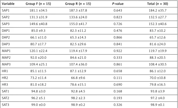
Between the pre-induction (T1), post-induction (T2), and post-intubation (T3) periods, no signifi-cant differences were found between groups for the demographic values of SAP, DAP, MAP, HR, or
SAT (p > 0.005) (Table I).
There were no significant differences found when comparing intergroup CVP values between T1 and T2, T1 and T3, or T2 and T3 for either the fentanil or remifentanil group (p > 0.05) (Table II). However, statistically significant differences were found when the same values were compared be-tween the two groups in all pre-induction (T1), post-induction (T2), and post-intubation (T3) pe-riods (p < 0.05) (Figure 1).
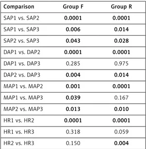
Statistically significant differences were found when intergroup MAP values were compared for both the fentanil and remifentanil groups between both T1 and T2, and T2 and T3 values (p < 0.05) (Table III). Additionally, while a significant differ-ence was found between T1 and T3 in the fentanil group (p < 0.05), no such difference was found in the remifentanil group over the same time points (p > 0.05) (Figure 2).
In both groups, there were significant differenc-es found for intergroup HRs between T1 and T2
(p < 0.05). However, no significant difference was observed for these groups between T1 and T3 (p > 0.05). While there was no significant difference observed in the fentanil group between T2 and T3 (p > 0.05), there was a significant difference ob-served in the remifentanil group (p < 0.05) (Table III, Figure 3).
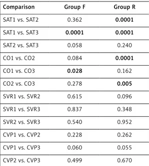
No significant difference was observed for CO values within the fentanil group between T1 and T2 (p > 0.05), although a significant difference was ob-served within the remifentanil group over the same time points (p < 0.05). While there was a significant difference observed in the fentanil group between T1 and T3 (p < 0.05), no such difference was ob-served in the remifentanil group (p > 0.05). Addi-tionally, there was no significant difference observed between T2 and T3 in the fentanil group (p > 0.05); however, a significant difference was observed in the remifentanil group (p < 0.05) (Table IV, Figure 4).
When CO values were compared between the two groups, there was a statistically significant difference found in the post-induction (T2) peri-od (p < 0.05). However, there were no statistically significant differences found for the pre-induction (T1) and post-intubation (T3) periods (p > 0.005) (Figure 4).
When CVR values were compared between the two groups, there were no statistically significant differences found in either the pre-induction (T1), post-induction (T2), or post-intubation (T3) peri-ods (p > 0.005) (Figure 5).
Table I. Comparison of systolic arterial pressure, diastolic arterial pressure, mean arterial pressure, heart rate, and arterial oxygen saturation between fentanil and remifentanil groups during pre-induction, post-induction, and post-intubation periods
Variable Group F (n = 15) Group R (n = 15) P-value Total (n = 30)
SAP1 181.1 ±34.5 187.3 ±37.8 0.643 184.2 ±35.7 SAP2 131.3 ±31.9 133.6 ±24.0 0.823 132.5 ±27.7 SAP3 149.6 ±40.8 155.0 ±41.7 0.726 152.3 ±40.6 DAP1 85.0 ±9.3 82.3 ±11.2 0.476 83.7 ±10.2 DAP2 66.1 ±11.0 65.3 ±14.3 0.866 65.7 ±12.6 DAP3 80.7 ±17.7 82.5 ±29.6 0.841 81.6 ±24.0 MAP1 120.1 ±22.4 119.4 ±17.9 0.922 119.7 ±19.9 MAP2 92.0 ±20.0 84.6 ±21.0 0.333 88.3 ±20.5 MAP3 109.4 ±25.1 107.4 ±36.0 0.861 108.4 ±30.5 HR1 85.1 ±11.5 87.1 ±12.9 0.658 86.1 ±12.0 HR2 73.2 ±11.4 66.8 ±9.6 0.111 70.0 ±10.8 HR3 81.0 ±18.2 78.6 ±15.1 0.690 79.8 ±16.5 SAT1 94.8 ±3.0 92.8 ±4.5 0.168 93.8 ±3.9 SAT2 96.2 ±5.1 98.2 ±2.3 0.193 97.2 ±4.0 SAT3 99.0 ±0.0 98.9 ±0.2 0.326 98.9 ±0.1
SAP – systolic arterial pressure (mm Hg), DAP – diastolic arterial pressure (mm Hg), MAP – mean arterial pressure (mm Hg), HR – heart rate (BPM), SAT – arterial oxygen saturation (%), 1 – pre-induction, 2 – post-induction, 3 – post-intubation, F – fentanil, R – remifentanil.
Measurements of the degree of chin relaxation, ease of laryngoscopy, vocal cord status, the pres-ence of cough, and the prespres-ence of movement in extremities during intubation were evaluated for each patient. In all patients, chin relaxation was
complete, laryngoscopy was easy, and vocal cords were completely immobile. Neither cough nor limb movements were observed in any of the patients during surgery.
Discussion
The pharmaceutical agents used for the induc-tion of anaesthesia commonly lead to decreases in blood pressure, while laryngoscopy and intuba-tion increase haemodynamic parameters.
A central goal during cardiac surgery is the maintenance of adequate tissue perfusion in the patient. A widely used indicator to monitor this adequacy, particularly in patients who are not haemodynamically stable, is the measurement of Table II. Comparison of cardiac output, systemic vascular resistance and central venous pressure between fentanil and remifentanil groups during pre-induction, post-induction, and post-intubation periods
Parameter Group F (n = 15) Group R (n = 15) P-value Total (n = 30)
CO1 6.8 ±1.8 7.4 ±2.2 0.359 7.1 ±2.0 CO2 6.2 ±2.0 3.4 ±0.9 0.0001 4.8 ±2.1 CO3 5.6 ±1.8 6.1 ±3.2 0.611 5.9 ±2.5 SVR1 1236.1 ±408.9 1281.7 ±448.2 0.773 1258.9 ±422.2 SVR2 1274.0 ±255.0 1433.2 ±406.1 0.209 1353.6 ±342.8 SVR3 1251.2 ±249.7 1424.4 ±613.5 0.320 1337.8 ±468.6 CVP1 9.9 ±3.6 7.3 ±1.1 0.014 8.6 ±2.9 CVP2 10.5 ±3.6 7.6 ±1.1 0.006 9.0 ±3.0 CVP3 10.6 ±3.7 7.6 ±1.1 0.007 9.1 ±3.1
CO – cardiac output (l/dk), SVR – systemic vascular resistance (dyne*s/cm5), CVP – central venous pressure (mm Hg), 1 – pre-induction,
2 – post-induction, 3 – post-intubation, F – fentanil, R – remifentanil.
Figure 1. Comparison of central venous pressure be-tween fentanil and remifentanil groups during pre-in-duction, post-inpre-in-duction, and post-intubation periods
Central venous pressure [mm Hg]
16 14 12 10 8 6 4 2 0 CVP1 p = 0.014 CVP2 p = 0.006 CVP3 p = 0.007 Group F Group R
Table III. Comparison of intergroup parametric values
Comparison Group F Group R
SAP1 vs. SAP2 0.0001 0.0001 SAP1 vs. SAP3 0.006 0.014 SAP2 vs. SAP3 0.043 0.028 DAP1 vs. DAP2 0.0001 0.0001 DAP1 vs. DAP3 0.285 0.975 DAP2 vs. DAP3 0.004 0.014 MAP1 vs. MAP2 0.001 0.0001 MAP1 vs. MAP3 0.039 0.167 MAP2 vs. MAP3 0.013 0.010 HR1 vs. HR2 0.0001 0.0001 HR1 vs. HR3 0.318 0.059 HR2 vs. HR3 0.150 0.004
SAP – systolic arterial pressure (mm Hg), DAP – diastolic arterial pressure (mm Hg), MAP – mean arterial pressure (mm Hg), HR – heart rate (BPM), 1 – pre-induction, 2 – post-induction, 3 – post-intubation, F – fentanil, R – remifentanil.
Figure 2. Comparison of mean arterial pressure be-tween fentanil and remifentanil groups during pre-in-duction, post-inpre-in-duction, and post-intubation periods
£Intergroup MAP comparison; MAB1(T1) vs. MAB2(T2)
p < 0.05.
αIntergroup MAP comparison; MAB2(T2) vs. MAB3(T3)
p < 0.05.
≈Intergroup MAP comparison; MAB1(T1) vs. MAB3(T3)
p < 0.05.
Mean arterial pressure [mm Hg]
160 140 120 100 80 60 40 20 0
MAP1 p = 0.922 MAP2 p = 0.333 MAP3 p = 0.861 Group F Group R
α
£ ≈
α £
cardiac output. In the past, measuring cardiac out-put involved insertion of a pulmonary artery cath-eter prior to surgery; however, cardiac output can now be measured using the more practical and less invasive Flotrack/Vigileo system. Breukers
et al. [9] found that measuring the rate of change
in cardiac output obtained over consecutive time periods is a more effective method for determin-ing patient treatment protocols than relydetermin-ing on static cardiac output values. Furthermore, mea-surements of this rate of change should be sim-ilar whether they are obtained using the FloTrac/ Vigileo system or by the thermo dilution method because a good correlation has been found be-tween these two techniques. Additionally, increas-es in vascular tonus, as obtained from measuring cardiac output values using FloTrac/Vigileo equip-ment, are reflected by small increases in these values; thus, the measurement algorithm retains adequate sensitivity to detect dynamics for this haemodynamic parameter.
Cengiz et al. [10] previously examined patient responses to various remifentanil doses following a 2.5 mg/kg intravenous bolus of propofol. Patients in this study were divided into three groups: group I was administered 0.5 µg/kg remifentanil, group II was administered 1 µg/kg remifentanil, and group III was administered 2 µg/kg remifentanil. While
heart rate measurements following intubation were significantly higher than pre-induction val-ues in groups I and II, in group III post-induction heart rates were found to be significantly less than those measured during pre-induction. Over-all, as the remifentanil dose was increased, a con-comitant decrease in patient heart rates was ob-served. In the current study, when a 1 µg/kg dose of remifentanil was administered, post-intubation heart rates were found to be significantly higher than post-induction heart rates, and no significant difference was found when comparing these val-ues to pre-induction heart rates.
Güneş et al. [11]compared the effects of remifentanil and fentanil administration in con-junction with desflurane on intracranial inter-Figure 3. Comparison of heart rate between
fent-anil and remifentfent-anil groups during pre-induction, post-induction, and post-intubation periods
H
eart rate [rate/min]
120 100 80 60 40 20 0 HR1 p = 0.658 HR2 p = 0.111 HR3 p = 0.690 Group F Group R
Table IV. Comparison of intergroup arterial oxygen saturation, cardiac output, systemic vascular resis-tance, and central venous pressure values
Comparison Group F Group R
SAT1 vs. SAT2 0.362 0.0001 SAT1 vs. SAT3 0.0001 0.0001 SAT2 vs. SAT3 0.058 0.240 CO1 vs. CO2 0.084 0.0001 CO1 vs. CO3 0.028 0.162 CO2 vs. CO3 0.278 0.005 SVR1 vs. SVR2 0.615 0.096 SVR1 vs. SVR3 0.837 0.348 SVR2 vs. SVR3 0.540 0.952 CVP1 vs. CVP2 0.228 0.262 CVP1 vs. CVP3 0.060 0.055 CVP2 vs. CVP3 0.499 0.670
SAT – arterial oxygen saturation, CO – cardiac output (l/dk), SVR – systemic vascular resistance (dyne*s)/cm5), CVP – central
venous pressure (mm Hg), 1 – pre-induction, 2 – post-induction, 3 – post-intubation, F – fentanil, R – remifentanil.
Figure 4. Comparison of cardiac output between fentanil and remifentanil groups during pre-induc-tion, post-inducpre-induc-tion, and post-intubation periods
Cardiac output [l/dk] 12 10 8 6 4 2 0
CO1 p = 0.359 CO2 p = 0.0001 CO3 p = 0.611 Group F Group R
Figure 5. Comparison of systemic vascular resis-tance (SVR) between fentanil and remifentanil groups during pre-induction, post-induction, and post-intubation periods
Systemic vascular resistance [dyn × s/cm
5] 2500 2000 1500 1000 500 0 SVR1 p = 0.773 SVR2 p = 0.209 SVR3 p = 0.320 Group F Group R
vention patients with regard to intubation, skin incision, haemodynamic changes following imple-mentation of the head holder, and eye opening and response time to verbal commands during extubation at the end of the surgery. While in-creased haemodynamic values were detected in the fentanil group during intubation, skin inci-sion, and head holder implementation, a more stable haemodynamic profile was observed in the remifentanil group (p < 0.05). In our study, in both the fentanil and remifentanil groups, SAP, DAP, MAP, and HR values decreased during induction and increased during intubation, but there were no significant differences detected between the two groups for these values (p > 0.05). Fluctua-tions in MAP and HR values were more severe in the remifentanil group; however, the significant decrease in cardiac output values measured for the remifentanil group may be related to the low ejection fraction values of our selected patient group. In patients with normal ejection fractions, Kazmaier et al. [12] found that a high dose of remifentanil significantly decreased heart rate, mean arterial pressure, myocardial blood flow, and systemic vascular resistance values.
Gezer et al. [13] compared patients’ cardiac
sta-bility in response to the administration of either remifentanil, alfentanil, or fentanil during anaes-thetic induction. Patients were divided into three groups: Group I intravenously received 1 µg/kg remifentanil, group II intravenously received 15 µg/kg alfentanil, and group III intravenously received 2 µg/kg fentanil. Following this, all three groups received 2 mg/kg propofol and 0.6 mg/kg rocuronium. Statistically significant decreases were observed in HR values in groups I and II when comparing time points taken immediately prior to laryngoscopy versus during pre-induction. When intergroup HR values were compared, the HR creases found in groups II and III at 1 min after in-tubation were found to be statistically significant. However, this study utilised a higher single dose of remifentanil than we used cumulatively over the entirety of the surgical procedure. This may explain the lack of significant difference found be-tween heart rate values in our study. Furthermore, administering etomidate during the induction of anaesthesia (as in our study) may lead to a more balanced induction than propofol in terms of hae-modynamic stability. In particular, considering that our patients had poor ventricular function, the use of etomidate may be even more effective at preserving cardiac stability than direct compari-son with previous studies might indicate.
Zhang and Sun [14]compared how the use of fentanil, remifentanil, or alfentanil in conjunc-tion with etomidate during anaesthetic induc-tion affected patients’ haemodynamic parame-ters during elective abdominal surgery. A total of
90 ASA I-II patients were divided into three groups: group F patients received 1 µg/kg fent-anil as a 60-second bolus dose, group R patients received 1 µg/kg remifentanil as a 60-second bo-lus dose, and group A patients received 0.1 µg/ kg sufentanil as a 60-second bolus dose. Doses were continued at 0.1, 0.1, and 0.01 µg/kg/min, respectively, as continuous infusions. Blood pres-sure and heart rate values were recorded at five different time points. While endotracheal intuba-tion led to significant increases in blood pressure and heart rate in groups F and S, there was no sig-nificant change observed in group R (p < 0.01), al-though greater haemodynamic changes occurred in group F than in the other groups (p < 0.01). In group R, there was an average heart rate decrease of more than 30% following induction (p < 0.01). Thus, remifentanil was observed to be the most effective choice for suppressing the development of cardiovascular response following endotrache-al intubation. In our study, however, though the use of fentanil and remifentanil led to equivalent post-induction heart rate decreases amongst pa-tients, any increases in heart rate that were mea-sured following intubation were not found to be significant in the fentanil group. However, these measurements were significant in the remifent-anil group, which suggests that remifentremifent-anil may not be as successful at providing haemodynamic stability as the Zhang study suggests.
There is controversy in the literature as to whether fentanil is effective for preventing the hypertension and tachycardia that develop during endotracheal intubation and laryngos-copy. For example, Splinter and Cervenko [15], Chung and Evans [16], and Chung et al. [17] all reported that fentanil is effective at preventing HR and SAP increases when administered at a range of doses (all at 5 µg/kg or less) either immediately prior to or during intubation. How-ever, Kautto [18] found that while 2 µg/kg of fentanil significantly controlled patients’ arterial pressure and heart rate measurements, a 6 µg/kg dose was unable to provide the same effect. In the current study, we administered fentanil at a dose of 1 µg/kg, 5 min prior to intubation. Sim-ilar to what was found in previous studies, we concluded that at this dose fentanil was able to prevent both hypertension and tachycardia from developing during endotracheal intubation and laryngoscopy.
Howie et al. [19] compared the effects of three different remifentanil doses on coronary artery bypass patients with poor left ventricle function. For this study, 72 patients were divided into three groups: group 1 patients (n = 23) received a 1 µg/ kg/min remifentanil infusion, group 2 patients (n = 24) received a 2 µg/kg/min remifentanil
in-fusion, and group 3 patients (n = 25) received a 3 µg/kg/min remifentanil infusion. If a suffi-cient depth of anaesthesia was not obtained us-ing these doses, then additional remifentanil was given in either 1–2 µg/kg bolus doses or infusion increases, and 0.5–1.0% isoflurane was adminis-tered. Remifentanil alone (whether by infusion or bolus administration) prevented haemodynamic reflex responses in 44% of the patients in group 3, 37% in group 2, and 9% in group 1. Hypoten-sion developed in 64–75% of patients. In coronary bypass surgery patients with poor left ventricle function who were premedicated with lorazepam, effective anaesthesia was obtained with a 2–4 µg/ kg/min remifentanil infusion and the administra-tion of occasional low concentraadministra-tion isoflurane. In our study, there were significant decreases in both mean arterial pressure and heart rate values following induction, and these values significant-ly increased after intubation following a 1 µg/kg bolus or a 0.1 µg/kg/min remifentanil infusion. Neither of these dosing strategies prevents hae-modynamic reflex responses.
In conclusion, in this study, we compared the effects on haemodynamics and intubation re-sponses following the use of either fentanil or remifentanil during anaesthetic induction in pa-tients with an ejection fraction of less than 50% while they underwent coronary bypass surgery. Decreased cardiac output levels and increased fluctuations in heart rate and mean arterial pres-sures were observed following the administration of remifentanil. It is important to note that, al-though achieving an appropriate dose titration is an important consideration when using opioid agents for anaesthesia, the proper selection of hypnotic agent to use in conjunction with a given opioid is extremely important as well. Although many studies that have employed remifentanil during the induction of anaesthesia have con-cluded that it is as safe as fentanil when appro-priately titrated to the patient, our study suggests that fentanil may be a more appropriate choice during this period in patients presenting with a low ejection fraction.
Conflict of interest
The authors declare no conflict of interest. R e f e r e n c e s
1. Hamaya Y, Dohi S. Differences in cardiovascular re-sponse to airway stimulation at different sites and blockade of the responses by lidocaine. Anesthesiology 2000; 93: 95-103.
2. Collins VJ. Intravenous anesthesia: narcotic and neu-roleptic agents. In: Principles of Anesthesiology. 3rd edn. Collins VJ (ed.). Lea & Febiger, Philadelphia 1993; 571-75.
3. Derbyshire DR, Chimelewski A, Fell D, Vater M, Achola K, Smith G. Plasma catecholamine responses to tracheal intubation. Br J Anaesth 1993; 55: 855-60.
4. Edwards ND, Alford AM, Dobson PMS, Peacock JE, Reil-ly CS. Myocardial ischaemia during tracheal intubation and extubation. Br J Anaesth 1994; 73: 537-9.
5. Kayhan Z. Pathophysiologic effects and complications of intubation. In: Clinical Anesthesia. 2nd edn. Logos Publications, Istanbul 1997; 239-40.
6. Kaplan JD, Schuster DP. Physiologic consequences of tracheal intubation. Clin Chest Med 1991; 12: 425-32. 7. Millar FA, Dally FG. Acute hypertension during induction
of anesthesia. Br J Anaesth 1970; 42: 618-24.
8. Kayhan Z. Endotracheal intubation. In: Clinical Anesthe-sia. Expanded 3rd ed. Logos Publications, Istanbul 2004; 243-306.
9. Breukers RM, Sepehrkhouy S, Spiegelenberg SR, Groeneveld AB. Cardiac output measured by a new arterial pressure waveform analysis method without calibration compared with thermodilution after cardiac surgery. J Cardiothorac Vasc Anesth 2007; 21: 632-5. 10. Cengiz M, Ganidağlı S, Ölmez G. Endotracheal
intuba-tion without muscular relaxants: comparison of three different remifentanil doses following Propofol induc-tion. J Turk Anesthesiol Reanim Soc 2004; 32: 356-61. 11. Güneş Y, Gündüz M, Ünlügenç H, Özcengiz D,
Balcıoğ-lu O, Işık G. Comparison of desfBalcıoğ-luran-remifentanil with desfluran-fentanil anesthesia in intracranial interven-tions on hemodynamics and recovery. J Turk Anesthesiol Reanim Soc 2003; 31: 37.
12. Kazmaier S, Hanekop GG, Buhre W, Weyland A, Busch T. Myocardial consequences of remifentanil in patients with coronary artery disease. Br J Anaesth 2000; 84: 578-83. 13. Gezer A, Arslan G, Berkel G, Sezen Ö, Gürsu T, Arıkan Z.
Comparison of remifentanil, alfentanil and fentanil for prevention of hemodynamic response develops for la-ryngoscopy and endotracheal intubation. Med J Kartal Training Res Hospital 2002; 1: 16.
14. Zhang GH, Sun L. Peri-intubation hemodynamic chang-es during low dose fentanil, remifentanil and sufentanil combined with etomidate for anesthetic induction. Chin Med J (Engl) 2009; 122: 2330-4.
15. Splinter W, Cervenko F. Haemodynamic responses to laryngoscopy and tracheal intubation in geriatric pa-tients: effects of fentanil, lidocaine and thiopentone. Can J Anaesth 1989; 36: 370-6.
16. Chung F, Evans D. Low-dose fentanil: haemodynamic response during induction and intubation in geriatric patients. Can Anaesth Soc J 1985; 32: 622-8.
17. Chung KS, Sinatra RS, Halevey JD, Paige D, Silverman DG. A comparison of fentanil, esmolol their combination for blunting the hemodynamic responses during rapid se-quence induction. Can J Anaesth 1992; 39: 774-9. 18. Kautto UM. Attenuation of the circulatory response to
laryngoscopy and intubation by fentanil. Acta Anaes-thesiol Scand 1982; 26: 217-21.
19. Howie MB, Michelsen LG, Hug CC Jr, et al. Comparison of three remifentanil dose-finding regimens for coro-nary artery surgery. J Cardiothorac Vasc Anesth 2003; 17: 51-9.