A rare cause of acute abdominal pain in adolescence:
Hydrosalpinx leading to isolated torsion of fallopian tube
Arzu Pampal
a,⁎
, G. Kaan Atac
b, Z. Safinur Nazli
b, I. Onur Ozen
c, Tansu Sipahi
da
Department of Pediatric Surgery, Ufuk University, Faculty of Medicine, Ankara, Turkey
b
Department of Radiology, Ufuk University, Faculty of Medicine, Ankara, Turkey
c
Department of Pediatric Surgery, Gazi University, Faculty of Medicine, Ankara, Turkey
d
Department of Pediatrics, Ufuk University, Faculty of Medicine, Ankara, Turkey Received 23 May 2012; revised 4 July 2012; accepted 23 August 2012
Key words:
Isolated torsion of the fallopian tube; Tubal torsion; Hydrosalpinx; Children
Abstract Torsion of the fallopian tube accompanying hydrosalpinx is a rare occurrence in the pediatric population. This report describes a 13 year old sexually inactive girl with isolated tubal torsion due to hydrosalpinx. The girl had lower left abdominal pain for two days. The physical examination revealed left lower quadrant tenderness with a firm round anterior mass on rectal examination. Abdominal ultrasound showed left tubal enlargement with free pelvic peritoneal fluid. Magnetic Resonance Imaging (MRI) showed engorgement and dilatation of the left fallopian tube without contrast enhancement suspicious of tubal torsion. At operation, torsion of the left tube on its longitudinal axis was observed, and a salpingectomy was performed.
Although rare, the diagnosis of torsion of the fallopian tube should be considered when evaluating acute abdominal pain. The earlier tubal torsion is diagnosed, the greater the likelihood of salvaging the fallopian tube.
© 2012 Elsevier Inc. All rights reserved.
Isolated torsion of the fallopian tube is a rare occurrence observed in women of reproductive age, with an annual prevalence of 1 in 1.5 million [1]. Despite the obscure etiology, several predisposing factors have been identified. These include, intrinsic factors like abnormal tubal peristal-sis, hydro/hematosalpinx, tubal ligation, tubal neoplasms or extrinsic factors like paraovarian/ paratubal masses, uterine enlargement, postoperative or postinfectious adhesions, and hemodynamic alterations causing congestion of the
meso-salpingeal veins [2]. In the pediatric age group, as most of these factors are unlikely to be present, the possible factors are preexisting congenital malformations[3].
This report describes a 13-year-old sexually inactive girl with isolated tubal torsion due to hydrosalpinx.
1. Case Report
A 13-year-old girl was admitted to the emergency room (ER) with pain in her lower left quadrant of two days’ duration. She described nausea and two episodes of vomiting when the pain began. She denied any history of sexual
⁎ Corresponding author. Ufuk Universitesi Tip Fakultesi, Dr Ridvan Ege Hastanesi, Cocuk Cerrahisi Bolumu, Konya Yolu No: 86-88 Balgat 06520, Ankara/Turkey. Tel.: +90 312 2044269; fax: +90 312 2026213.
E-mail address:[email protected](A. Pampal).
www.elsevier.com/locate/jpedsurg
0022-3468/$– see front matter © 2012 Elsevier Inc. All rights reserved.
http://dx.doi.org/10.1016/j.jpedsurg.2012.08.040
activity. She had regular menstrual periods over the pre-vious eight months and was on the 14th day of her 28 day cycle. Her physical examination revealed tenderness in the left lower quadrant with mild guarding. A digital rectal examination revealed a firm round mass in the anterior region of the rectum. Blood and urine tests were within normal limits.
The patient had been seen at another ER because of this pain the previous day. She told that she was examined by both a gynecologist and a pediatric surgeon and was evaluated by an abdominal ultrasound (US) and computed tomography (CT). Her radiological evaluation revealed a 4×6 cm cystic mass free from the left ovary with pelvic free fluid. She was discharged with a prescription for nonste-roidal anti-inflammatory drugs and a scheduled follow-up visit for a presumed ovarian cyst.
Her pain had worsened, and her physical examination revealed tenderness and guarding in the left lower quadrant. She was reevaluated with a US which showed left tubal enlargement with increased free peritoneal fluid around the internal genitalia and bladder. The left fallopian tube was 16 mm in the largest portion at the ampullary tip, undulated, and longer than the right. The uterus and both ovaries were normal in shape, location and size. A Color Doppler US of the left ovary and fallopian tube revealed high resistance arterial blood flow with an increased resistance index (RI) of 0.72 (peak systolic arterial blood flow is 35 cm/s and end-diastolic blood flow is 6 cm/s). Magnetic Resonance Imaging (MRI) of the lower abdomen was performed, with a torso coil in a supine position using a 1.5 T superconductive magnet (GE Signa, General Electric, Milwaukee, USA). The MRI showed similar findings, including engorgement, dilation of the fallopian tube, and increased peritubal fluid. The am-pulla of the left tube was closed and more dilated than the
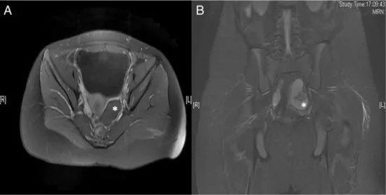
body. Internal signals of the tube were hyperintense in the T1 and T2-weighted images. The STIR and fat-suppression T1-weighted images revealed increased intensity of this fluid, compatible with the bloody content of hydrosalpinx (hematosalpinx). (Fig. 1A, B) The wall of the tube was not enhanced after an intravenous (IV) paramagnetic contrast material injection (Gadovist, Schering, Germany) confirm-ing decreased perfusion of the tissue. As the tubal torsion with hematosalpinx was diagnosed, the patient underwent an emergent exploration. A 10 mm trocar was inserted via umbilicus, and the exploration of the pelvic cavity revealed a dilated and twisted plum-colored salpinx on the left side. Both ovaries and the right salpinx appeared normal. A conversion to laparotomy was planned in order to be sure of the tubal viability. Torsion of the left tube on the longi-tudinal axis was untwisted to its neutral position twice (Fig. 2). But with time, as no change in color was observed, the salpingectomy was performed. The postoperative course was uneventful and the patient was discharged on the second postoperative day.
The pathology of the tube revealed hematosalpinx with hemorrhagic necrosis in all sections of the specimen.
2. Discussion
Hydrosalpinx is a distally blocked and dilated fallopian tube filled with serous or clear fluid, generally seen after pelvic inflammatory disease as a result of ascending infec-tions of C. trachomatis, N. gonorrhoeae, or as the conse-quences of peritoneal drains or postoperative adhesions. It can also be seen as a result of endometriosis, tubal preg-nancies, and tubal cancers that occur during reproductive
Fig. 1 A: Post-contrast T1 weighted axial picture crossing through the bladder reveals the enhancing soft tissue besides the enlarged left ovarian tube (*) indicating decreased perfusion of the tubal wall. Higher internal intensity of the tube comparing to the bladder signify the bloody content of it. B: Coronal T2 weighted MRI image shows enlarged and tortuous left ovarian tube (*) with free peritoneal fluid around internal genitalia.
age. Hydrosalpinx generally results in increased mobility of the fallopian tube on the wide ligamentum latum. In the absence of predisposing ovarian or tubal pathologies, torsion of the hydrosalpinx is generally due to abnormal motility. Moreover, during early puberty the elevated FSH due to the stimulated hypothalamic–pituitary–gonadal axis triggers the ovarian and tubal motility that causes a silent hydrosalpinx to become symptomatic[3,4].
Isolated tubal torsion may occur without any concomitant abnormality, and at that point it is difficult to diagnose without the aid of imaging technologies. Imaging findings usually show the abnormal side and dilatation of the tube. Ultrasound is the primary diagnostic tool for young patients with suspected pregnancy. Free peritoneal fluid and tubal dilatation in addition to triggering pathology may be easily observed. A Color Doppler US may show increased vas-cularity and decreased resistance index (RI) for when infec-tions and tumors are causative. An increased RI and decreased vascularity may not indicate the torsion since arterial blood supply may not be hampered due to the dual vascular supply of the ovary and fallopian tube. Computed Tomography (CT) with or without IV contrast material may show similar findings, but increased density of the fluid should be analyzed for bloody content. In this case, hyperintense signals in fat-suppressed T1-weighted images in the MRI helped confirm the bloody contents of the tube and lack of enhancement of the tubal wall supported the lack of arterial perfusion[4,5].
Isolated torsion of the fallopian tube secondary to hy-drosalpinx is very rare in children. In a recent review by Boukaldi et al., 13 cases of isolated torsion of fallopian tube with hydrosalpinx were presented [3]. There are in fact several more cases in the English literature[1,2,6–11]. The majority of the cases presented with left sided pain, nausea, and vomiting. No specific laboratory findings were noted. Even though the radiological findings were helpful, only a few of the patients underwent surgery with an accurate preoperative diagnosis. Most of the cases underwent sal-pingectomy. There is no further information in the literature about the fertility properties of these patients.
Right-sided tubo-ovarian pathologies are more commonly diagnosed for two reasons. The first is the preventive effect
of anatomic positioning of the left side like cushioning of the sigmoid colon, dextrorotated uterus and less congestion due to relatively increased venous flow of the left fallopian tube. The second reason is that the right-sided pathology is evaluated more often as it is part of the differential diag-nosis of acute appendicitis. Despite these observations, the review by Boukaldi et al., and the other cases in the literature point out the dominance of left-sided torsions. It is a fact that awareness of left-sided tubo-ovarian patho-logies has increased, and they are more often searched. But the interval from the start of pain until surgery is still not short enough to salvage the fallopian tubes. Harmon et al. presented a mean of 1.67 days (between 0 and 5 days) from initial imaging (not the start of pain) till surgery for patients with isolated tubal torsion [11]. All the cases required salpingectomy.
Despite the fact that most of the presented cases underwent salpingectomy, Boukaldi et al. suggest a staged surgical intervention for the torsion of hydrosalpinx. They recommend first untwisting the tube with or without puncturing, and second, performing a salpingoneostomy in another surgical session. These researchers were able to follow 6 cases of hydrosalpinx with torsion. In 3 of the 4 patients with whom they followed this algorithm, they had to excise the same tube due to recurrence of torsion during the follow-up period, with a median interval of 78 days (43–83 days). The unidentified congenital factors that existed after detorsion probably resulted in recurrence of the torsion. Even though tubal preservation is the primary goal, partial salpingectomy is not a viable option, as the left part of the tube is a future candidate for an ectopic preg-nancy or a recurrence of hydrosalpinx [3].
Clinicians should be alert while evaluating left lower quadrant pain, as the clinical findings are generally related to many nonsurgical diagnoses like constipation, urinary tract infection, ureterolithiasis or a ruptured ovarian cyst. Even if tubal torsion is a rare occurrence in pubertal chil-dren experiencing lower quadrant pain, it should be con-sidered in differential diagnosis. Ultrasound and contrast enhanced MRI are helpful at revealing anatomical and functional information for decreased perfusion of tissue. Tubal preservation depends on a low threshold for surgical intervention, and the decision to perform surgery should be made as soon as the suspicion is raised in order to salvage the fallopian tubes.
References
[1] Lima M, Libri M, Aquino A, et al. Bilateral hydrosalpinx with asyn-chronous tubal torsion: an exceptional finding in a premenarcheal girl. J Pediatr Surg 2011;46:e27-9.
[2] Harmon JC, Binkovitz LA, Binkovitz LE. Isolated fallopian tube torsion: sonographic and CT features. Pediatr Radiol 2008;38:175-9. [3] Boukaidi SA, Delotte J, Steyaert H, et al. Thirteen cases of isolated
tubal torsions associated with hydrosalpinx in children and
Fig. 2 Perioperative findings of normal left ovary (*) with fluid filled plum-colored left salpinx with the closed tip of the ampulla.
E33 Hydrosalpinx leading to isolated torsion of fallopian tube
adolescents, proposal for conservative management: retrospective review and literature survey. J Pediatr Surg 2011;46:1425-31. [4] Merlini L, Anooshiravani M, Vunda A, et al. Noninflammatory
fallo-pian tube pathology in children. Pediatr Radiol 2008;38:1330-7. [5] Kim MY, Rha SE, Oh SN, et al. MR imaging findings of hydrosalpinx:
a comprehensive review. Radiographics 2009;29:495-507.
[6] Jalaguier-Coudray A, Thomassin-Naggara I, Chereau E, et al. A case of isolated torsion of the fallopian tube: added value of magnetic resonance imaging. Eur J Radiol Extra 2011;79:e1-4.
[7] Markhardt BK, Jones L, Drugas GT. Isolated torsion of the fallopian tube in a menarchal 11-year-old girl. Pediatr Emerg Care 2008;24: 374-6.
[8] Lineberry TD, Rodriguez H. Isolated torsion of the fallopian tube in an adolescent: a case report. J Pediatr Adolesc Gynecol 2000;13: 135-7.
[9] Lim WH, Roex AJ. Laparoscopic management of a fallopian tubal torsion complicated by a large hydrosalpinx. Int J Womens Health 2011;3:381-4.
[10] Thomas JM. Torsion of hydrosalpinx at 14 years of age. Br Med J 1954;2:1271-2.
[11] Kopec M, Rosenkrantz AB, Rivera R, et al. Case report: MRI ap-pearance of isolated fallopian tube torsion in an adolescent with a congenital Müllerian duct anomaly and ispilateral renal agenesis. Clin Radiol 2010;65:89-93.