The effect of Nd:YAG laser irradiation on root surface: shear-bond
strength and SEM
–EDX study
Melek Akmana* , Kezban Celikb, Betül Ozçopurcand Arslan Terlemeza aFaculty of Dentistry, Department of Endodontics, University of Necmettin Erbakan, Konya,
Turkey;bFaculty of Dentistry, Department of Endodontics, University of Medipol, Istanbul,
Turkey;cPrivate Clinician, Antalya, Turkey
(Received 29 November 2014;final version received 8 March 2015; accepted 2 April 2015) To investigate the effects of Nd:YAG laser and 17% EDTA treatment on root-dentin mineral content using scanning electron microscope–energy dispersive X-ray spec-troscopy (SEM–EDX) and on shear-bond strength of epoxy-resin-based sealer (AH Plus) to root dentin. Twelve extracted premolars were decoronated and roots were sectioned, so that 24 two-root halves were obtained. Element levels of each half were examined by SEM/EDX, and AH Plus build-ups were created. After shear-testing, the test surfaces were reground and subjected to a 5.25% NaOCl. Two subgroups were created according to the surface treatment (n = 12): G1, with 17% EDTA for 5 min; G2, with the Nd:YAG laser. The element level analysis and shear-bond strength test were repeated for each half; the data were recorded (MPa) and analyzed (paired samples t-test). The EDTA treatment increased the O, C, Ca/P ratio (p < 0.001), decreased Ca, P content (p = 0.000), but did not change Na, Mg content (p > 0.05). The Nd:YAG laser increased O, Ca/P ratio (p < 0.001), and decreased the Ca, P content (p = 0.000). The C, Mg, Na content did not change with the Nd:YAG laser (p > 0.05). Both 17% EDTA and Nd:YAG laser had an effect on the mineral content of roots. The 17% EDTA treatment decreased the shear-bond strength of AH Plus to root dentin (p = 0.000); however, the Nd:YAG laser did not affect the bond strength (p = 0.238). Thus, an Nd:YAG laser can be used for disinfection of the root canal when AH Plus is used as a sealer.
Keywords: Nd:YAG laser; EDTA; SEM/EDX spectroscopy; mineral content; shear-bond strength
1. Introduction
The success of root canal therapy depends on the technique and the quality of instru-mentation, irrigation, disinfection, and three-dimensional obturation of the root canal system.[1] The adhesion of a root canal sealer may be affected by many factors, among these being the presence of a smear layer.[2] Removal of the smear layer created during the root canal treatment has been recommended because of its low resistance against the leakage between dentin and the filling material.[3] Different irrigant solutions have been used to remove the smear layer, especially because of their antibacterial effects and their ability to dissolve necrotic tissues. Although NaOCl is the most commonly used endodontic irrigant, an effective method for removing the organic and inorganic remnants is to irrigate the root canal with EDTA, followed by NaOCl.[4] However,
*Corresponding author. Email:[email protected]
© 2015 Taylor & Francis
their effects on dentin are also important. Researchers have reported that some chemical agents cause significant alterations in the mineral content of root dentin.[5,6] In particu-lar, chelating agents may affect the physical and mechanical properties of dentin. The alterations in the Ca/P ratio may change the original proportion of organic and inor-ganic components, which in turn can change the permeability and solubility characteris-tics of the dentin.[6,7] As dentin adhesion partially depends on the presence of residual Ca2+on the bonding surface area,[8,9] alterations in the Ca/P ratio may affect the adhe-sion of dental materials to root dentin.[6,8]
Recently developed techniques involving laser applications, including vaporization of tissue, removal of smear layer, and melting, have gained popularity in endodontic treatment.[10] In particular, Nd:YAG and Er:YAG laser systems have been widely used. Researchers have demonstrated that these lasers are effective in smear layer and debris removal, antibacterial activity, and sealer adhesion.[11] Camorgo et al. [12] has reported that Nd:YAG laser irradiation applied within the root canal with circular move-ments parallel to the surface of the tooth produces limited morphologic changes in the dentin wall of the root canal. Gürbüz et al. [13] has recommended using a Nd:YAG laser, because of its cleaning efficacy and low tendency to cause mineral loss from the root-canal dentin. Several studies have been performed in which the mineral content of root dentin after various surface treatments has been measured. However, to our knowl-edge, SEM/EDX spectroscopy was not used in any of these studies. The purposes of the current study were:
• to investigate the effects of Nd:YAG laser and EDTA treatments on root-dentin mineral content using SEM/EDX analysis,
• to evaluate the comparative effects of EDTA and a Nd:YAG laser on the shear-bond strength between an epoxy-resin-based sealer, AH Plus (Dentsply, De Trey, Konstanz, Germany), and root dentin.
The null hypotheses of this study were that Nd:YAG laser and EDTA treatments do not affect the mineral content of root dentin and do not change the shear-bond strength of AH Plus.
2. Materials and methods
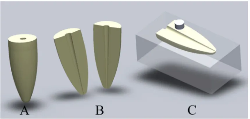
Twelve, extracted, human, single-rooted, premolar teeth were selected. The soft-tissue covering the surfaces was scaled using a hand scaler (Gracey Curetta SG 17/18; Hu-Friedy, Chicago, IL, USA). The crowns of all teeth were removed at the cement-enamel junction with a high-speed diamond saw (Isomet, Buehler, Lake Bluff, IL, USA) under water cooling. The roots were then longitudinally sectioned using a diamond saw under water cooling to obtain two root halves from each tooth (Figure 1(B)). Each half was embedded horizontally in a self-curing polymethyl-methacrylate resin (Vertex, Vertex Dental, Zeist, Netherlands) with the root surfaces exposed. In this way, 24 samples were obtained.
2.1. SEM/EDX analysis
SEM/EDX spectroscopic analyses were done at Selcuk University, Advanced Technology Research and Application Center, Konya, Turkey. Before the SEM/EDX examination, specimens were dried at 50 °C in an incubator (EN 120 Incubator, Nüve,
Ankara, Turkey) for 24 h. After sputter coating the specimens with gold, the mineral content of each specimen was measured using a scanning electron microscope (Evo LS10, Carl Zeiss, Oberkochen, Germany) operating at 20 kV, between the 8 and 11 mm working distance, and at 50 Pa.
The samples were numbered from 1 to 24 and the mineral content of each sample was evaluated. A surface analysis was performed during this procedure, with the coro-nal third of the roots chosen for examination, as this is the most widened part of the root and therefore provides an adequate surface for the shear-bond strength test. The distribution of the elements, calcium (Ca), oxygen (O), phosphorus (P), carbon (C), magnesium (Mg), and sodium (Na), as well as the Ca/P levels, were examined for each sample and recorded in tables and graphs (EHT: 20.00 kV).
2.2. Shear-bond strength test
Three 3 mm-high build-ups, each with a constant surface area of 3.45 mm2, were cre-ated in the coronal third of the roots using an epoxy-resin-based sealer (AH Plus, Dentsply, De Trey, Konstanz, Germany), and were prepared according to the manufac-turer’s instructions. Polyethylene tubes were used to create the build-ups (Figure 1(C)), and were allowed to set at 37 °C and 100% humidity for 72 h. The samples were retained in a universal testing machine (Instron, Norwood, MA) and subjected to shear stress at a crosshead speed of 1 mm/min. A custom-made constructed attachment was used to enable easy and accuratefixation. This custom fabrication allowed the chisel to travel parallel to the root dentin surface and contact the sealer cylinder at its interface with the root dentin. The maximum load (in newton (N)) for specimen debonding was recorded and divided by the contact surface area to determine the shear-bond strength, in megapascal (MPa). After testing, the fracture modes were examined under a stere-omicroscope with 22× magnification (SZ-PT, Olympus, Japan).
2.3. Irrigation regimen
The root surfaces were reground with 800 grit emery paper after the SEM–EDX analy-sis, and then subjected to a 5.25% NaOCl solution for 5 min. Two subgroups were cre-ated and then trecre-ated with the irrigation solution or laser irradiation as follows:
G1: 17% EDTA for 5 min (n = 12);
G2: Nd:YAG laser at the standard settings (power output = 1.5 W; energy = 100 mJ; pulse frequency = 10 pps for 10 s, and light frequency = 15 Hz with 200 µfiber tip) (n = 12).
The root surfaces were then re-examined by SEM/EDX spectroscopy and the ele-ment levels were recorded. AH Plus build-ups were recreated on the treated surfaces following the same procedures described above, allowed to set (37 °C and 100% humidity for 72 h) and tested to failure for shear-bond strength.
2.4. Statistical analysis
The initial SEM/EDX analysis for each sample was assumed as the control of that sam-ple. The mineral content of the root surfaces and shear-bond strength study results before and after the irrigation regimen was statistically analyzed using a paired-t test with a significance set at p < 0.05.
3. Results
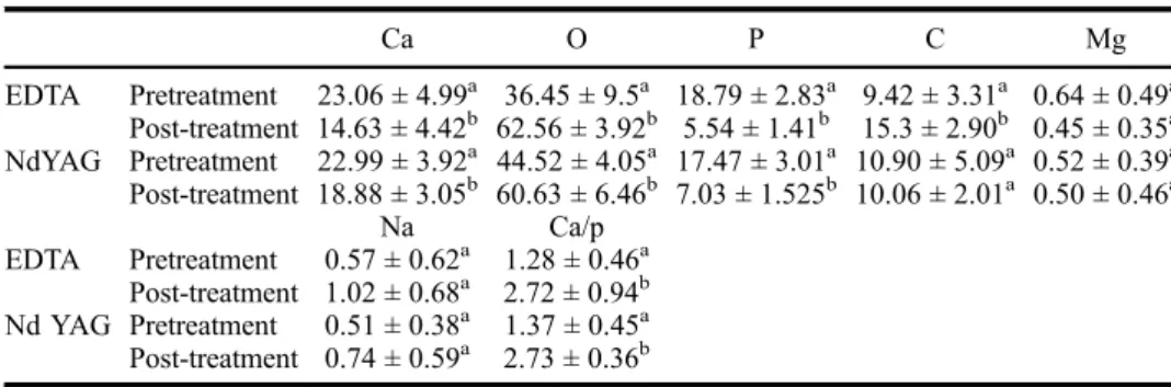
Table1 presents the mineral content of the experimental groups. EDTA irrigation treat-ment affected the mineral content of root dentin. As seen in Table 1, quantities of O, C, and the Ca/P ratio increased after EDTA treatment, and statistical analysis showed that this increase was significant (p < 0.001). Ca and P levels decreased with EDTA treatment (p = 0.000). SEM/EDX analysis also showed that 17% EDTA application for 5 min did not affect Na and Mg content (p > 0.05).
When root surfaces were treated with the Nd:YAG laser, O levels and Ca/P ratios increased significantly (p < 0.001), while element levels of Ca and P decreased (p = 0.000). As with the 17% EDTA treatment, Mg and Na content of root dentin sam-ples did not change with the Nd:YAG laser (p > 0.05). Additionally, no change was observed in C content of the root dentin after the laser treatment (p > 0.05).
The mean shear-bond strength measurements and failure patterns of the tested sam-ples are shown in Table2. The Nd:YAG laser treatment did not change the shear-bond strength of AH Plus (p = 0.238), whereas the EDTA treatment decreased the sealer’s shear-bond strength (p = 0.000). The EDTA treatment groups showed mostly adhesive
Table 1. Mineral content of the samples in the experimental groups.
Ca O P C Mg EDTA Pretreatment 23.06 ± 4.99a 36.45 ± 9.5a 18.79 ± 2.83a 9.42 ± 3.31a 0.64 ± 0.49a Post-treatment 14.63 ± 4.42b 62.56 ± 3.92b 5.54 ± 1.41b 15.3 ± 2.90b 0.45 ± 0.35a NdYAG Pretreatment 22.99 ± 3.92a 44.52 ± 4.05a 17.47 ± 3.01a 10.90 ± 5.09a 0.52 ± 0.39a Post-treatment 18.88 ± 3.05b 60.63 ± 6.46b 7.03 ± 1.525b 10.06 ± 2.01a 0.50 ± 0.46a Na Ca/p EDTA Pretreatment 0.57 ± 0.62a 1.28 ± 0.46a Post-treatment 1.02 ± 0.68a 2.72 ± 0.94b Nd YAG Pretreatment 0.51 ± 0.38a 1.37 ± 0.45a Post-treatment 0.74 ± 0.59a 2.73 ± 0.36b
Notes: The same superscript letters are not significantly different (p > 0.05). Different letters identify signifi-cantly different (p < 0.05).
failure patterns, whereas the Nd:YAG laser treatment groups showed mostly mixed failure patterns.
4. Discussion
The cleaning effectiveness of different instrumentation techniques and irrigation solu-tions on the smear layer has been investigated by several researchers. They found that none of the investigated techniques and irrigation solutions totally debrided the entire root-canal system.[14–16] On the other hand, studies have shown that a combination of NaOCl and EDTA removed the smear layer only partially.[1,17] The results of previous investigations have shown that the combined use of irrigating solutions decreases the effectiveness of chelating agents, might enhance the destruction of the dentinal surface [18] and has negative effects on the bond strength of adhesive cement to root-canal dentin.[19]
The shear-bond strength test method has been advocated as a more suitable test for evaluating the bond strengths of intracanal filling materials.[20] Rahimi et al. [21] reported high standard deviations with push-out test results, as compared to shearbond values. The type of dentin, number of tubules, and presence or absence of sclerotic dentin were listed as highly variable with the push-out test, in addition to the great variability in canal shape and size. To avoid frictional stresses on the canal walls [22] and to control the testing conditions, the shear-bond strength test was chosen.
Earlier studies reported that irrigating solutions significantly changed the mineral content of the root dentin.[13,18,19] Confirming these studies, SEM/EDX spectroscopy results of this in vitro study indicated that irrigating solutions altered the mineral con-tent of root dentin. Thus, the first null hypothesis that the Nd:YAG laser and EDTA treatments do not affect the mineral content of root dentin was rejected.
One attractive system used for the determination of trace elements in dentin is inductively coupled plasma-atomic emission spectrometry (ICP-AES).[19] ICP–AES can detect elements at the parts per billion (micrograms per liter) level. In addition, multiple elements can be measured at the same time by ICP-AES.[19] SEM/EDX spec-troscopy is ideally suited for detailing surface morphology and identifying surface composition.[23] However, as the porosity of tissues may produce secondary diffrac-tions, this instrument requires perfectly polished surfaces.[18] SEM/EDX spectroscopy
Table 2. Mean shear-bond strength and standard deviation values (before and after Nd:YAG Laser, 17% EDTA) and distribution of failure pattern.
Groups
Nd:YAG laser (MPa) ± SD (p = 0.238) Failure pattern (%) 17% EDTA (MPa) ± SD (p = 0.000) Failure pattern (%) Pretreatment 4.52 ± 1.28 83.3 A 4.63 ± 0.72 83.3 A 0 C 16.6 M 0 C 16.6 M Post-treatment 4.32 ± 1.01 16.6 A 2.16 ± 1.23 75 A 0 C 25 M 0 C 83.3 M
Notes: A: adhesive failure along the material–dentin interface; C: cohesive failure within the material; M: mixed failure.
can be used also to evaluate the mineral content of dentin.[6] Yet variations of the EDX results in different sample areas may exist. This is a typical behavior for the SEM/EDX technique, as this is a surface analysis method that shows only a part of the whole analytical information. For this study, surface analysis of a specific sample area was needed. This was possible with the SEM/EDX technique.
Arı and Erdemir [19] found that Ca and P levels in dentin decreased after treatment with all irrigating solutions. Similarly, the result of this present study showed that Ca and P levels decreased with NaOCl solution irrigation following the 17% EDTA treat-ment. The Mg level was not affected by the irrigating solution in the previously reported study.[19] In the present study, the irrigating solution also did not change Mg content.
Recently, lasers have been introduced in root canal therapy as an aid in disinfection and removal of debris and smear layer to improve sealer adhesion to the root canal walls.[24,25] Ultrastructural changes caused by various types of lasers and irrigating solutions that are potentially suitable for endodontic application have been described previously.[14,26,27] Camargo et al. [12] reported that intracanal laser application with circular movements produced limited morphologic changes in the dentin wall of the root canal. Kaitsas et al. [28] demonstrated that the root canal walls of teeth irradiated with an Nd:YAG laser showed a clear glazed surface, some open dentinal tubules, and some surface craters with cracks. Such observations confirmed that the smear layer and debris can be removed by using the Nd:YAG laser.[13]
Lin et al. [29] reported that during laser irradiation, the evaporation of organic components might lead to an increase in the relative Ca or P content of the dentin. These findings are in accordance with the results of a study reported by Altundasar et al. [27], who demonstrated an increase in Ca, P, and Mg levels after Er, Cr:YSGG laser irradiation. In the present study, SEM/EDX analysis of Nd:YAG laser-irradiated samples demonstrated increases in the O levels and Ca/P ratio, as well as decreases in the Ca and P content of the root dentin. The C, Mg, and Na levels did not change with Nd:YAG laser treatment. However, this discrepancy may be due to the difference in the angle of the laser application. Laser irradiation produces different effects on the same tissue for different parameters, such as power, mode of the energy-delivery system, and irradiation time.[30]
Türkmen et al. [31] and Sousa-Neto et al. [24] reported that the application of Nd: YAG and Er:YAG lasers in root canals increased the surface area through the creation of micro-irregularities on the dentin. In addition, AH Plus sealer penetrates deeper into these micro-irregularities because of its high flow rate and longer setting time.[32,33] The favorable diffusion of the sealer through the dentinal tubules, allied with the cohe-sion between the sealer molecules,[34] increases the resistance to removal and/or dis-placement from the dentin surface. In addition, for laser-irradiated surfaces the absence of the smear layer and exposure of dentin tubules [11] can facilitate the adaptation of the endodontic materials to the root canal walls and the resistance of these materials to shear forces, thus improving the bond strength.[24] Sousa-Neto et al. [24] reported that increasing the frequency of the Nd:YAG and Er:YAG lasers increased adhesion of an epoxy-based root canal sealer. In the present study, the specimens were irradiated with a Nd:YAG laser at 15 Hz, and this did not change the shear-bond strength of AH Plus. On the other hand, the 17% EDTA treatment decreased the shear-bond strength of AH Plus. The second null hypothesis that the Nd:YAG laser and EDTA treatments do not affect shear-bond strength of AH Plus had to be accepted for the Nd:YAG laser treat-ment, but was rejected for the 17% EDTA treatment. Similar findings have been
reported by Saleh et al. and Wennberg and Orstavik [32,35]. These authors explained the decreased bond strength by the weak demineralization created by EDTA. According to Saleh et al., EDTA leaves a relatively smooth surface on the organic dentin structure, which does not offer an increased surface area for adhesion.[32] Confirming these find-ings, Buzoğlu et al. reported that single or combined use of EDTA significantly decreased the surface free energy of root dentin,[36] and according to Milosevic, the lower the surface free energy, the lower the adhesion would be.[37]
In conclusion, the results obtained under the experimental conditions of this study suggest that Nd:YAG laser treatment does not affect the shear-bond strength of AH Plus to root dentin, and produces limited changes in the dentin wall of the root canal. Therefore, it can be used for disinfection of the root canal.
Disclosure statement
No potential conflict of interest was reported by the authors.
ORCID
Melek Akman http://orcid.org/0000-0003-0433-7869
Arslan Terlemez http://orcid.org/0000-0002-6092-4817
References
[1] Şen BH, Wesselınk PR, Türkün M. The smear layer: a phenomenon in root canal therapy. Int. Endod. J. 1995;28:141–148.
[2] Kennedy WA, Walker WA 3rd, Gough RW. Smear layer removal effects on apical leakage. J. Endod. 1986;12:21–27.
[3] Economides N, Liolios E, Kolokuris I, et al. Long-term evaluation of the influence of smear layer removal on the sealing ability of different sealers. J. Endod. 1999;25:123–125. [4] Yamada RS, Armas A, Goldman M, et al. A scanning electron microscopic comparison of a
high volumefinal flush with several irrigating solutions: part 3. J. Endod. 1983;9:137–142. [5] Hennequin M, Douillard Y. Effects of citric acid treatment on the Ca, P and Mg contents of
human dental roots. J. Clin. Periodontol. 1995;22:550–557.
[6] Rotstein I, Dankner E, Goldman A, et al. Histochemical analysis of dental hard tissues fol-lowing bleaching. J. Endod. 1996;22:23–26.
[7] Mello I, Coil J, Antoniazzi JH. Does afinal rinse to remove smear layer interfere on dentin permeability of root canals? Oral Surg. Oral Med. Oral Pathol. Oral Radiol. Endod. 2009;107:47–51.
[8] Perdigão J, Eiriksson S, Rosa BT, et al. Effect of calcium removal on dentin bond strengths. Quintessence Int. 2001;32:142–146.
[9] Panighi M, G’Sell C. Influence of calcium concentration on the dentin wettability by an adhesive. J. Biomed. Mater. Res. 1992;26:1081–1089.
[10] Sulewski JG. Historical survey of laser dentistry. Dent. Clin. North Am. 2000;44:717–752. [11] Takeda FH, Harashima T, Kimura Y, et al. A comparative study of the removal of smear
layer by three endodontic irrigants and two types of laser. Int. Endod. J. 1999;32:32–39. [12] Camargo SE, Valera MC, Camargo CH, et al. Effects of Nd:YAG laser irradiation on root
canal dentin wall: a scanning electron microscopic study. Photomed. Laser Surg. 2005;23:399–404.
[13] Gurbuz T, Ozdemir Y, Kara N, et al. Evaluation of root canal dentin after Nd:YAG laser irradiation and treatment with five different irrigation solutions: a preliminary study. J. Endod. 2008;34:318–321.
[14] Takeda FH, Harashima T, Kimura Y, et al. Efficacy of Er:YAG laser irradiation in removing debris and smear layer on root canal walls. J. Endod. 1998;24:548–551.
[15] Anić I, Segović S, Katanec D, et al. Scanning electron microscopic study of dentin lased with argon, CO2, and Nd:YAG laser. J. Endod. 1998;24:77–81.
[16] Khan MA, Khan MF, Khan MW, et al. Effect of laser treatment on the root canal of human teeth. Dent. Traumatol. 1997;13:139–145.
[17] Cıucchı B, Khettabı M, Holz J. The effectiveness of different endodontic irrigation proce-dures on the removal of the smear layer: a scanning electron microscopic study. Int. Endod. J. 1989;22:21–28.
[18] Doğan H, Çalt S. Effects of chelating agents and sodium hypochlorite on mineral content of root dentin. J. Endod. 2001;27:578–580.
[19] Arı H, Erdemır A. Effects of endodontic irrigation solutions on mineral content of root canal dentin using ICP-AES technique. J. Endod. 2005;31:187–189.
[20] Goracci C, Tavares AU, Fabianelli A, et al. The adhesion betweenfiber posts and root canal walls: comparison between microtensile and push-out bond strength measurements. Eur. J. Oral. Sci. 2004;112:353–361.
[21] Rahimi M, Jainaen A, Parashos P, et al. Enhancing the bond of a resin-based sealer to root dentine. Int. Endod. J. 2012;45:1141–1147.
[22] Üreyen Kaya B, Keçeci AD. Micropush-out bond strengths of gutta-percha versus thermo-plastic synthetic polymer-based systems– an ex vivo study. Int. Endod. J. 2008;41:211–218. [23] Gwinnett AJ. Smear layer: morphological considerations. Oper. Dent. 1984;3:2–12.
[24] Sousa-Neto MD, Silva Coelho FI, Marchesan MA, et al. Ex vivo study of the adhesion of an epoxy-based sealer to human dentine submitted to irradiation with Er:YAG and Nd:YAG lasers. Int. Endod. J. 2005;38:866–870.
[25] Harashima T, Takeda FH, Zhang C, et al. Effect of argon laser irradiation on instrumented root canal walls. Dent. Traumatol. 1998;14:26–30.
[26] Takeda FH, Harashima T, Eto JN, et al. Effect of Er:YAG laser treatment on the root canal walls of human teeth: an SEM study. Dent. Traumatol. 1998;14:270–273.
[27] Altundasar E, Özçelik B, Cehreli ZC, et al. Ultramorphological and histochemical changes after ER, CR:YSGG laser ırradiation and two different ırrigation regimes. J. Endod. 2006;32:465–468.
[28] Kaitsas V, Signore A, Fonzi L, et al. Effects of Nd:YAG laser irradiation on the root canal wall dentin of human teeth: a SEM study. Bull Group Int. Rech. Sci. Stomatol. Odontol. 2001;43:87–92.
[29] Lın CP, Lee BS, Lın FH, et al. Phase, compositional, and morphological changes of human dentin after Nd:YAG laser treatment. J. Endod. 2001;27:389–393.
[30] Goya C, Yamazaki R, Tomita Y, et al. Effects of pulsed Nd:YAG laser irradiation on smear layer at the apical stop and apical leakage after obturation. Int. Endod. J. 2000;33:266–271. [31] Türkmen C, Günday M, Karaçorlu M, et al. Effect of CO2, Nd:YAG, and ArF excimer
lasers on dentin morphology and pulp chamber temperature: an in vitro study. J. Endod. 2000;26:644–648.
[32] Saleh IM, Ruyter IE, Haapasalo M, et al. The effects of dentine pretreatment on the adhe-sion of root-canal sealers. Int. Endod. J. 2002;35:859–866.
[33] Saleh IM, Ruyter IE, Haapasalo MP, et al. Adhesion of endodontic sealers: scanning elec-tron microscopy and energy dispersive spectroscopy. J. Endod. 2003;29:595–601.
[34] Sousaneto MD, Marchesan MA, Pecora JD, et al. Effect of Er:YAG laser on adhesion of root canal sealers. J. Endod. 2002;28:185–187.
[35] Wennberg A, Orstavik D. Adhesion of root canal sealers to bovine dentine and gutta-percha. Int. Endod. J. 1990;23:13–19.
[36] Dogan Buzoglu H, Calt S, Gümüsderelioglu M. Evaluation of the surface free energy on root canal dentine walls treated with chelating agents and NaOCl. Int. Endod. J. 2007;40:18–24.
[37] Mılosevıc A. The influence of surface finish and in-vitro pellicle on contact-angle measure-ment and surface morphology of three commercially available composite restoratives. J. Oral Rehabil. 1992;19:85–97.