Autopsy and Case Reports. ISSN 2236-1960. Copyright © 2015. This is an Open Access article distributed
a Department of Orthopedics and Traumatology - School of Medicine - Istanbul Medipol University, Istanbul – Turkey. b Department of Radiology - School of Medicine - Mevlana University, Konya – Turkey.
c Department of Orthopedics and Traumatology - School of Medicine - Necmettin Erbakan University, Konya – Turkey.
Ganglion cysts in the lateral portal region of the knee after
arthroscopy: report of two cases
Cem Sever
a, Melih Malkoc
a, Turker Acar
b, Faik Turkmen
c, Ozgur Korkmaz
a,
Onur Oto
aSever C, Malkoc M, Acar T, Turkmen F, Korkmaz O, Oto O. Ganglion cysts in the lateral portal region of the knee after arthroscopy: report of two cases. Autopsy Case Rep [Internet]. 2015;5(4):71-76. http://dx.doi.org/10.4322/acr.2015.023
ABSTRACT
A ganglion cyst (GC) is a mucinous or gelatinous-filled benign tumor overlying a joint or tendon sheath, which commonly arises in the dorsal and volar wrist side but may occur anywhere in the body. Although cystic lesions around the knee are common, the occurrence of GCs are rare. Ganglia may arise from intra or extra-articular, soft tissue, intraosseous, or periosteal location. Symptoms may vary according to the size and location. After the more frequent performance of magnetic resonance imaging for the assessment of a knee joint, the number of incidental, asymptomatic lesions have been increasingly diagnosed. The etiology of GC remains unclear; however, trauma and a flaw in the joint tissues may explain its appearance. The authors report two cases of women aged 47 years and 37 years who presented pain and swelling in their left knees following arthroscopic partial medial meniscectomy. The former was surgically treated and had a favorable outcome, while the latter had conservative treatment and a gloomy outcome.
Keywords
Knee; Arthroscopy; Ganglion cysts.
INTRODUCTION
Intra-articular ganglion cysts (GCs) are relatively rare and can be related to other intra-articular pathologies. Although the actual cause is unknown, synovial herniation from ligament fibers, ectopic settlement of synovial tissue, post-traumatic mucinous connectival degeneration, and totipotent mesenchymal cell proliferation are possible etiological factors contributing to GC formation.1-4 GCs in the knee can be detected bilaterally5 and may originate congenitally or post-traumatically.1,4,6-9 Trauma seems to be a major predisposing factor because 67% of ganglia have been shown to be associated with trauma.6 Intra-articular cysts
originate mostly from the anterior cruciate ligament, posterior cruciate ligament, menisci, popliteus tendon, chondral fractures, subchondral bone cysts, alar folds, and Hoffa’s infrapatellar fat pad.2,4,6,8,10,11 Many patients present with non-specific symptoms; therefore, a clinical diagnosis is very difficult without performing magnetic resonance imaging (MRI).6,9,10 Cysts in the portal inlet region after arthroscopic intervention are very rare as they are normally due to incomplete repair. In this case study, we present two cases of GCs originating from the lateral portal region after arthroscopic intervention in the knee.
CASE 1
A 47-year-old woman sought medical care complaining of pain and swelling in the left knee. She had been submitted to a partial meniscectomy, due to grade 3 tears in the medial meniscus, which had been detected by MRI 2 years before. During the early post-operative period, she presented a generalized swelling in the knee, which was concomitant with premature full-load walking. On the fifteenth day after surgery, a progressive subcutaneous swelling of the
lateral aspect of the knee ensued close to the incision line. Physical examination revealed an old incisional scar at the lateral portal inlet, where a 2 × 2 cm painful, semi-mobile, supple mass covered by normal skin was palpated.
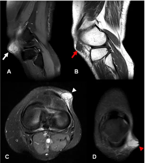
The imaging work-up performed by MRI disclosed a well-circumscribed, multiloculated cystic mass lesion originating from the joint capsule on the lateral aspect of the knee. The lesion was hypointense on T1-weighted MR images and hyperintense on T2-weighted MR images – findings that are consistent
Figure 1. MRI of the left knee. A - Sagittal fat-sat T2-weighted image that demonstrates a 2-cm hyperintense cystic lesion in the subcutaneous fat tissue close to the outer part of the lateral retinaculum (white arrow); B - Sagittal T1-weighted MR image shows a homogeneous hypointense cystic lesion (red arrow); C and D - Axial and coronal views, respectively. Fat-sat T2-weighted MR images demonstrate cystic formation adjacent to skin causing a tumefaction. Note the internal septa within the cyst (white and red arrow heads).
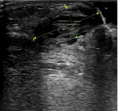
with the GC diagnosis (Figure 1). Sonographic examination showed a 20 × 9 mm mixed echogenic and septated cystic mass (Figure 2).
After the MRI evaluation, total excision of the mass was scheduled. A 3-cm oblique incision was made to approach the cystic mass on the left knee
(Figures 3A and 3B). During the surgery, a pedicle connecting the cystic mass and the joint capsule was observed (Figure 3C). After the mass and capsule excision, the defect in the primary excision region was sutured (Figures 3D and 3E).
Histopathological examination of the excised mass was reported as dense fibrous tissue without synovial or epithelial lining, which was consistent with the diagnosis of GC. No recurrence was detected during 12 months of follow-up and the patient recovered the full range of knee motion.
CASE 2
A 37-year-old woman was admitted to hospital because of pain and swelling in her left knee. Similar to Case 1, she had been submitted to a partial medial arthroscopic meniscectomy due to a grade 3 tear 5 years ago. No elastic bandage was used in the post-operative period, and she was able to walk with a full load. However, a diffuse swelling in the surgical site ensued soon after. During physical examination, an old incisional scar at the lateral portal inlet was detected where a 3 × 2 cm painful, semi-mobile, supple mass was palpable. The skin overlying the mass was completely normal. The patient was referred for MRI examination, which showed a well-circumscribed, multiloculated, cystic mass lesion under the joint Figure 2. Ultrasound of the left knee. Transvers view of
the lateral portal entry of the knee demonstrates a cystic complex mass with echogenic contents. Note that the posterior acoustic enhancement helps to distinguish the cystic nature of the mass.
Figure 3. Surgical procedure. A - Note the tumor mass on the lateral surface of the knee; B - An oblique incision was performed; C and D - GC was excised with its pedicle; E - Reconstruction of the capsule.
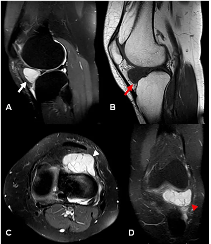
capsule on the lateral aspect of the knee. The lesion was hypointense on T1-weighted MR images and hyperintense on T2-weighted MR images – findings that were consistent with a GC (Figure 4). Sonographic examination showed a 26 × 14 mm echogenic and
septated cystic mass (Figure 5). In contrast to Case 1, the GC was under the joint capsule and more painful. The range of motion of the knee was 10°-120° (normal range is 0°-135°).
Figure 4. MRI of the left knee. A - Sagittal fat-sat T2-weighted image demonstrates a 3.5 × 2 cm hyperintense cystic lesion in the infrapatellar fossa compatible with ganglion cyst (white arrow); B - Sagittal T1-weighted image showing homogeneous hypointense cystic lesion (red arrow); C and D - Axial and coronal views, respectively. Fat-sat T2-weighted images demonstrate cystic mass adjacent to lateral retinaculum. Note that there are some internal septa within the cyst (white arrow in C and red arrow head in D).
After MRI evaluation, total excision of the mass was planned. However, the patient refused surgical treatment, so the condition was managed conservatively, which consisted of an elastic bandage. At the 1-year follow-up, the clinical findings remained unchanged.
DISCUSSION
Intra-articular GC is a relatively rare pathology and frequently develops in association with other intra-articular lesions. Although it may arise in any joint, the wrist is mostly involved. Many diagnoses are incidentally done, but when symptomatic the clinical features comprise joint tenderness, effusion, swelling, palpable mass, and pain, especially when the knee is involved.1,2,12 Even though the development of GCs are thought to have been caused by the deterioration of joint structures, intra-articular GC is frequently Figure 5. Ultrasound of the left knee. Transverse view of the lateral aspect of the knee demonstrates a cystic echogenic and septated mass (white arrow).
acquired after trauma or irritation of the synovia. Mucinous substances and hyaluronic acid release leads to cyst formation as a tissue and cellular response to trauma.13 Intra-articular GCs of the knee typically originate from infrapatellar fat pads or alar folds,and sometimes the cruciate ligaments.14 However, GCs localized in the anterolateral region of the knee and cysts in the portal inlet region are rarely described due to incomplete repair after arthroscopy.15 We report two cases in which the first GC occurred in the lateral entry region after arthroscopic intervention.
Synovial sarcoma, pigmented villonodular synovitis, hemangioma, lipoma arborescens, post-operative changes, infrapatellar fat pads, horizontal slits, and meniscal cysts extending to infrapatellar fat pads should be considered in the differential diagnosis.2
Therapeutic options for GC are varied and include both conservative and surgical approaches. Ultrasonography-guided aspiration, which has both diagnostic and therapeutic features, is the preferred method when patients refuse open surgery or arthroscopy. Open excision is recommended to remove the entire cyst wall.1
Although Salzler et al.16 reported many different complications following knee arthroscopy in their extensive series, no post-operative GC development was described in the arthroscopic portal entry region. GC development can be another complication following knee arthroscopy.
CONCLUSION
We believe that appropriate anatomical closure of entry sites that completely evacuate intra-articular fluid accumulation to lower pressure after arthroscopic procedures, and the use of elastic bandages after arthroscopic surgery, are practical and effective solutions for avoiding this type of complication.
REFERENCES
1. David KS, Korula RJ. Intra-articular ganglion cyst of the knee. Knee Surg Sports Traumatol Arthrosc. 2004;12(4):335-7. http://dx.doi.org/10.1007/s00167-003-0476-2. PMid:14985949.
2. Krudwig WK, Schulte KK, Heinemann C. Intra-articular ganglion cysts of the knee joint: a report of 85 cases and review of the literature. Knee Surg Sports Traumatol
Autopsy and Case Reports 2015;5(4):71-76
76
Arthrosc. 2004;12(2):123-9. http://dx.doi.org/10.1007/ s00167-003-0372-9. PMid:14504717.
3. Stäbler A, Glaser C, Reiser M. Musculoskeletal MR: knee. Eur Radiol. 2000;10(2):230-41. http://dx.doi. org/10.1007/s003300050039. PMid:10663752.
4. Yilmaz E, Karakurt L, Ozercan I, Ozdemir H. A ganglion cyst that developed from the infrapatellar fat pad of the knee. Arthroscopy. 2004;20(7):e65-8. http://dx.doi. org/10.1016/S0749-8063(04)00608-5. PMid:15346126. 5. Noda M, Kurosaka M, Maeno K, Mizuno K. Case
report ganglion cysts of the bilateral cruciate ligaments. Arthroscopy. 1999;15(8):867-70. http://dx.doi. org/10.1053/ar.1999.v15.0150862. PMid:10564867. 6. Sarimo J, Rantanen J, Helttula I, Orava S. Intra-articular
cysts and ganglia of the knee: a report of nine patients. Knee Surg Sports Traumatol Arthrosc. 2005;13(1):44-7. http://dx.doi.org/10.1007/s00167-004-0519-3. PMid:15654646.
7. Bui-Mansfield LT, Youngberg RA. Intraarticular ganglia of the knee: prevalence, presentation, aetiology, and management. AJR Am J Roentgenol. 1997;168(1):123-7. http://dx.doi.org/10.2214/ajr.168.1.8976934. PMid:8976934.
8. Kang CN, Kim DW, Kim DJ, Kim SJ. Intra-articular ganglion cysts of the knee. Arthroscopy. 1999;15(4):373-8. http://dx.doi.org/10.1016/S0749-8063(99)70054-X. PMid:10355712.
9. Kim RS, Kim KT, Lee JY, Lee KY. Ganglion cysts of the posterior cruciate ligament. Arthroscopy.
2003;19(6):E36-40. http://dx.doi.org/10.1016/S0749-8063(03)00386-4. PMid:12861224.
10. Kim MG, Kim BH, Choi JA, et al. Intra-articular ganglion cysts of the knee: clinical and MR imaging features. Eur Radiol. 2001;11(5):834-40. http://dx.doi.org/10.1007/ s003300000713. PMid:11372618.
11. Yasuda K, Majima T. Intra-articular ganglion blocking extension of the knee: brief report. J Bone Joint Surg Br. 1988;70(5):837. PMid:3192590.
12. Jacobson JA, Lenchik L, Ruhoy MK, Schweitzer ME, Resnick D. MR imaging of the infrapatellar fat pad of Hoffa. Radiographics. 1997;17(3):675-91. http:// dx.doi.org/10.1148/radiographics.17.3.9153705. PMid:9153705.
13. Roeser WM, Tsai E. Ganglion cysts of the anterior cruciate ligament. Arthroscopy. 1994;10(5):574-5. http://dx.doi. org/10.1016/S0749-8063(05)80017-9. PMid:7999170. 14. Resnick D. Internal derangements of joints. In: Resnick
D. Diagnosis of bone and joint disorders. Philadelphia: Saunders; 1995. p. 3061-3.
15. Franceschi F, Rizzello G, Maffei MV, Papalia R, Denaro V. Arthroscopic ganglion cyst excision in the anterolateral aspect of the knee. Arthroscopy. 2003;19(4):E32. http:// dx.doi.org/10.1053/jars.2003.50121. PMid:12671607. 16. Salzler MJ, Lin A, Miller CD, Herold S, Irrgang JJ, Harner
CD. Complications after arthroscopic knee surgery. Am J Sports Med. 2014;42(2):292-6. http://dx.doi. org/10.1177/0363546513510677. PMid:24284049.
Conflict of interest: None
Submitted on: August 29th, 2015
Accepted on: November 8th, 2015
Correspondence
Ozgur Korkmaz
Department of Orthopedic and Traumatology - School of Medicine - Istanbul Medipol University Tem Avrupa Otoyolu Goztepe Cikisi No:1 Bagcilar 34214 – Istanbul – Turkey
Phone: 00 (90) 212-4607777