DOI 10.5195/d3000.2018.83
New articles in this journal are licensed under a Creative Commons Attribution 4.0 United States License.
This journal is published by the University Library System, University of Pittsburgh as part of its D-‐Scribe Digital Publishing Program and is cosponored by the University of Pittsburgh Press.
Comparison of root canal length measurement methods in primary
teeth
Mine Koruyucu1, Merve Bayram2, Yelda Kasımoğlu1, Figen Seymen3
1Research Assistant, Istanbul University, Faculty of Den9stry, Department of Pedodon9cs, Istanbul, Turkey 2Assistant Professor, Istanbul Medipol University, Faculty of Den;stry, Department of Pedodon;cs, Istanbul, Turkey 3Professor, Istanbul University, Faculty of Den.stry, Department of Pedodon.cs, Istanbul, Turkey
Abstract
Objec&ves: The purpose of this study was to evaluate the accuracy of conven5onal radiography, intraoral digital radi-‐ ovisiography and electronic apex locator in determining the working length of root canals in primary teeth (in-‐vivo) and to compare the results with scanning electron microscopy measurements (ex-‐vivo). Materials and Methods: This study was conducted on 50 primary molar teeth. Standard endodon7c access cavity prepara7ons were prepared and the actual length was calculated by calibrated inves3gators. Working lengths were determined by using conven5onal radiography, RVG and apex locator in a total of 116 root canals. A;er the extrac<on of the teeth, each canal length was determined by using SEM. Data were analysed sta6s6cally by using Oneway Anova, Tukey HDS, Student t test and Bonferroni tests. Results: The mean root length measurements with conven3onal radiography were significantly higher than apex locator, RVG and SEM. The accuracy of apex locator and RVG were higher than conven>onal radiog-‐ raphy in determining the working length in primary teeth. Conclusions: The electronic apex locators provide an ac-‐ ceptable level of accuracy in determining root canal length in primary teeth. Clinical Relevance: The purpose of this study was to evaluate the accuracy of conven,onal radiography, intraoral digital radiovisiography and electronic apex locator in determining the working length of root canals in primary teeth and,to compare the results with scanning electron microscopy measurements.
Cita%on: Koruyucu M, et al. (2018) Comparison
of root canal length measurement methods in primary teeth. Den$stry 3000. 1:a001
doi:10.5195/d3000.2018.83
Received: March 2, 2018 Accepted: April 23, 2018 Published: July 13, 2018
Copyright: ©2018 Koruyucu M, et al. This is an
open access ar!cle licensed under a Crea!ve Commons A"ribu%on Work 4.0 United States License.
Email: [email protected]
Introduction
The primary objective of pulpectomy of primary teeth is to retain teeth with irreversible pulp pathosis in a symptom free state and maintain the integrity of the arch form [1, 2]. However, a num-‐ ber of reasons, such as the com-‐ plex anatomic morphology of the root canal system in primary teeth, innate physiological root resorption, the close proximity of the permanent successor tooth and the difficulty of producing satisfactory radiological images of primary tooth apices makes it dif-‐ ficult to achieve proper treatment [3].
Radiography is among the most common and widely used techniques for root canal length determination [4, 5]. Use of radi-‐ ography to calculate root canal length may not always lead to ac-‐ curate results especially in case of physiological resorption of primary teeth. Instrumentation and/or overfilling becomes much more possible if there is a mistake in measurement technique, thus the germ of a permanent tooth might get damaged [4, 6, 7]. The over-‐ filled material can retain after natural exfoliation of the primary tooth [8]. Furthermore, poor co-‐ operation of children makes it dif-‐ ficult to take a radiograph with acceptable diagnostic value [4, 5].
Proper detection of the working length is very important before pulpectomy in primary teeth. Due to limitations of radio-‐ graphic interpretation and high possibility of over-‐instrumentation of the unevenly resorbed roots and subsequent overfilling, the use of electronic apex locators is recommended regardless of the stage of root resorption [7].
Working length determina-‐ tion is an extremely relevant fac-‐ tor for the success of root canal treatments. To achieve best re-‐ sults, the narrowest part of the root canal where the width of blood vessels are smaller and the possibility of healing is highest, namely apical constriction or api-‐
cal foramen are prepared [9]. In theory, the canal terminus is re-‐ garded as the cemento-‐dentinal junction, even though it is not
possible to detect it clinically [10].
Therefore, the apical terminus used is minor foramen whose lo-‐ cation is around 0.5–1.0 mm other than the radiographic apex [11-‐ 13].
Generally, the convention-‐ al radiographic method has been used to determine the root canal length [12, 14]. However, this method has some disadvantages, such as the superposition of ana-‐ tomical structures and image dis-‐ tortions, and the film processing time [15]. While producing imag-‐ es, not a radiographic film but a sensor is used in the digital radio-‐ graphic method. This method is more advantageous than the con-‐ ventional one, as it is faster in im-‐ age acqusition, its amount of ra-‐ diation is less and it allows image editing, making it easy to examine the details [16, 17].
The apical foramen can be evaluated with an electronic apex locator, but only the radiographic apex can slightly be displayed by radiographs [5, 18]. Electronic apex locator, which helps to locate the file position in the canal, has been used clinically for more than 40 years [19-‐21]. Several genera-‐
tions of electronic apex loca-‐ tors have evolved [20]. There is no indication in the literature whether there is any difference in utilization the electronic apex locators in permanent or primary teeth, and as they prevent using radiation more than needed, electronic apex locators are re-‐ garded as safe, painless, and help-‐ ful. As a result, its use in primary teeth is recommended [5].
The objective of this study was to evaluate the accuracy of conventional radiography, in-‐ traoral digital radiovisiography and electronic apex locator in deter-‐ mining the working length of root canals in primary teeth (in-‐vivo) and to compare the results with scanning electron microscopy measurements (ex-‐vivo).
Materials and Methods
The study was approved by the Ethics Committee of the Istan-‐ bul University, Medical Faculty (No:2012/1738-‐1298). Fifty ex-‐ tracted human primary molar teeth (116 root canals) referred for extraction to Istanbul Universi-‐ ty, Faculty of Dentistry, Depart-‐ ment of Pedodontics because it is not possible to
restore them after caries are removed or when there is a root with re-‐ sorption great-‐ er than two-‐ thirds were se-‐ lected follow-‐ ing clinical and
radiographic examination. Primary molars which had abnormal root morphology and evidence of frac-‐ tures were excluded from the study.
Two calibrated pediatric dentist (MK, MB) carried out den-‐ tal examinations. Intra-‐examiner calibration was performed by re-‐ peating examinations of 10 teeth after an interval of 1 week. Kappa values for intra-‐examiner con-‐ sistency were 95.5% for teeth. The inclusion criteria for teeth selec-‐ tion were as follows: teeth have to demonstrate extensive caries, show the presence of two-‐thirds of the root length radiographically, and have adequate tooth struc-‐ ture to be restored. Also there should be no radiographic and clinical evidence of pulp pathology (resorption, spontaneous pain, tenderness to percussion or palpa-‐ tion, swelling, sinus tract, patho-‐ logic mobility etc.). During the treatment, the teeth were anes-‐ thetized and the endodontic ac-‐ cess was performed using high speed diamond burs. The canals were initially explored with #10 K-‐ files (Mani, Tochigi, Japan).
After extirpating the pulp with a barbed broach, the canals
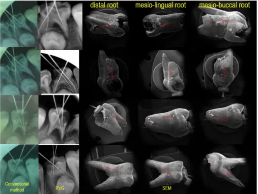
were irrigated with saline solution then dried with cotton pellets without any attempt to dry the canal. The length of the root ca-‐ nals were measured with one of the fitting K-‐type file, sized be-‐ tween 15-‐40 (Mani, Tochigi, Ja-‐ pan). After that, a silicone stop was adjusted to the reference, and the distance between stop and file tip was measured with endodontic ruler by magnifying loupes. The reference point was marked on the coronal portion of the roots with a fine paint marker to facili-‐ tate accurate reinsertion of the files. Root canal length for each tooth was measured with a scale in milimeters by all methods (in-‐ traoral periapical radiography, RVG and electronic apex locater before extraction, SEM after ex-‐ traction) (Picture 1). The measur-‐ ing precision was set to 0.5 mm and the measurements were re-‐ calculated three times for each canal by the same dentist and av-‐ erage value was recorded.
For conventional radiog-‐ raphy groups; intraoral periapical radiographs were taken by X-‐ray device operating 8 mA and 70 Kvp
(Kodak, Tokyo, Japan) using paral-‐ leling technique The films were then placed parallel to the X ray tube (Trophy, Tokyo, Japan).
The apex locator was used for the electronic measurement (Endo Master, EMS, Switzerland). The labial clip was inserted and K-‐ file attached to the file holder was smoothly inserted till “apex” ap-‐ peared on the screen. Then, the rubber stop was placed at the cor-‐ onal reference, and the measure-‐ ment of root canal length was done electronically. For RVG groups, after all intraoral prepara-‐ tions RVG (Kodak, Tokyo, Japan) were used to assess the length of the root canal.
When the eruption of permanent teeth start, the teeth that were mobility were extracted. After the teeth had been extract-‐ ed, they were preserved in 10% formaldehyde solution. They were numbered and immersed in 5.25% sodium hypochlorite solution for 15 minutes to get rid of organic residues in root surfaces. Then they were scaled to remove any stain and calculus. In accordance with electron microscopy instruc-‐
tions, the Snonputter technique (Polaron Sputter Coater) for electron microscopy images was used to coat the specimens with gold (150 seconds). SEM under x50 magnification was used to calculate every canal length (Jeol JSM-‐5600, SEM).
SPSS for Windows, ver-‐ sion 10.0 was run for all statisti-‐ cal analyses (SPSS, Chicago, USA). Data were analysed statis-‐ tically by using Oneway Anova, Tukey HDS, Student t test and Bonferroni tests post-‐hoc analysis. A P-‐value <0.05 was considered statistically significant.
Results
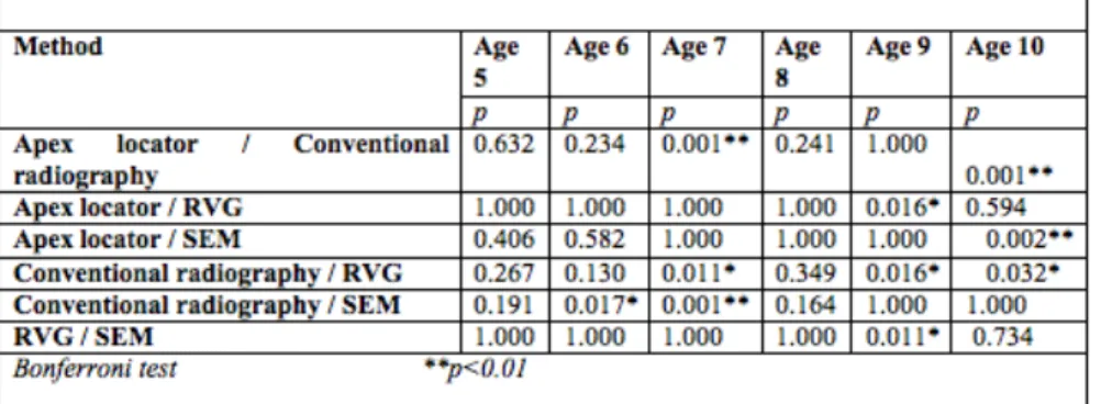
A statistically significant difference was found between the mean root canal length of the methods (p <0.01) (Table 1). The root canal measurement average of the conventional method was found significantly higher than the apex locator (p: 0.001), RVG (p: 0.001) and SEM (p: 0.001) meas-‐ urements (p <0.01). There is no statistically significant difference between the mean apex locator, RVG and SEM measurement, (p> 0.05) (Table 2).
There is a statistically sig-‐ nificant difference between apex locator, conventional method, RVG and SEM averages according to age results (p<0.001). The apex locator averages of ages 6 and 8 were found significantly higher in the 7 years (p: 0.008) and 9 years (p: 0.028) (p <0.05, p <0.01). The conventional methods of 9 and 7 years were significantly lower than
5 years (p:0.008), 6 years (p: 0.001) and 8 years (p: 0.004). The conventional method avetage of the 7 years was significantly lower than that of 6 years (p: 0.048) (p<0.05). RVG averages of 6 and 8 years were significantly higher than 7 years (p:0.004) and 9 years (p:0.002) (p<0.01). The SEM aver-‐ age of the 7 year was significantly lower than the 6 year (p:0.033) and the 8 year (p:0.022) (Table 3).
The root canal measure-‐ ment average of the conventional method was significantly higher than the SEM (p:0.017) measure-‐ ments for 6 years old children (p<0.05). The root canal meas-‐ urement average of the conven-‐ tional method was found signifi-‐ cantly higher than the apex locator (p:0.001), RVG (p: 0.011) and SEM (p:0.001) measurements for 7 years old children (p<0.01). The root canal measurement averages of RVG were significantly lower than the apex locator (p:0.016), conventional method (p:0.016) and SEM (p:0.011) for 9 years old children (p<0.05). The root canal measurement average of the con-‐ ventional method was found sig-‐ nificantly higher than the apex locator (p:0.001) and RVG
(p:0.032) for 10 years old children
(p <0.01; p<0.05). The root canal measurement average of the SEM method was found significantly higher than the apex locator (p: 0.002) (p <0.01) for 10 years old children (Table 3).
There is a statistically sig-‐ nificant difference between the root canal averages of the meth-‐ ods according to upper and lower jaw (p<0.01) (Table 5). The root canal measurement average of the SEM method was found to be sig-‐ nificantly lower than the apex locator (p: 0.004), conventional method (p: 0.005) and RVG in the upper jaw (p: 0.036) (p <0.05; p<0.01). The root canal measure-‐ ment average of the conventional method was found significantly higher than the apex locator (p: 0.001), RVG (p: 0.001) and SEM (p: 0.001) measurements in the lower jaw (p <0.01) (Table 6).
Discussion
One of the most significant steps to get successful results in root canal treatment is to determine
the length of root canal accurate-‐ ly. Root resorption generally makes apex of root canals in pri-‐ mary teeth ambigious.
In determination of length in a clinical setting, the only prac-‐ tice that is approved globally, ac-‐ cessible and trustworthy is radiog-‐ raphy. However, repeated radio-‐ graphic exposure of pediatric pa-‐ tients before, during, and immedi-‐ ately after the endodontic treat-‐ ment may cause high concerns. Nevertheless radiography can give misleading results in resorption [5].
Some studies have report-‐ ed that while measuring mean working length, both digital radi-‐ ography and apex locator demon-‐ strated almost similar results with the conventional radiographic method in primary molars [5, 22]. Likewise, Krishnan and Sreedharan study how precisely electronic apex locators and conventional radiographic technique determine the root canal length of primary teeth by comparing with the ana-‐ tomic root canal length of these teeth. The results of this study from conventional radiography and electronic apex locator were similar; intra‑class correlation co-‐ efficient also showed that both
the radiographic and electronic methods are reliable in determin-‐ ing the root canal length and there is no need to use them together [23].
In the study of Subrama-‐ niam et al, measured root canal lengths in primary teeth obtained from four different techniques are compared. The conclusion of the study is that reliable and precise root canal length assessment in primary teeth is possible by the digital radiographic and apex loca-‐ tor technology. Moreover, these methods increase both the safety of endodontic treatment and com-‐ fort in children [24].
In one laboratory study, Mello-‐Moura et al compared the accuracy of four different methods in determining root canal length. The study demonstrated that among all the methods the elec-‐ tronic apex locators provided the most accurate root canal length results, however in case electronic resources are unavailable the combination of radiographic and tactile sense methods might be an alternative as the results were sat-‐ isfactory [25].
The existence of root re-‐ sorption is an important charac-‐
teristic of pulpec-‐ tomy in primary teeth. It is hard to radiologically asses the small areas of resorp-‐ tion especially if the resorption is on buccal/lingual aspects of the root. To discard the disadvantages of radiographic assessment in the-‐ se cases, electrical root length de-‐ termination may be used.
Mente et al. evaluated the accuracy of an electronic apex lo-‐ cater device in initial resorption of primary incisors and concluded that whether there was resorption in primary teeth did not affect how accurate the electrical meas-‐ urement of root canal length in-‐
vitro [26]. Another study compar-‐
ing two different root canal meas-‐ uring device in primary teeth with and without resorption concluded that these devices did not provide reliable data. However, these apex locators may be useful in primary root canal therapy in case other diagnostic measures support them. Additional in vivo assess-‐ ments of them in primary teeth need to be performed [27].
Apex locator was more likely to miscalculate root length in primary molars with root re-‐ sorption than direct canal meas-‐ urement, yet Root ZX (Morita, USA) type apex locator calculated accurately in cases in which root resorption was less than one third of root length in primary molar teeth [28]. In the study of Bel-‐
trame et al, the Root ZX apex loca-‐ tor was studied in primary molar teeth (with or without root re-‐ sorption) to analyze its in vivo and ex vivo accuracy. As a conclusion, it was not significant that whether there was root resorption or not, as in both cases the Root ZX apex locator measured the root canal working length ±1 mm in primary molars, in vivo and ex vivo [29]. This conclusion backs up other ex vivo studies as its results con-‐ firmed that electronic apex loca-‐ tors can be used to measure canal length in primary teeth [25, 50]. In addition to that, Bahrololoomi et al. showed that the accuracy of Root ZX electronic apex locator was high in the primary anterior teeth despite root resorption. Therefore, using this device as an adjunct is recommended for root canal length measurements in primary anterior teeth [4].
Leonardo et al. evaluated ex vivo accuracy of two different root canal measuring devices in measuring root canal length of primary incisors and molar teeth that have physiological root re-‐ sorption. They concluded that electronic apex locators are effec-‐ tive and correct when root canal length of primary incisor or prima-‐ ry molar teeth (whether or not they have root resorption) are de-‐ termined because of the results of the electronic ones were almost perfectly parallel to the actual root canal length measurements [30].
In addition to clinical and laboratory studies in primary teeth, studies have been carried out on permanent teeth and dif-‐ ferent results have been obtained. Esmaeili et al. aimed to compare the accuracy of digital and conven-‐ tional radiographic techniques in the assessment of the endodontic working length in premolar teeth and concluded that the accuracy of digital and conventional radiog-‐ raphy techniques were similar in determination of working length
[31]. According to another study
findings showed that although there was a significant difference between experimental method and actual working length but electronic apex locator showed the most accurate reading when compared to actual working length in premolar teeth [32].
Orosco et al compared conventional and digital radio-‐
graphic methods in measurement of the root canal working length of maxillary incisor or canine teeth. They concluded that the root ca-‐ nal working length measured with conventional radiographic method were more accurate than the ones obtained with the digital radio-‐ graphic method [15]. Diwanji et al. compared radiographic method and electronic method with apex locator in determining exact work-‐ ing length of young permanent root canal. Then, the results from these methods were compared to the actual length from histological sections under stereomicroscope. In the light of these comparisons, apex locator was decided to be more reliable and precise than the
digital radiography [33].
Mittal et al. compared the accuracy of digital radiograph ex-‐ posed with paralleling technique, 6th generation apex locator, tac-‐
tile method, paper point method, combined electronic and radio-‐ graphic working length and com-‐ bined electronic, tactile and paper point working length method to determine the working length in permanent teeth with open apex. In the end they found that a com-‐ bination of electronic tactile and paper point methods in open apex increases the accuracy of working
length determination [34].
Martins et al. published a review of compared the radio-‐ graphic apex locators and radio-‐ graphs in permanent dentition. They reported that electronic apex locators were mostly more useful than radiography when there is a respective anatomic reference point (for example, the apical con-‐ striction or apical foramen) and when radiography was only meth-‐ od in determining working length, most of the times more radio-‐ graphs were needed than using electronic apex locator. This study shows that in primary teeth (with and without initial root resorption) interexaminer reproducibility of electrical assessment of root canal length in vitro was high. In com-‐ parison with the radiographic re-‐ sults, the accuracy of the working
length was higher [35].
This study evaluates the accuracy of conventional radiog-‐ raphy, intraoral digital radiovisiog-‐ raphy and electronic apex locator in determining the working length of root canals in primary teeth (in
vivo) and to compare the results
with scanning electron microscopy measurements (ex vivo). The dif-‐ ference of this study from the
other studies is that there is no study which is compared with SEM in the literature. In addition, the findings of comparison of methods according to age and jaws were not found in other literature.
Besides different results are obtained according to the methodological differences in the research it can be concluded that electronic apex locators provide an acceptable level of accuracy in determining root canal length in primary teeth. In this study the accuracy of apex locator and RVG were higher than conventional radiography in determining the working length in primary teeth.
Conclusion
The results confirm that electronic apex locaters can accu-‐ rately determine the root canal length in primary teeth.
Acknowledgements
We would like to thank the Dt. Pelin Barlak for his contribu-‐ tion to the research.
References
1. American Academy of Pe-‐ diatric Dentistry Clinical Af-‐ fairs Committee-‐-‐Pulp Therapy Subcommit-‐ tee; American Academy of Pediatric Dentistry Council on Clinical Affairs (2008-‐ 2009) Guideline on Pulp Therapy for Primary and Immature Permanent Teeth. Pediatr
Dent 30(7):170-‐4.
2. Rodd HD, Waterhouse PJ, Fuks AB, Fayle
SA, Moffat MA; British So-‐ ciety of Paediatric Dentis-‐ try (2006) Pulp therapy for primary molars. Int J Pae-‐ diatr Dent 16(1):15-‐23. 3. Carotte P (2005) Endodon-‐
tic Treatment for Children. British Dental Journal 198(1):9-‐15.
4. Bahrololoomil Z, Soleymani AA, Modaresi J, Imanian M, Lotfian M (2015) Accuracy of an electronic apex loca-‐ tor for working length de-‐ termination in primary an-‐ terior teeth. Jornal of Den-‐ tistry, Tehran University of Medical Sciences
12(4):243-‐8.
5. Oznurhan F, Unal M, Kapdan A, Ozturk C, Aksoy S (2015) Clinical evaluation of apex locator and radiog-‐ raphy in primary teeth. Int J Paediatr Dent 25:199-‐ 203.
6. Jerrel RG, Ronk SL (1982) Develeopmental arrest of succedaneous tooth fol-‐ lowing pulpectomy in a primary tooth. J Pedod 6(4):337-‐42.
7. Ahmed HMA (2013) Ana-‐ tomical challenges, elec-‐ tronic working length de-‐ termination and current developments in root canal preparation of primary mo-‐ lar teeth. Int Endod J 46:1011-‐1022.
8. Chawla HS, Setia S, Gupta N, Gauba K, Goyal A (2008) Evaluation of a mixture of zinc oxide, calcium hydrox-‐ ide, and sodium fluoride as a new root canal filling ma-‐ terial for primary teeth. J Indian Soc Pedod Prev Dent 26(2):53-‐8. 9. Ricucci D, Langeland K
(1998) Apical limit of root canal instrumentation and obturation, part 2. A histo-‐ logical study. Int Endod J 31:394–409.
10. Burch JG, Hulen S (1972) The relationship of the api-‐ cal foramen to the anatom-‐ ic apex of the tooth root. Oral Surg Oral Med Oral Pathol 34:262–268.
11. Kuttler Y (1958) A precision and biologic root canal fill-‐ ing technic. J Am Dent As-‐ soc 56: 38–50.
12. Katz A, Tamse A, Kaufman AY (1991) Tooth length de-‐ termination: a review. Oral Surg Oral Med Oral Pathol 72:238–242.
13. Morfis A, Sylaras SN, Geor-‐ gopoulou M, Kernani M, Prountzos F (1994) Study of the apices of human permanent teeth with the use of a scanning electron microscope. Oral Surg Oral Med Oral Pathol 77:172– 176.
14. Stein TJ, Corcoran JF (1992) Radiographic ‘working length’ revisited. Oral Surg
Oral Med Oral Pathol 74:796–800.
15. Orosco FA, Bernardineli N, Garcia RB, Bramante CM, Duarte MAH, Moraes IG (2012) In vivo accuracy of conventional and digital radiographic methods in confirming root canal working length determina-‐ tion by Root ZX. J Appl Oral Sci 20(5):522-‐525.
16. Ellingsen MA, Hollender LG, Harrington GW (1995) Radiovisiography versus conventional radiography for detection of small in-‐ struments in endodontic length determination. II. In vivo evaluation. J Endod 21(10):516-‐20.
17. Shearer AC, Horner K, Wil-‐ son NH
(1990) Radiovisiography for imaging root canals: an in vitro comparison with conventional radiog-‐ raphy. Quintessence Int 21(10):789-‐94.
18. Kobayashi C. Electronic ca-‐ nal length measurement (1995) Oral Surg Oral Med Oral Pathol Oral Radiol En-‐ dod 79:226–231.
19. Alves AM, Felippe MC, Fe-‐ lippe WT, Rocha MJ (2005) Ex vivo evaluation of the capacity of the Tri Auto ZX to locate the apical fora-‐ men during root canal re-‐ treatment. Int Endod J 38:718–724.
20. Ebrahim AK, Wadachgi R, Suda H (2007) Electronic apex locators-‐review. J Med Dent Sci 54:125–136. 21. Goldberg F, Marroquin BB, Frajlich S, Dreyer C (2005) In vitro evaluation of the ability of three apex loca-‐ tors to determine the working length during re-‐ treatment. J Endod 31:676–678.
22. Neena IE, Ananthraj A, Praveen P, Karthik V, Rani P (2011) Comparison of digital radiography and apex locator with the con-‐ ventional method in root length determination of primary teeth. J Indian Soc Pedod Prev Dent
29(4):300-‐304.
23. Krishnan IS, Sreedharan S (2012) A comparative eval-‐ uation of electronic and radiographic determination of root canal length in pri-‐ mary teeth: An in vitro study. Contemporary Clini-‐ cal Dentistry 3(4):416-‐420. 24. Subramaniam P, Konde S,
Mandanna DK (2005). An in vitro comparison of root canal measurement in pri-‐ mary teeth. J Indian Soc Pedod Prev Dent
23(3):124-‐125.
25. Mello-‐Moura ACV, Moura-‐ Netto C, Araki AT, Guedes-‐ Pinto AC, Mendes FM (2010) Ex vivo performance of five methods for root canal length determination
in primary anterior teeth. Int Endod J 43:142–147. 26. Mente J, Seidel J, Buchalla
W, Koch MJ (2002) Elec-‐ tronic determination of root canal length in prima-‐ ry teeth with and without root resorption. Int Endod J 35:447–452.
27. Bodur H, Odabaş M, Tulu-‐ noglu O, Tinaz AC (2008). Accuracy of two different apex locators in primary teeth with and without root resorption. Clin Oral Invest 12:137-‐141.
28. Angwaravong O, Panitvisai P (2009) Accuracy of an electronic apex locator in primary teeth with root re-‐ sorption. Int Endod J 42:115-‐121.
29. Beltrame APCA, Triches TC, Sartori N, Bolan M (2011) Electronic determination of root canal working length in primary molar teeth: an in vivo and ex vivo study. Int Endod J 44:402–406. 30. Leonardo MR, Silva LAB, Nelson-‐Filho P, Silva RAB, Raffaini MSGG (2008) Ex vivo evaluation of the ac-‐ curacy of two electronic apex locators during root canal length determination in primary teeth. Int Endod J 41:317–321.
31. Esmaeili F, Akbari FA, Zarandi A (2016) Compari-‐ son of accuracy of digital and conventional radiog-‐
raphies in determining en-‐ dodontic working length. SADJ 71(9): 395-‐397. 32. Mittal R, Singla MG, Sood
A, Singla A (2015) Compar-‐ ative evaluation of working length determination by using conventional radiog-‐ raphy, digital radiography and electronic apex loca-‐ tor. Journal of Restorative Dentistry 3(3):70-‐75. 33. Diwanji A, Rathore AS, Aro-‐
ra R, Dhar V, Madhusudan A, Doshi J (2014) Working length determination of root canal of young per-‐ manent tooth: An invitro study. Annals of Medical and Health Sciences Re-‐ search 4(4):554-‐558
34. Mittal P, Jadhav GR, Logani A (2016) Accuracy of dif-‐ ferent methods to deter-‐ mine working length in teeth with open apex-‐an ex vivo comparative study. J Dent Specialities 4(1):39-‐ 45.
35. Martins JNR, Marques M, Mata A, Carames J (2014) Clinical efficacy of electron-‐ ic apex locators: Systematic review. J Endod 40(6):759– 77.