Investigating the Effects of DNA Demethylation in
Chemotherapy Resistance by ChIP-on-Chip Method
Received: March 03, 2019 Accepted: March 04, 2019 Online: May 28, 2019 Accessible online at: www.onkder.org
Meltem DEMİREL KARS,1 Gözde KOYGUN2
1Program of Medicinal and Aromatic Plants, Necmettin Erbakan University Meram Vocational School, Konya-Turkey
2Department of Nanotechnology and Advanced Materials, Selçuk University, Institute of Science and Technology, Konya-Turkey
OBJECTIVE
This study aimed to determine the epigenetic basis of drug resistance mechanisms developed in MCF-7 breast cancer cell line that is resistant to an anticancer agent paclitaxel. Thus, we investigated the effects of the changes in DNA level on gene expression profile and proposed methods of inhibiting resistance by DNA modifications.
METHODS
We investigated the epigenetic basis of acquired drug resistance in whole genome by comparing chro-mosome immunoprecipitation in paclitaxel-resistant MCF-7 (MCF-7/Pac) cells and in drug-sensitive (MCF-7/S) cells. For this analysis, DNA samples from both cell lines were immunoprecipitated and labeled with Cy3 and Cy5 fluorescent dyes. Hybridization and array scanning was performed with Agi-lent all-Genome Microarray platform that was designed to detect DNA methylation. The obtained high-throughput information was analyzed with a bioinformatics analysis program.
RESULTS
The results showed that demethylation and epigenetic modulation of the DNA regions encoding 90 genes are significant in the development of multiple drug resistance (MDR) in breast cancer. Some of these genes, ICAM4, COX6B2, ITGB8, SLC39A4, TUBB2C, COL6A1, DAPK1, RUNX3, SLC35F3, and MAP6, are important players in the development of drug resistance and cancer stem cells.
CONCLUSION
Studies on reversing multidrug resistance can be carried out by DNA modification or methylation of target genes regions on DNA. The results presented in this study may shed light on drug development studies to make DNA modifications.
Keywords: Demethylation; epigenetics; multidrug resistance; MDR; MCF-7; paclitaxel.
Copyright © 2019, Turkish Society for Radiation Oncology
Introduction
Stable and inherited changes in gene expression with-out any change in DNA sequences are described as epi-genetic alterations. Generally, modulation occurs on DNA or in chromatin by covalent binding and mod-ifications. Epigenetics causes the expression of genes
in genetically identical cells and organisms in different forms and show phenotypic differences.[1]
The most emphasized mechanisms is the DNA methylation, which is the hereditary change caused by the addition of methyl group to the 5′ end of the cytosine followed by the guanine nucleotide by the catalysis of enzyme DNA methyl transferase.[2] DNA Dr. Meltem DEMİREL KARS
Necmettin Erbakan Üniversitesi, Meram Meslek Yüksek Okulu, Tıbbi ve Aromatik Bitkiler Programı, Konya-Turkey
E-mail: [email protected]
OPEN ACCESS This work is licensed under a Creative Commons
as the culture medium of the cells. To prevent micro-bial infection, gentamycin (1 mg/ml) was added to the medium, and the cells were incubated at 37°C, with 5% CO2 incubator. Adhesive cells were transferred into
new culture medium with trypsin-EDTA when they covered 70% of the cell culture vessel. Paclitaxel-resis-tant cells were established by adding stepwise increas-ing paclitaxel in to the culture medium in two years. This cell line was used as rational model for drug resis-tance in breast cancer.[9]
DNA Isolation and Immunoprecipitation
The experiments were conducted according to Agilent G4170-90012_Methylation_Protocol_v.2.0 in the labo-ratories of Selçuk University ILTEK. The genomic DNA isolation from MCF-7/S and MCF-7/Pac cells (1×10⁶
cells from each) was performed using the genomic DNA isolation kit (QIAamp DNA mini kit, QIAGEN). The quality control procedure was performed by spec-trophotometric measurements (OD260nm/OD280nm, Nanodrop-Thermo) and agarose gel electrophoresis. Genomic DNA (5 µg from each) was fragmented by ultrasonication by ultrasonicator (using micro probe, Heidolph) that was set output power to 70%. The DNA fragmentation was ensured by sonicating the DNA samples for 5 s, five cycles on ice by holding 5 s between each cycle intervals (to prevent heating and foaming). For immunoprecipitation procedure, Dynabeads Pan Mouse IgG magnetic beads were precipitated by mag-netic stand (DynaMag-2 Magnet, Life Technologies), and the beads were bound to ChIP grade anti-5-Methyl Cytidine antibody (Abcam) by incubating in a rotator (VWR) at 4°C incubator. The fragmented DNA samples were then hybridized with the magnetic bead bound 5-methylcytidine antibody in immunoprecipitation buffer (Triton X, yeast t-RNA, PBS) to let the binding of methylated DNA fragments on to the magnetic an-tibodies (in rotator, overnight at 4°C incubator). Fi-nally, the methylated DNA fragments were sorted on magnetic stand. The methylated DNA samples and reference DNA samples (that were not immunoprecip-itated) were eluted by elution in TE and SDS solution. DNA fragments were extracted through phenol-chlo-roform extraction method by use of MaXtract High Density tubes (QIAGEN). The aqueous phase was col-lected, and DNA was precipitated by adding the precip-itation solution that constitutes NaCl (200 mM), glyco-gen 20 µg/mL, and pure ethanol. DNA was precipitated by incubating the mixture at 4°C and centrifugation at 12.000 g for 3 min (Hettich). Finally, the DNA pellets were air dried. The quality of eluted methylated DNA demethylation occurs by non-methylating the DNA
strand after replication or during development by a replication-independent process. The impaired sig-naling in cancer cells may result in stable silencing of downstream targets regulated by epigenetic mecha-nisms.[3] Methods such as methylation-specific PCR, HELP (HpaII tiny fragment Enrichment by Ligation-mediated PCR) assay can detect DNA methylation. Another approach in the determination of methy-lation level named “ChIP-on-chip” was developed by ChIP (chromatin immunoprecipitation) method. [4] In this method, the double-stranded DNA is sep-arated and treated with methyl cytosine antibodies. Once the DNA has been labeled, it is hybridized with the microarray platform affixed with specific probes covering the whole human genome.
Drug resistance, which is acquired or intrinsic in the patient post-treatment or pre-treatment period, se-verely prevents success in cancer chemotherapy. This case is called multidrug resistance (MDR).[5] Resis-tance against chemotherapy prevents many anticancer drugs to show the expected effects on the patients and causes progression of the disease. Increased drug doses lead to increased side effects and limited treatment.
This study aimed to determine the effects of DNA demethylation and epigenetic bases of drug resis-tance mechanisms developed in MCF-7 breast cancer cell line that is resistant to anticancer agent paclitaxel (MCF-7/Pac). The resistant cell line was developed by treatment of MCF-7 breast cancer cells with paclitaxel by dose increments. Paclitaxel is an anticancer drug that inhibits cell division by interfering microtubules during mitosis. Changes in gene expression profiles of paclitaxel-resistant MCF-7 (MCF-7/Pac) cells com-pared to drug-sensitive (MCF-7/S) cells have been pre-sented in previous publications.[6,7] Recently, it was reported that MCF-7/Pac cells exhibit the major fea-tures of breast cancer stem cells.[8] Therefore, determi-nation of DNA methylation levels of MCF-7/Pac cells with respect to MCF-7/S cells will reveal the effects of DNA methylation in breast cancer stem cells. In this study, epigenetic bases of developing resistance were proposed with preliminary findings.
Materials and Methods Cell Culture Conditions
MCF-7/S cell line sensitive to anticancer drugs and pa-clitaxel-resistant MCF-7/Pac cell line was used in the study.[9] RPMI 1640 containing 10% (v/v) serum (fetal bovine serum, FBS) and 2 mM L-glutamine was used
and reference DNA samples of 7/Pac and MCF-7/S were measured by Nanodrop (Thermo).
Fluorescent Labeling of DNA Samples
Methylated and reference DNA fragments were labeled using the SureTag DNA Labeling Kit (Agilent). Briefly, DNA samples were mixed with random primers and incubated at 95°C for 3 min on heating-cooling dry block (Biosan). While DNA samples were denatured, they were labeled by Exo Klenow enzyme, dNTPs. and fluorescently labeled dUTPs (Cy-3 and Cy-5 labeled) at 37°C for 2 h in PCR machine (Biorad). After the reac-tion was stopped at 65°C, labeled DNA fragments were purified by purification columns, and fluorescence binding success was determined by spectrophotomet-ric measurements (Nanodrop, Thermo). Specific activ-ity and labeled DNA amounts were calculated using the following formulas:
Specific activity=pmol dye per μL of dye/μg per μL DNA Yield (μg)=DNA concentration (ng/μL)×sample vol-ume (μL)/1000 ng/μg
Hybridization of Labeled DNA on Arrays
Since DNA labeling quality was good (yield >2.5 µg and appropriate specific activity), the labeled DNA frag-ments were hybridized on the array platforms (Human DNA Methylation Microarray, 1×244K (HD)). Briefly, the hybridization master mix was prepared for four samples (duplicates for each cell line). Hybridization master mix constituted Human Cot-1 DNA for inter-nal control, CGH (chromosomal genomic hybridiza-tion) blocking agent, HI-RPM hybridization buffer, and deionized formamide. Hybridization master mix (420 µL for 1-pack microarray format) was combined with Cy3- and Cy5-labeled DNA samples (40 µL each) in one tube for each cell line. Two replicates of hy-bridization mixtures were prepared for MCF-7/S and MCF-7/Pac cell lines and incubated at 95°C for 3 min and 37°C for 30 min in thermal cycler (Biorad). The hybridization solution should be kept at 37°C until it is loaded on to the array slides. Hybridization solution was slowly dispensed on to the gasket slide carefully preventing the overflow of the solution out of the gas-ket chamber. Then the printed microarray slide (Agi-lent, Unrestricted Amadid Chip-on-Chip 1×244K) was put on to the gasket slide (barcode number should be outside). Finally, the microarray slide was assembled in the slide chamber. The hybridization oven was set to 67°C, and the chambers were placed in to the ar-ray holders in a balanced way. The hybridization
pro-ceeded for 40 h at 20 rpm rotation speed. Two repeats were performed for each group (MCF-7/S and MCF-7/ Pac), (n=2).
Washing and Scanning
The washing equipments were washed with acetonitrile and ultrapure water previously. ChIP-on-chip wash buf-fer 2 was prewarmed in a coplin jar before disassembling the slide chambers (37°C). The slides were disassembled in ChIP-on-chip wash buffer 1, in to a separate coplin jar. Then the slides were put into another jar that contains wash buffer 1 for 5 min at room temperature (a mag-netic bead was put at the basement of the jar to provide continuous stirring by magnetic stirrer). Arrays were immediately air dried, and ozone covers were put on to prevent adverse effects of the ozone on to the slides. During the whole procedure, slides should be kept by forceps. Sure Scan Microarray Scanner (Agilent), which was in the laboratory of Selçuk University ILTEK, was turned on to warm up the lasers. The G4900DA SureS-can Microarray SSureS-canner System Microarray SSureS-can Con-trol Software 9.1 (Agilent) was run, and Protocol Agilent HD-CGH was selected. The slides were immediately in-serted into the scanner as the scanner was ready to scan the arrays. The machine recognized the barcode of the array. The green and red lasers excited the fluorescent dyes, and then photomultiplier tube detector detected the data. After scan protocol was completed, the Feature Extraction software v11.0.1.1 was operated for quality control (QC). The QC reports were generated using the grid Human DNA Methylation Microarray 244k-023795-D-F-20111018.
Advanced Analysis and Statistics
The scan data of each cells were analyzed by Genomic Workbench ver6.0 (Agilent) program. Z-score algo-rithm was used for calculations. Gaussians were fit-ted to the bimodal log ratio distribution. Significant changes were determined between the groups (MCF-7/S and MCF-7/Pac) by Student’s t-test (p<0.05). Finally, the significant results were listed, and the demethylation levels were calculated by calculating the fold change values and presented as the logarithm of the fold change ratios (MCF-7/Pac/MCF-7).
Results
Proliferation of the Cells and Preparation of DNA Fragments
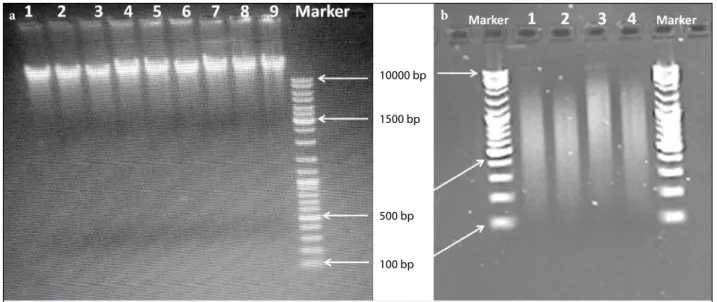
The cell lines were cultured, and DNA isolation and QC of DNA samples were performed. Figure 1
ex-Hybridization, Washing, and Scanning Protocols
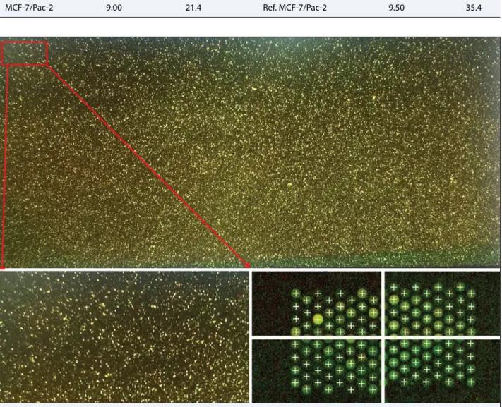
The hybridization, washing, and scanning protocols were completed; and the QC reports were created for each arrays with the Feature Extraction program (Agi-lent). The results obtained for all the scanning protocols were reported as “good.” The scan image and grid results obtained after scanning are presented in Figure 3. The quality analysis of the repetitive arrays for each group yielded favorable results for further analysis (Figs. 4, 5).
Advanced Analysis by Genomic Workbench Program
The raw data obtained after scanning the arrays were analyzed by Genomic Workbench ver6.0 (Agilent). The fluorescence values were analyzed, and array re-sults for MCF-7/S and MCF-7/Pac were compared. As a result, when the drug resistance was developed, de-creases in the methylation level of the gene regions on DNA (demethylation) were identified. The fold change values were calculated to reveal the demethylation lev-els after acquired drug resistance. The logarithms of hibits the image of the MCF-7/Pac cells under an
inverted phase contrast microscope (Leica). In Fig-ure 2, agarose gel electrophoresis images of the ge-nomic DNA samples and fragmented DNA smears are presented. The purity, concentration, and quality of the DNA samples isolated from the cell lines were found to be suitable for the continuation of the mi-croarray protocol (Table 1). Table 2 presents the re-sults of the specific activity and yield calculations of fluorescently labeled methylated and reference DNA fragments. These results showed that it was possible to continue experiments with the labeled DNA frag-ments of suitable concentrations. According to these results, microarray hybridization and scanning were performed.
Fig. 1. Invert microscope image of MCF-7/Pac cells.
10000 bp
1500 bp
500 bp
100 bp
a b
Fig. 2. (a) Agarose gel electrophoresis image of genomic DNA from cells; 1–5: MCF-7/S, 6–9: MCF-7/Pac, Marker: High
range DNA ladder: 100 bp–10 kb. (b) Fragmented DNA smear from DNA samples; 1,2: MCF-7/S, 3–4: MCF-7/ Pac, Marker: High range DNA ladder: 100 bp–10 kb.
Table 1 Quality and yield of genomic DNA samples from cell lines
Concentration Yield Quality
ng/µL µg 260/280 260/230
MCF7/S1 143.60 7.90 1.82 2.23 MCF7/S2 141.50 7.78 1.88 2.17
MCF7/Pac1 158.10 8.69 1.81 2.04
the results were calculated and significant results were listed (p<0.05) (Table 3). At the end of the analysis, we identified significant demethylation in 90 gene regions in paclitaxel-resistant breast cancer cells with respect to drug-sensitive breast cancer cell line. The demethy-lation levels were in range of 10.35–2.14 values. The demethylated ICAM4, COX6B2, ITGB8, SLC39A4, TUBB2C, COL6A1, DAPK1, RUNX3, SLC35F3, and MAP6 gene regions are related to cancer metastasis and cancer stem cell development.
Discussion
Cancer is a clinical problem, and it seriously affects hu-man health and life. Although important studies have been carried out in the development of new chemother-apeutic agents, cancer still affects millions of patients worldwide. Chemotherapy-resistant breast cancer stem cells are known to make the treatment of the disease dif-ficult.[8,10] Recent studies focus on epigenetic events and MDR1 transcription changes [11,12,13] due to epi-Table 2 Cy5- and Cy3-labeled DNA concentrations and yields
Cy 5 labeled ChIP DNA Cy 3 labeled reference DNA
Specific activity (pmol/µg) Yield (µg) Specific activity (pmol/µg) Yield (µg)
MCF-7/S-1 9.80 27.1 Ref. MCF-7/S-1 11.0 41.1
MCF-7/S-2 9.60 28.3 Ref. MCF-7/S-2 12.0 43.9
MCF-7/Pac-1 7.50 22.5 Ref. MCF-7/Pac-1 10.5 38.4
MCF-7/Pac-2 9.00 21.4 Ref. MCF-7/Pac-2 9.50 35.4
genetic alterations. It is known that methylation causes decrease in gene expression level, and demethylation may be a way to turn the gene on. Baker and El-Osta reported that epigenetic changes of ZFP (zinc finger
protein-encoding gene regions) may be important in the development of drug resistance.[11] Our findings also show that different ZFP (zinc finger protein) pro-teins are demethylated in the development of resistance Table 3 Significant demethylation levels of the gene regions in MCF-7/Pac cells (MCF/Pac vs. MCF-7/S ratio of signal
logarithms, p<0.05, genes were annotated according to probe numbers.)
Probe name Gene Demethylation level Probe name Gene Demethylation level
A_17_P17107441 ICAM4 10.35 A_17_P16147607 EPPK1 2.97
A_17_P16897418 IRX3 9.36 A_17_P02636320 CHST2 2.91
A_17_P11002493 COX6B2 8.71 A_17_P07432271 TLX1 2.89
A_17_P10977774 PNMAL1 6.74 A_17_P16278514 PTGDS 2.88
A_17_P17028063 FASN 6.50 A_17_P16760152 SNRPN 2.87
A_17_P11852323 HNRNPH2 5.77 A_17_P24840452 HLA-DRB5 2.86
A_17_P15009776 MMP23A 5.75 A_17_P10865025 UHRF1 2.86
A_17_P27241394 LPAR1 5.59 A_17_P01898996 WNT6 2.85
A_17_P15599963 PAPSS1 5.27 A_17_P01281692 LBX2 2.79
A_17_P16269780 SETX 5.21 A_17_P23515292 NEUROG2 2.78
A_17_P15046194 TXLNA 5.13 A_17_P31459414 GPX4 2.71
A_17_P09170423 FAM158A 5.12 A_17_P26552644 REXO1L2P 2.69
A_17_P04865469 TBX18 5.02 A_17_P01844528 FZD5 2.69
A_17_P27336397 NTNG2 4.95 A_17_P16047954 PTPRN2 2.67
A_17_P10810700 NETO1 4.52 A_17_P16468290 CCND1 2.64
A_17_P15033838 KLHDC7A 4.49 A_17_P16631901 NUFIP1 2.58
A_17_P01379445 POU3F3 4.45 A_17_P16607147 ULK1 2.56
A_17_P05346160 ITGB8 4.44 A_17_P15358368 HOXD9 2.55
A_17_P09888740 SYNGR3 4.28 A_17_P30001382 TMEM121 2.52
A_17_P16149974 SLC39A4 4.21 A_17_P17093035 ARID3A 2.52
A_17_P10485826 CBX4 3.81 A_17_P16242726 HIATL1 2.51
A_17_P17152705 NOSIP 3.76 A_17_P17094542 DAZAP1 2.50
A_17_P16982751 SC65 3.73 A_17_P01110105 CDC42EP3 2.50
A_17_P16047426 PTPRN2 3.67 A_17_P31464357 MOBKL2A 2.46
A_17_P03114655 HOPX 3.60 A_17_P11375307 SIM2 2.45
A_17_P10175143 ZCCHC14 3.57 A_17_P20003247 SAMD11 2.42
A_17_P17276338 PISD 3.45 A_17_P09986653 COX6A2 2.37
A_17_P17194832 SYS1-DBNDD2 3.45 A_17_P15011388 SKI 2.35
A_17_P15587902 CDS1 3.44 A_17_P05171451 AKAP12 2.31
A_17_P31623096 PPFIA3 3.32 A_17_P10345122 TBKBP1 2.29
A_17_P07845095 DRAP1 3.31 A_17_P16933459 NXN 2.29
A_17_P17157815 ZNF816A 3.25 A_17_P00110295 TMEM200B 2.28
A_17_P04646304 PRRT1 3.20 A_17_P09533565 AHNAK2 2.28
A_17_P20005955 MXRA8 3.20 A_17_P00096239 RUNX3 2.28
A_17_P31345484 ZNF532 3.19 A_17_P00870602 SLC35F3 2.26
A_17_P12033051 ZNF275 3.19 A_17_P27342502 ADAMTS13 2.25
A_17_P17199320 KCNG1 3.13 A_17_P11430491 GP1BB 2.24
A_17_P17234564 SIM2 3.13 A_17_P16283000 DIP2C 2.23
A_17_P16279555 TUBB2C 3.13 A_17_P17191219 PPP1R16B 2.23
A_17_P17412386 GABRE 3.11 A_17_P07881315 MAP6 2.23
A_17_P16615763 ZDHHC20 3.11 A_17_P27143285 NXNL2 2.22
A_17_P23032802 GAK 3.11 A_17_P16364294 DYDC2 2.20
A_17_P32082386 COL6A1 3.02 A_17_P17235496 KCNJ6 2.19
A_17_P09889457 CASKIN1 3.00 A_17_P16463611 CD248 2.15
(Table 3). Demethylated ICAM4, COX6B2, ITGB8, SLC39A4, TUBB2C, COL6A1, DAPK1, RUNX3, SLC35F3, and MAP6 gene regions may attract attention during selection of target genes.
It was previously claimed that intercellular adhesion protein-coding gene ICAM4 may be a breast cancer susceptibility gene, and genetic variants in the DNA loci was correlated with disease severity and
metasta-Fig. 4. Results from quality control report after Feature Extraction for an MCF-7/Pac array.
sis.[14] COX6B2 encodes the subunitVIb polypeptide 2 of cytochrome-C-oxidase enzyme that functions in respiratory chain. Ayyasamy et al. demonstrated that COX6B2 was downregulated in breast cancer.[15] Here we found that COX6B2 was demethylated about 8.71 folds. Therefore, this result needs further investi-gation. Integrin beta-8 is an important player of drug resistance [16] in parallel, microarray results revealed that ITG8B coding DNA was demethylated 4.4 folds in paclitaxel-resistant breast cancer cells. Solute carrier protein (SLC39A4) was proved to be a biomarker and an important protein in tumor development.[17] Here we found that SLC39A4 was demethylated about four folds in drug-resistant breast cancer cell line. Here, we can propose that SLC39A4 may be a target protein in MCF-7/Pac cell line that expresses the features of breast cancer stem cells. Tubulin beta family members are the targets of paclitaxel that inhibits mitotic divi-sion. We previously reported that expression of TUBB genes is upregulated in MCF-7/Pac cells.[18] We found here that TUBB2C gene coding DNA was demethy-lated about three folds. We also previously found that DAPK1 and COL6A1 genes were over-expressed in MCF-7/Pac cell line.[19] In this study, we confirmed that upregulation of those genes may be due to the demethylation process. In this study, the findings of our previous cDNA microarray analysis are confirmed by ChIP-on-chip microarray method.[20]
Conclusion
The listed 90 genes have merit to be further inves-tigated to eliminate the uncertainty about some of them. New research questions may be asked for fur-ther research: can the clinical course of chemofur-therapy affect demethylation? What is the effect of an active DNA demethylase enzyme associated with the MDR1 promoter on the development of drug resistance? An-swers to these questions and the results derived from this paper will allow identifying the association be-tween demethylation and alterations in gene expres-sion levels in drug resistance in breast cancer and for developing new methylating or demethylating thera-peutic agents.
Acknowledgment: We acknowledge support from Selçuk
University BAP.
Peer-review: Externally peer-reviewed.
Conflict of Interest: The authors have no conflict of interest
pertaining to this manuscript.
Ethics Committee Approval: Since cell lines were used in
this study, ethics committee permission is not required.
Financial Support: Selçuk University BAP project (number
14401033).
Authorship contributions: Concept – M.D.K.; Design –
M.D.K.; Supervision – M.D.K.; Materials – G.K.; Data col-lection &/or processing – G.K.; Analysis and/or interpreta-tion – G.K.; Literature search – M.D.K.; Writing – M.D.K.; Critical review – M.D.K.
References
1. Jones PA, Baylin SB. The epigenomics of cancer. Cell 2007;128(4):683–92.
2. Jaenisch R, Bird A. Epigenetic regulation of gene ex-pression: how the genome integrates intrinsic and en-vironmental signals. Nat Genet 2003;33 Suppl:245–54. 3. Dworkin AM, Huang TH, Toland AE. Epigenetic al-terations in the breast: Implications for breast cancer detection, prognosis and treatment. Semin Cancer Biol 2009;19(3):165–71.
4. Gilchrist DA, Fargo DC, Adelman K. Using ChIP-chip and ChIP-seq to study the regulation of gene expres-sion: genome-wide localization studies reveal wide-spread regulation of transcription elongation. Meth-ods 2009;48(4):398–408.
5. Ueda K, Cardarelli C, Gottesman MM, Pastan I. Expres-sion of a full-length cDNA for the human "MDR1" gene confers resistance to colchicine, doxorubicin, and vin-blastine. Proc Natl Acad Sci USA 1987;84(9):3004–8. 6. Kars MD, Işeri OD, Gündüz U. A microarray based
expression profiling of paclitaxel and vincristine re-sistant MCF-7 cells. Eur J Pharmacol 2011;657(1-3):4–9.
7. Işeri OD, Kars MD, Arpaci F, Atalay C, Pak I, Gündüz U. Drug resistant MCF-7 cells exhibit epithelial-mes-enchymal transition gene expression pattern. Biomed Pharmacother 2011;65(1):40–5.
8. Kars MD, Yıldırım G. Determination of the target proteins in chemotherapy resistant breast cancer stem cell-like cells by protein array. Eur J Pharmacol 2019;848:23–9.
9. Kars MD, Işeri OD, Gunduz U, Molnar J. Rever-sal of multidrug resistance by synthetic and natural compounds in drug-resistant MCF-7 cell lines. Che-motherapy 2008;54(3):194–200.
10. Chen K, Huang YH, Chen JL. Understanding and tar-geting cancer stem cells: therapeutic implications and challenges. Acta Pharmacol Sin 2013;34(6):732–40. 11. Baker EK, El-Osta A. The rise of DNA methylation
and the importance of chromatin on multidrug resis-tance in cancer. Exp Cell Res 2003;290(2):177–94.
12. Bhattacharya SK, Ramchandani S, Cervoni N, Szyf M. A mammalian protein with specific demethylase activity for mCpG DNA. Nature 1999;397(6720):579–83.
13. Jovanovic J, Rønneberg JA, Tost J, Kristensen V. The epigenetics of breast cancer. Mol Oncol 2010;4(3):242–54.
14. Rosette C, Roth RB, Oeth P, Braun A, Kammerer S, Ekblom J, et al. Role of ICAM1 in invasion of human breast cancer cells. Carcinogenesis 2005;26(5):943– 50.
15. Ayyasamy V, Owens KM, Desouki MM, Liang P, Bakin A, Thangaraj K, et al. Cellular model of Warburg ef-fect identifies tumor promoting function of UCP2 in breast cancer and its suppression by genipin. PLoS One 2011;6(9):e24792.
16. Wang WW, Wang YB, Wang DQ, Lin Z, Sun RJ. Inte-grin beta-8 (ITGB8) silencing reverses gefitinib
resis-tance of human hepatic cancer HepG2/G cell line. Int J Clin Exp Med 2015;8(2):3063–71.
17. Cui XB, Shen YY, Jin TT, Li S, Li TT, Zhang SM, et al. SLC39A6: a potential target for diagnosis and ther-apy of esophageal carcinoma. J Transl Med 2015 Oct 6;13:321.
18. Iseri ÖD, Kars MD, Gündüz U, Drug Resistant MCF-7 Cells have Altered Expression Levels of ß-Tubulin Iso-types and Mutations in TUBB Gene. Int J Hematology Oncol 2010;20:70–83.
19. Kars MD, Molecular mechanisms of vincristine and paclitaxel resistance in MCF-7 cell line. PhD Thesis; METU: Ankara, 2008.
20. Kars MD, İşeri ÖD, Arpacı F, Gündüz U. Determi-nation of mechanisms of multiple drug resistance in MCF-7 cell line developed against paclitaxel and vin-cristine by microarray analysis. Turkish Journal of On-cology (TOD) 2009;24(4):153–8.