Images of Spine Care
Spinal brucellosis diagnosed with
positron emission tomography
combined with computed
tomography (PET/CT)
A 48-year-old female patient was referred to our clinic for sudden-onset right-sided waist pain that radiated down both legs. The pain, which was also present along her waist cur-vature, inhibited her ability to walk, sit, or stand. Associated symptoms were fever, night chills, and a complaint of 5-kg weight loss within the last month. The patient reported a pre-vious surgical history of vertebral instrumentation (intramedullary stud) that was inserted after a traffic acci-dent approximately 20 years ago. Further interrogation revealed a history of consumption of non-pasteurized dairy products. Laboratory findings were as follows: white blood cell count was 13,400/mm3(4,500–10,000/mm3), C-reactive protein was
130 mg/L (0–5 mg/L), and erythrocyte sedimentation rate was 89 mm/h (0–20 mm/h). Blood biochemistry was normal. Com-puted tomography (CT) imaging of the lumbar spine and leukocyte marked scintigraphy were normal. A positron emis-sion tomography combined with computed tomography (PET/ CT) study, which was performed because of the presence of a magnetic resonance imaging-incompatible prosthesis, showed findings (contrast involvement at the L2 and L3 vertebrae ex-tending toward the disc and its vicinity) that were consistent with an epidural abscess (Figs. 1 and 2). Rose Bengal and Wright tests of the patient were negative, leading to a repeat Brucella serology 10 days later; Coombs and Wright test titers were found to be positive (1/160). The patient was started on a triple combination therapy of doxycycline, rifampicin, and streptomycin. She showed a complete response to therapy and treatment was stopped after 6 months.
In conclusion, the current case demonstrates the safety and use of PET/CT scans in diagnosing prosthetic bone and joint infections. A PET/CT scan can serve as a useful tool for non-invasive diagnosis of brucellosis in cases where other radiological and nuclear methods are restricted.
Ferhat Arslan, MDa
Ergenekon Karagöz, MDb
Birsen Yig˘it Arslan, MDc
Bahadir Ceylan, MDa
Ali Mert, Professora
a
Department of Infectious Diseases and Clinical Microbiology Istanbul Medipol University Hospital Ataturk Street, 34083, Fatih Istanbul, Turkey
bDepartment of Infectious Diseases and Clinical
Microbiology Van Military Hospital Altıntepe Street, 65040, Tusba Van, Turkey
cDepartment of Anesthesiology and Reanimation
Esenyurt State Hospital 19 Mayis Street, 34517, Esenyurt Istanbul, Turkey
FDA device/drug status: Not applicable.
Author disclosures: FA: Nothing to disclose. EK: Nothing to disclose.
BYA: Nothing to disclose. BC: Nothing to disclose. AM: Nothing to
disclose.
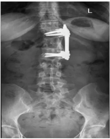
Fig. 1. Posteroanterior (PA) lumbar x-ray radiograph showing internal fix-ation between the left side of the T12 and L2 vertebrae.
http://dx.doi.org/10.1016/j.spinee.2015.12.016
1529-9430/© 2016 Published by Elsevier Inc.
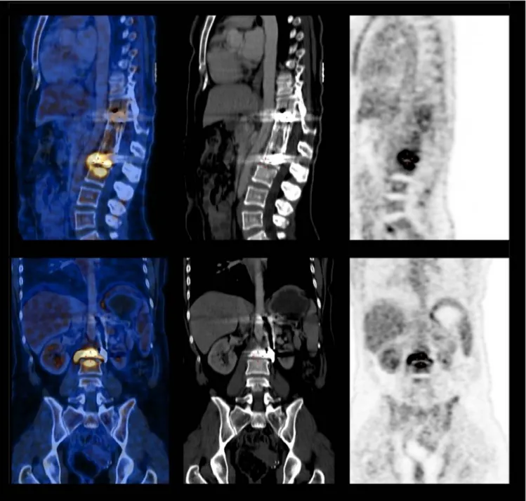
Fig. 2. Sagittal and coronal slices of fluorodeoxyglucose (FDG) positron emission tomography combined with computed tomography (PET/CT) images. Ar-tifact due to internal fixator is seen on the CT images (Middle) of the patient. Attenuation-corrected PET images (Right) and fusion images (Left) show increased glucose metabolism.