An Analysis of Xanthogranulomatous Cholecystitis Cases
Ksantogranülomatöz Kolesistit Vakalarının AnaliziSerhat Tokgöz
1, Muzaffer Akkoca
1, Demet Yılmazer
2, Șener Balas
1, Cem Azılı
1,
Kerim Bora Yılmaz
1, Ahmet Oğuz Hasdemir
11 University of Health Science Dışkapı Yıldırım Beyazıt Reseach and Training Hospital Department of General Surgery
2 University of Health Science, Dışkapı Yıldırım Beyazıt Research and Training Hospital, Department of Pathology, Ankara, Turkey
Aim: Xanthogranulomatous cholecystitis (XGC) is usually an inflammatory condition due to gallstones and bile stasis. Pathological, radiological and clinical features are similar to gallbladder cancer, leading to erroneous treatments such as excessive or inadequate surgery. The aim of this study was to identify the clinical and surgical characteristics of XGC cases.
Materials and Methods: The medical records were reviewed of 55 patients diagnosed as XGC from 4818 patients who underwent cholecystectomy between 2008 and 2015.
Results: The most common clinical finding (90%) was right upper quadrant pain, followed by acute cholecystitis (30%). Biliary wall thickening was found in 64% of patients. Conversion to open cholecystectomy rate was 39%, which was significantly higher than that of all cholecystectomies conversion rates 3.4%. Postoperative biliary fistula was determined at 9% and total morbidity at 23%.
Conclusion: XGC is a rare form of cholecystitis. Although it is histologically benign, preoperative and intraoperative diagnosis is difficult and complicated due to its aggressive course. Although carcinoma may be suspected during surgery in XGC, the association is not very high. It is quite useful to study frozen samples to avoid extensive surgery. The definitive treatment is surgery, but it should not be forgotten that there are high morbidity rates during and after the operation
Key Words: xanthogranulomatosis, cholecystitis, conversion cholecystectomy
Amaç: Ksantogranulomatöz kolesistit (XGC), safra tașı ve safra stazı bağlı gelișen inflamatuar bir durumdur. Patolojik, radyolojik ve klinik özellikleri safra kesesi kanserine benzediğinden, așırı veya yetersiz cerrahi gibi yanlıș tedavilere neden olmaktadır. Bu çalıșmada XGC olgularının klinik ve cerrahi özelliklerini belirlemek amaçlanmıștır.
Gereç ve Yöntem: Kliniğimizde 2008 ile 2015 yılları arasında kolesistektomi yapılan 4818 hastadan XGC tanısı alan 55 hastanın tıbbi kayıtları incelendi.
Bulgular: En sık rastlanan klinik bulgu (% 90) sağ üst kadran ağrısıydı. Hastaların % 30’unda ise akut kolesistit tablosu mevcuttu. Bilier duvar kalınlașması hastaların% 64'ünde bulundu. Laparaskopik kolesistektomiden açığa dönüș oranı ise %39 olarak bulundu ve bu oran tüm kolesistektomilerin konversiyon oranı %3.4 ile karșılaștırıldığında oldukça yüksekti. Postoperatif bilier fistül % 9 ve toplam morbidite ise % 23 olarak tespit edildi.
Sonuç: XGC nadir bir kolesistit formudur. Histolojik olarak benign olmasına rağmen, preoperatif ve intraoperatif tanı agresif seyri nedeniyle zor ve karmașıktır. XGC'de ameliyat sırasında karsinom șüphesi olmasına rağmen ilișkisi çok yüksek değildir. Daha kapsamlı cerrahiden kaçınmak için frozen incelenmesi oldukça yararlıdır. Kesin tedavi ameliyattır ancak ameliyat sırasında ve sonrasında yüksek morbidite oranları olduğu unutulmamalıdır.
Anahtar Sözcükler: Ksantogranülomatöz, Kolesistit, Konversiyon kolesistektomi
Xanthogranulomatous cholecystitis (XGC), is a rare form of cholecystitis with incidence varying from 0.7% to 9% (1). It is characterized by wall thickening due to nodules on the wall. (2) An acute or chronic cholecystitis-like clinical picture can also be seen. In XGC, adhesion because of intense inflammation in the bile duct and surrounding tissues, makes the operation very difficult.
The pathogenesis has not yet been elucidated. Mediators and cytokines secreted by biliary extravasation and delayed cellular immunity have been reported to be responsible for the pathogenesis of biliary stricture and biliary stasis which develops (3,4). Biliary extravasation and lipid-laden histiocytes to the biliary tree muscle layer are also microscopically responsible for bile stapling foci and fibrosis formation (5,6).
Received: Oct. 11, 2017 Accepted: Nov. 02, 2017 Corresponding Author:
Serhat Tokgöz
E-mail: [email protected] GSM: +90 (505) 907 96 66
University of Health Science, Dıșkapı Yıldırım Beyazıt Research and Training Hospital, Șehit Ömer Halisdemir Caddesi Altındağ, Ankara / Turkey
XGC usually occurs clinically as chronic cholecystitis and is difficult to diagnose preoperatively. XGC may be clinically confused with carcinoma due to the presence of inflammatory processes in neighboring organs and the release of serum Carbohydrate Antigen 19-9 (CA 19-9) levels (7,8). Macroscopically, it may be confused with bile duct carcinomas because it can be palpated as a hard mass on the bile duct. As all these findings may suggest malignancy originating from the bile duct or bile ducts, intraoperative frozen examination may be required for differential diagnosis (9). In literature, an association of bile duct carcinoma and XGC has also been reported at rates of 2%-15% (10,11). Up to 25% of XGC can be diagnosed incorrectly, leading to inappropriate treatment such as inadequate surgery (12). Although laparoscopic cholecystectomy is the gold standard for biliary surgery, open cholecystectomy is more prominent in XGC due to the prolonged operation time, high conversion rates (5%-80%) and complications (31%) (13,14) In this article, clinical data and literature
information are presented. It was aimed to give information about XGC which should always be kept in mind in clinical practice because it can be easily diagnosed and in addition to the diagnosis, the difficulties in the treatment process and bile duct carcinoma.
Material and methods
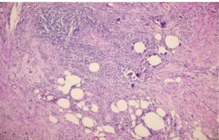
Following the required approval of the Ethics Committee, the pathology records were reviewed of 4818 patients undergoing cholecystectomy between 2008 and 2015. A total of 55 patients with XGC diagnoses were included in the study. A histopathological diagnosis was made from mucosal ulceration in a limited part of the bile duct showing mixed types of inflammatory cells consisting of fibrosis, macrophages, lymphocytes, plasma cells, eosinophil leukocytes, neutrophil leukocytes (Figures 1,2), and cholesterol clefts (Figure 3) body type and Touton type giant cells.
Figure 1) Histopathology findings included severe mucosal ulceration and submucosal bile pigment, Aschoff Rokitansky sinus, macrophages and
inflammatory cells.(H&E X 100).
Figure 2) Histopathology findings included macrophages in the gallbladder wall.(CD68 X 100)
Figure 3) Histopathology findings included inflammation in the gallbladder wall cholesterol clefts Body type and Touton type giant cells. (H&EX400)
Age, gender, and clinical findings (abdominal right upper quadrant pain, acute cholecystitis, nausea-vomiting, obstructive jaundice, carcinoma suspicion) were recorded. Laboratory values White Blood Cells (WBC ), aspartate and alanine aminotransferase (AST,ALT), alkaline phosphatase (ALP) , bilirubin (BİL), radiological findings (bile wall thickness, stone presence and size, fluid presence around the pouch, gall bladder dilatation), endoscopic retrograde cholangiopancreatography (ERCP) , operative form, intraoperative information, postoperative complications, duration
of hospitalization and postoperative morbidity and mortality for acute exacerbation and general status impairment requiring operation were investigated.
Results
In the 4818 cholecystectomy cases performed between 2008 and 2015, the conversion rate was found to be 3.4%. A total of 55 patients (1.2%) were diagnosed with XGC. There were no patients with XGC and carcinoma together. The patients comprised 28 (51%) females and 27 males (49%) with a mean age of 53.8 years (range, 24-85 years). The most common clinical finding in the patients was right upper quadrant pain in the abdomen. Patients have biliary pain attacks that have been passed 3 times in 4 patient, 2 times in 9 patient and once in 37 patient. Acute
cholecystitis was determined in 30%, nausea and vomiting in 25%, and jaundice in 7% of patients (Tables 1 and 2).
Table 1: Clinical findings Abdominal pain
(biliary pain) 50/55 (90%) Acute cholecystitis 17/55 (30%) Nause and vomiting 14/55 (25%) Obstructive jaundice 4/55 (7%) Carcinoma suspicion 2/55 (3.6%)
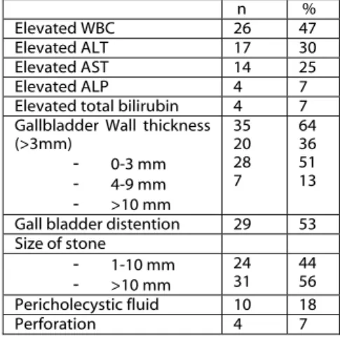
Table 2: Biochemical and USG findings n %
Elevated WBC 26 47
Elevated ALT 17 30
Elevated AST 14 25
Elevated ALP 4 7
Elevated total bilirubin 4 7 Gallbladder Wall thickness
(>3mm) - 0-3 mm - 4-9 mm - >10 mm 35 20 28 7 64 36 51 13 Gall bladder distention 29 53 Size of stone - 1-10 mm - >10 mm 24 31 44 56 Pericholecystic fluid 10 18 Perforation 4 7
WBC elevation in laboratory parameters was the most common finding in 26 patients (47%), followed by ALT in 17 patients, AST in 14 patients and Alkaline phosphatase (ALP) and total bilirubin in 4 patients. Abdominal ultrasonography (USG) was performed on all patients and all were determined with bile stones. Only one patient was suspected with XGC with USG findings (nodule in the gallbladder wall) and XGC preliminary diagnosis could be made. Wall thickness was 0-3 mm in 20 patients, 4-9 mm in 28 patients, and 10 mm in 7 patients (Table 2). (Figures 4,5).
Figure 4-5) Imaging from patient with xanthogranulomatous cholecystitis. Gallbladder diffuse wall thickening
Jaundice was determined in 4 patients during the operation and ERCP in 8 patients because of jaundice due to biliary tract stone. Percutaneous cholecystostomy was applied to 3 patients due to acute cholecystitis, additional disease and deterioration in general condition impairment. Open cholecystectomy and frozen sampling was performed on 2 patients with suspicion of carcinoma due to a mass on the gallbladder wall. Free fluid around the gall bladder was also present in 10 patients, 4 of whom had suspected perforation. Open cholecystectomy was performed on these patients. Gall bladder size was increased in 29 patients. Magnetic Resonance Cholangiopancreaticography (MRCP) was applied to 2 patients with jaundice A total of 15 patients were applied with abdominal computed tomography (CT). Abdominal CT in 2 patients was reported as bile duct carcinoma so these patients underwent open cholecystectomy. Open cholecystectomy was applied to 9
patients, laparoscopic cholecystectomy to 28, conversion cholecystectomy to 18. Of these, 3 had previous surgery, 2 had carcinoma suspicion, and 4 had acute abdomen related to perforation. The conversion cholecystectomy rate in patients with XGC was significantly higher than the conversion rate in all cases (XGC conversion rate: 39%, conversion rate in all cholecystectomies: 3.4%). The causes of conversion were callot dissection difficulty in 11 cases, adherence to the surrounding tissue in 5 cases and choledochal injuries in 2 cases. In the 2 patients with choledochal injuries, cholecystectomy was performed and primary repair and t-tube insertion were applied. (Table 3)
Table 3. Type of surgery
Type of surgery (n)
open 9 Lap converted to open
-obscure calot’s triangle anatomy - adhesion
- bile duct injury
18 11 5 2
As complications, a total of 5 patients developed biliary fistula postoperatively. While this resolved spontaneously in 3 cases, ERCP was performed on 2 patients, 1 of whom died due to sepsis (mortality 1/55). In 3 patients, hospitalization and intravenous antibiotic therapy were required because of infection, and in 5 patients medical treatment was required for infection (pneumonia, wound infection) (Morbidity 13/55). The mean hospital stay of the patients was 6.4 days.
Discussion
Xanthogranulomatous cholecystitis (XGC), is a rare form of chronic cholecystitis and is usually associated with fibrosis. Although the pathogenesis is unknown, there is a view that it is a reactive process that develops through the penetration of bile into the ruptured sac wall from an ulcer of the mucous membrane, or the Aschoff-Rokitansky sinus. It has been reported that Enterobacter can be isolated in biliary culture in 50% of patients and incidence ranges from 0.7 to 9% (1). In the current study, incidence was 1.2%, the female to male ratio was almost equal at F/M: 28/27 , mean age was 53.8 years and the age range was 24-85 years. In previous studies, gender has not been a predictive value for XGC, and mean age has been reported as 58.3 years in the range of 44-63 years (15).
Clinical findings of XGC are nonspecific, and abdominal pain is usually the most common symptom. Abdominal pain has been reported in 84.6% of cases (16,17,18) with the second most common clinical table being acute cholecystitis (53%) (2). XGC is typically indistinguishable from clinical cholecystitis (2,12)
Biliary stones and XGC associations have been reported at rates of 92%-100% (19,20) and similar results were seen in the ccurrent study. It is believed that gallstones cause obstruction in the cystic duct, resulting in bile stasis,
mucosal ulceration and extravasation of the bile causing XGC (2). Wall thickness on USG and the presence of intramural hypoechoic nodules support the XGC diagnosis in the preoperative period. In the current study, only 1 patient had an intramural hypoechoic nodule. USG was performed preoperatively in all cases. The increase in wall thickness was 63%, the increase in gall bladder size was 53%, and fluid around the gall bladder was 18% preoperatively.
However, preoperative ultrasonography was not found to be
useful enough to make XGC diagnosis in this series. Abdominal CT was performed on 15 patients in total. Although 2 were suspected with bile-duct carcinoma, there was no pre-diagnosis of XGC.
An association of bile duct carcinoma and XGC has been reported at 0.09%-1.1% in European literature (21,22) and at 0.3%-4.7% in Far Eastern literature (23,24), but no case was determined with carcinoma in the current study. There is no conclusive evidence that XGC is associated with biliary cancer or an increased risk of biliary cancer in current literature. (25) Despite being histologically benign, excessive or inadequate surgery may be performed due to an aggressive clinical course such as in cancer. The radiological findings of XGC are
non-specific. Compared with the radiological features of carcinoma, bile duct diffuse wall thickening, continuous mucosal line enhancement, submucosal hypoattenuation nodules or bands are
more frequent in XGC (26). Biliary stones and bile duct stones are also more common in XGC.
The positive result of fine needle aspiration biopsy performed in conjunction with endoscopic USG in the preoperative period is important in the diagnosis of cancer and in directing treatment. In a previous study, tissue sampling with endoscopic USG demonstrated that
biliary tree carcinoma could be diagnosed with 93% accuracy (27). However, a negative outcome leads to uncertainty in diagnosis, as well as risks of tumor seeding and fistulae (28).
Although several studies have reported that fluoro-18 fluorodeoxyglucose positron emission tomography (18F-FDG PET) is effective in distinguishing malignant and benign lesions from bile ducts, positive results are not specific, as 18F FDG accumulation is also present in inflammatory cells (29). Again, many studies have shown levels of up to 54% CA19-9 in XGC (30). However, intraoperative frozen sampling is considered the most effective way to prevent unnecessary or excessive surgery (2.20-75). In the current study, 2 patients were evaluated with frozen sampling and consequently, surgical procedures with high morbidity such as radical cholecystectomy, liver bed excision, or lymphadenectomy were avoided.
In XGC, intense inflammation and fibrosis may cause adhesions with neighboring organs, resulting in stricture, fistula, and external pressure (31). External pressure-related jaundice was determined in 4 patients in the current series.
A total of 9 patients underwent open cholecystectomy, including 3 patients with previous surgery, 2 with carcinoma suspicion, and 4 patients with acute abdomen findings due to
perforation. Conversion cholecystectomy was performed in 18
patients (39%) despite the laparoscopic initiation of operation in 46 patients. This rate of conversions was quite high when compared with the conversion rates of all cases. Highly variable conversion rates have been reported in literature ranging from 19% to 80% (13,32). The reasons for conversion were difficulty of callot dissection in 11 cases, adherence to surrounding tissue in 5 cases, and choledochal injuries in 2
cases. In the 2 patients with choledochal injuries, cholecystectomy was performed and primary repair and t-tube were applied. Conversion cholecystectomy was not performed on any patient due to bleeding. Of 9 patients who underwent open cholecystectomy, 2 developed wound infection which resolved with antibiotherapy. In XGC, morbidities such as intraoperative biliary injury and postoperative biliary fistulae have been reported as 15%-28% (14). In the current series, 7 of 55 (12%) developed these morbidities, 2 of which were biliary injuries and 5 were bile fistula. Conversion rates were found to be lower in the current study
compared to rates in literature. Conversion rates decrease with the use of advanced laparoscopic technique and the long learning curve, which suggests that laparoscopic cholecystectomy should be continued despite the higher morbidity rates in XGC than in standard cholecystectomies.
Conclusion
XGC is a rare form of cholecystitis. Although it is histologically benign, preoperative and intraoperative diagnosis is difficult and complicated due to its aggressive course. Even if
carcinoma is suspected during surgery in XGC, the association is not very high. It is quite useful to study frozen samples to avoid extensive surgery. The definitive treatment is surgery but it should not be forgotten that there is a high morbidity rate during and after the operation.
Disclosure of conflict of
interest
The authors declare no conflict of interests regarding the publication of this paper.
REFERENCES
1) Dixit VK, Parakash A, Gupta A, Pandey M, Gautam A, Kumar M, Shukla VK. Xanthogranulomatous cholecystitis. Dig Dis Sci. 1998 May;43(5):940–942. 2) Gilberto Guzman-Valdivia.
Xanthogranulomatous Cholecystitis: 15 years Experience. World J Surg. 2004;28: 254–257.
3) Mori M, Watanabe M, Sakuma M, Tsutsumi Y. Infectious etiology of xanthogranulomatous cholecystitis: Immunohistochemical identification of bacterial antigens in the xanthogranulomatous lesions. Pathol Int. 1999;49:849–852
4) Seiko Sawada, Kenichi Harada, Kumiko Isse, Yasunori Sato, Motoko sasaki, Yasuharu Kaizaki, Yasi Nakanuma. Involvement of Escherichia coli in pathogenesis of xanthogranulomatous cholecystitis with scavenger receptor class A and CXCL16-CXCR6 interaction. Pathol Int. 2007;57:652–663. 5) Goodman Z, Ishak K. (1981) Xanthogranulomatous cholecystitis. Am J Surg Pathol 5:653–659. 6) Roberts K, Parsons M. (1987) Xanthogranulomatous cholecystitis:clinico-pathological study of 13 cases. J Clin Pathol 40:412–417 7) Yoshida J, Chijiiwa K, Shimura H, et al.
Xanthogranulomatous cholecystitis versus gallbladder cancer: Clinical differentiating factors. Am Surg. 1997;63(4):367–371.
8) Adachi Y, Iso Y, Moriyama M, et al. Increased serum CA 19-9 in patients with xanthogranulomatous cholecystitis. Hepatogastroenterology. 1998;45(19):77–80. 9) L.-F.Zhang,C.-S.Hou, J.-Y.Liuet al.,
“Strategies for diagnosisof xanthogranulomatous cholecystitis masquerading as gallbladder cancer,” ChineseMedical Journal, vol. 125, no. 1, pp. 109–113, 2012.
10) Krishnani N, Dhingra S, Kapoor S, Pandey R. (2007) Cytopathologic diagnosis of xanthogranulomatous cholecystitis and coexistent lesions. A prospective study of 31 cases. Acta Cytol 51:37–41.
11) Lee HS, Joo KR, Kim DH, Park NH, Jeong YK, Suh JH et al. (2003) A case of simultaneous xanthogranulomatous cholecystitis and carcinoma of thegallbladder. Korean J Intern Med 18:53–56.
12) Yang T, Zhang B, Zhang J, Zhang Y, Jiang X, Wu M. (2007) Surgical treatment of xanthogranulomatous cholecystitis: experience in 33 cases. Hepatobiliary Pancreat Dis Int 6:504–508.
13) Kwon AH, Matsui Y, Uemura Y. Surgical Procedures and Histopathologic Findings for Patients with Xanthogranulomatous Cholecystitis. J Am Coll Surg. 2004;199:204–210.
14) Guzman- Valdivia G:
Xanthogranulomatous cholecystitis in laparoscopic surgery. J Gastrointest Surg 2005; Apr. 9 (4) : 494–497.
15) Qasaimeh, Ghazi Raji, et al. "Xanthogranulomatous Cholecystitis in the Laparoscopic Era Is Still a Challenging Disease." Journal of Gastrointestinal Surgery 19.6 (2015): 1036-1042.
16) Kwon A-H, Matsui Y, Uemura Y. (2004) Surgical procedures and histopathologic findings for patients with xanthogranulomatous cholecystitis. J Am Coll Surg 199:204–210.
17) Chang BJ, Kim SH, Park HY, Lim SW, Kim J, Lee KH et al. (2010) Distinguishing xanthogranulomatous cholecystitis from the wallthickening type of early-stage gallbladder cancer. Gut Liver 4:518–523. 18) Krishna RP, Kumar A, Singh RK, Sikora
S, Saxena R, Kapoor VK. (2008) Xanthogranulomatous inflammatory strictures of extrahepatic biliary tract: presentation and surgical management. J Gastrointest Surg 12:836–841
19) Ueda J, Yoshıda H, Arıma Y, Mamada Y, Tanıaı N,Mıneta S, Yoshıoka M, Kawano Y, Naıto Z, Uchıda E.A case of xanthogranulomatous cholecystitis preoperativelydiagnosed with contrast-enhanced ultrasonography.J Nihon Med Sch 2011; 78: 194-198.
20) Sharma D, Babu R, Sood G, Kapoor G, Solankı RS, Thomas S. Xanthogranulomatous cholecystitis masquerading as malignancy with liver metastasis.ANZ J Surg 2009; 79: 946-947.
21) Hale, Matthew David, et al. "Xanthogranulomatous cholecystitis: a European and global perspective." HPB 16.5 (2014): 448-458.
22) Morera Ocón FJ, Ballestín Vicente J, Ripoll Orts F, Landete Molina F, García-Granero Ximénez M, Millán Tarín J et al. (2009) [Gallbladder cancer in a regional hospital]. Cir Esp 86:219–223.
23) Choi SB, Han HJ, Kim CY, Kim WB, Song T-J, Suh SO et al. (2009) Incidental gallbladder cancer diagnosed following laparoscopic cholecystectomy. World J Surg 33: 2657–2663.
24) Yamamoto H, Hayakawa N, Kitagawa Y, Katohno Y, Sasaya T, Takara D et al. (2005) Unsuspected gallbladder carcinoma after laparoscopic cholecystectomy. J Hepatobiliary Pancreat Surg 12:391–398. 25) Benbow EW, Taylor PM: Simultaneous
xanthogranulomatous cholecystitis and primary adenocarcinoma of the gallbladder. Histopathology 1988; 12(6): 672–675.
26) Rammohan, Ashwin, et al. "Xanthogranulomatous cholecystitis masquerading as gallbladder cancer: can it be diagnosed preoperatively?." Gastroenterology research and practice 2014 (2014).
27) Hijioka S, Mekky MA, Bhatia V, Sawaki A, Mizuno N, Hara K et al. (2010) Can EUS-guided FNA distinguish between gallbladder cancer and xanthogranulomatous cholecystitis? Gastrointest Endosc 72:622–627.
28) Heimbach JK, Sanchez W, Rosen CB, Gores GJ. Trans-peritoneal fine needle aspiration biopsy of hilar cholangiocarcinoma is associated with disease dissemination. HPB (Oxford). 2011;13:356–360.
29) Sawada, Shigeaki, et al. "Expression of GLUT-1 and GLUT-3 in xanthogranulomatous cholecystitis induced a positive result on 18F-FDG PET: report of a case." International surgery 98.4 (2013): 372-378.
30) Yu H, Yu TN, Caı XJ. Tumor biomarkers: help or mislead in the diagnosis of xanthogranulomatous cholecystitis?-analysis of serum CA 19-9, carcinoembryonic antigen, and CA 12-5. Chin Med J (Engl) 2013; 126: 3044-3047. 31) Krishna RP, Kumar A, Singh RK, Sikora
S, Saxena R, Kapoor VK. Xanthogranulomatous inflamantio stricture of extrahepatic biliary tract: Presentation and surgical management. J Gastrointest Surg. 2008;12:836–841. 32) Srikanth G, Kumar A, Khare R, Siddappa
L, Gupta A, Sikora SS, et al. Should Laparoscopic Cholecystectomy be performed in patients with thick-walled gallbladder? J Hepatobiliary Pancreat Surg. 2004;11:40–44.