DOI 10.1007/s00068-015-0579-8 ORIGINAL ARTICLE
Evaluation of hematological markers in minor head trauma
in the emergency room
E. Acar1 · A. Demir2 · Ö. D. Alatas1 · H. Beydilli1 · B. Yıldırım1 · U. Kırlı3 ·
D. B. Hazer4 · M. R. Kılınç5 · Ü. Karagöz1 · S. Derin6
Received: 28 April 2015 / Accepted: 5 October 2015 / Published online: 19 October 2015 © Springer-Verlag Berlin Heidelberg 2015
tests (Mann–Whitney U test) were used with non-normally distributed data. A p ≤ 0.05 was considered significant. Finding When we divided the cases into two groups based on the presence of pathologies on CT scan, we deter-mined that there were significant differences between the groups in terms of white blood cell (WBC), hemoglobin (Hb), mean platelet volume (MPV), neutrophil (neu), tro-ponin T, neutrophil/lymphocyte ratio (NLR) and platelet/ lymphocyte ratio (PLR). When differentiating the patients that had brain pathologies on CT scan from patients that had normal CT scans, the troponin T cut-off value of 6.16 lead to 90 % specificity, and setting the NLR cut-off value at 4.29 resulted in a specificity of 90 %.
Conclusion MPV, NLR, and troponin T can be used as parameters that indicate brain pathologies on CT scans of patients presenting to the emergency department with iso-lated minor head trauma and GCS of 15 when the necessity of a CT scan is otherwise unclear.
Keywords Minor head trauma · CT requirements ·
Hematological markers · Emergency room
Introduction
Minor head trauma is one of the most common injury types seen in the emergency department. Every 15 s a head injury is sustained, and every 12 min there is a death due to head trauma [1, 2]. Therefore, emergency physicians are faced with head trauma very often. Patients with minor head trauma constitute approximately 80 % of all head trauma patients admitted to the emergency room and are character-ized as having a Glasgow Coma Score (GCS) of 15, loss of consciousness for less than 20 min, and no neurological deficits [3].
Abstract
Introduction A rule exists regarding the use of computed tomography (CT) for patients presenting to the emergency department with head trauma and a Glasgow coma score (GCS) of 15; however, it can be difficult to make this deci-sion due to overcrowded emergency rooms or exaggerated patients complaints. We evaluated patients who presented to the emergency room with minor head trauma, and we aimed to investigate the relationship between brain pathol-ogy on CT and hematological markers in order to find markers that help us identify brain pathology in patients with a GCS of 15.
Methods This retrospective study included 100 patients with pathologies present on their CT scans and a control group consisting of 100 patients with a normal CT. All data obtained from this study were recorded and evaluated using “Statistical Package for Social Sciences for Windows 20” program. Parametric tests (independent samples test) were used with normally distributed data, while non-parametric
* E. Acar
1 Department of Emergency Medicine, Mugla Sitki Kocman
Universty Medicine School, Mugla, Turkey
2 Sultanbeyli State Hospital, Emergency Service, Istanbul,
Turkey
3 Department of Pediatry, Mugla Sitki Kocman Universty,
Mugla, Turkey
4 Department of Neurosurgery, Mugla Sitki Kocman Universty,
Mugla, Turkey
5 Department of Radyology, Mugla Sitki Kocman Universty,
Mugla, Turkey
6 Department of Otolaryngology, Mugla Sitki Kocman
When patients are admitted to the emergency room, they are classified according to the GCS, and CT scans are taken for patients whose GC scores are less than 15. Although there is a variety of data regarding the requirement for a CT scan in patients whose GCS is 15—for example, head-ache, suspected open or depressed skull fracture, any sign of basal skull fracture (hemotympanum, “panda” eyes, cerebrospinal fluid otorrhoea, Battle’s sign), drug or alco-hol intoxication, vomiting more than once, persistent anterograde amnesia (deficits in short-term memory), age >65 years, dangerous mechanism of injury (pedestrian struck by vehicle, ejection from vehicle, fall from greater than three feet or five stairs [4, 5]—emergency room physi-cians face a difficult decision due to exaggerated patients complaints and overcrowded emergency rooms [6]. Phy-sicians are often inclined to order CT scans for patients, which, if unnecessary, can lead to excessive radiation expo-sure for patients and can also lead to misuse of economic resources. On the other hand, bypassing the CT scan may result in missing pathologies and therefore not obtaining a diagnosis, which in turn could lead to the formation of per-manent brain damage or even death [7, 8].
Neu, lymphocytes (lym), NLR, PLR, and red cell distri-bution width (RDW) are known to indicate an inflamma-tory process [9, 10]. In the pathophysiology of intracranial events in head traumas, physical damage that occurs imme-diately following head trauma is followed by secondary tis-sue damage. The posttraumatic inflammatory response is one of the links in the chain of events that lead to tissue damage [1].
Our study focused on patients that were admitted to the emergency room with isolated head trauma and had a GCS of 15. We wanted to identify whether or not there is a connection between computed tomography requirement and hematological markers in minor head trauma. Taking inflammation into consideration, we investigated whether inflammatory markers such as neu, lym, NL), PLR, and RDW could serve as objective indicators for CT scan requirement for suspected cases in patients with a GCS of 15.
Methods
Patients, setting, and inclusion–exclusion criteria
This retrospective study was conducted between 1 January 2013 and 31 December 2014 in our university research hos-pital emergency department with the approval of the local ethics committee. We retrospectively examined the files of patients over a 2-year period. A total of 200 patients with minor head trauma that presented to the emergency room with isolated head trauma were included in the study. The
case group consisted of 100 patients who had some patho-logical condition on CT scan, while the control group con-sisted of 100 patients with normal CT scans.
When reviewing the files of patients, detailed neurologi-cal examination, the causes of trauma, and patients’ com-plaints (headache, nausea, vomiting, loss of consciousness, and duration of seizures) were recorded in the patient his-tory. We selected patients in whom a CT scan was per-formed that had presented with isolated head trauma, did not have focal neurological symptoms, and had a GCS of 15. Patients CT were examined by a neurosurgeon and a radiologist and an otolaryngologist to select the isolated head trauma. The results of the routine complete blood count and biochemical tests taken from these patients, the CT scan results, whether the patient was hospitalized, the provided treatment, and the final status of the patients were recorded. Unenhanced CT scanning was performed with 16-slice Toshiba Asteion TSX-021B (Toshiba Medi-cal Systems Corporation) within 30–60 min of emergency department admission. When evaluating a CT scan, epi-dural hematoma, subepi-dural hematoma, intracranial contu-sion and hematomas, subarachnoid hemorrhage, cerebral edema, pneumocephalus, and skull fractures were consid-ered pathologies. Hematomas and cuts outside the skull and intracranial structures were not accepted as patholo-gies. Patients whose GCS scores were below 15, patients who had multiple traumas, chest pain, anemia, or chronic renal failure were excluded from the study. For statistical analysis, epidural hematoma, subdural hematoma, intracra-nial contusions and hematomas, and subarachnoid hemor-rhage were considered intracranial hemorhemor-rhage, while the other pathologies were considered skull fracture.
Laboratory methods
Blood test results of the patients at their first admission to the emergency service of our hospital were reviewed. Dur-ing the study period, blood samples were drawn into tubes containing sodium citrate, and while blood samples for tro-ponin T were drawn into pure, red-capped tubes and ana-lyzed under room temperature in the biochemistry labora-tory using Pentra DF Nexus, Hariba Medical device, and troponin T was measured with Roche Hitachi Cobas E 601 device. The patients’ WBC (4.5–11.0 × 10³/µL), Hb (13.5–
16.0 g/dL), neu (2–12 K/mL), lym (1–4.9 K/mL), mean cor-puscular volume (MCV) (80–100 fL), RDW (11.8–14.3 %), MPV (6.9–10.8 fL), platelet count (plt) (156–373 × 103/
µL), and troponin T (14–100 pg/mL) were evaluated.
Statistical analysis
All data obtained from this study were recorded and evaluated using “Statistical Package for Social Sciences
for Windows 20” program. Quantitative variables were given as mean ± SD (standard deviation), while the cat-egorical variables were given as numbers and percent-ages. A frequency analysis was also performed. The normalcy of distribution was evaluated when compar-ing the groups. Parametric tests (independent samples test) were used with normally distributed data, while non-parametric tests (Mann–Whitney U test) were used with non-normally distributed data. The Chi-square test was used to compare two percentage data. The relation-ship between the demographic and hematological param-eters between the two groups was examined by backward logistic regression analysis. A p ≤ 0.05 was considered significant.
Results
A total of 200 patients who presented with isolated minor head trauma were included in our study. The mean age of the patients was 35.25 ± 20.25, and among these patients, 151 (75.5 %) were men and 49 (24.5 %) were women. It was noted that 169 (84.5 %) of the patients presented to the emergency department within 1 h of the accident. Evalu-ation of the CT scans showed that 100 patients had brain pathologies, while the CT scans of 100 patients were normal. Twenty-eight of the patients (14 %) with brain pathologies had skull fractures and 72 (36 %) had intrac-ranial hemorrhage (27 patients with epidural hematoma, 24 patients with subdural hematoma, 15 patients with subarachnoid hemorrhage, and 6 patients with intracer-ebral hematoma). Ninety-six patients (48 %) required hospitalization, and 41 patients (20.5 %) required surgical treatment. By the end of the study, 167 patients (83.5 %) were discharged and 17 patients (8.5 %) were transferred to another institution for some reason.
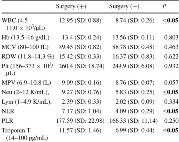
When we divided the cases into two groups based on the presence or absence of pathology on CT scan, we deter-mined that WBC, Hb, MPV, neu, troponin T NLR and PLR were statistically significant variables (Table 1). Moreover, the evaluation of the cases in terms of treatments revealed that WBC, neu, NLR and troponin T were statistically sig-nificant variables (Table 2).
The areas under the curve (AUC) of MPV and NLR were lower than the AUC of troponin T (the AUC for MPV, NLR, troponin T were 0.636, 0.765 and 0.873, respec-tively) (Fig. 1). When compared in terms of diagnostic value, WBC, MPV, neu, NLR, PLR, and troponin T were found to be statistically significant in detecting pathologies on CT scans of patients with minor head injury (Table 1).
Table 1 The relationship between hematological markers and CT imaging pathology in minor head trauma
Bold values are statistically significant
Pathology (+) Pathology (−) P
Age 37.39 (SD: 21.3) 33.1 (SD: 19.0) 0.140
Sex 75 % male, 25 % female 76 % male, 24 % female 0.870
WBC (4.5–11.0 × 10³/µL) 11.6 (SD: 2.1) 7.56 (SD: 2.34) ≤0.05 Hb (13.5–16 g/dL) 13.2 (SD: 0.15) 13.87 (SD: 0.13) ≤0.05 MCV (80–100 fL) 89.3 (SD: 0.56) 88.54 (SD: 0.5) 0.174 RDW (11.8–14.3 %) 15.48 (SD: 0.18) 16.84 (SD: 1.32) 0.264 Plt (156–373 × 103/µL) 247.81 (SD: 9.59) 255.43 (SD: 7.6) 0.265 MPV (6.9–10.8 fL) 9.07 (SD: 0.1) 8.6 (SD: 0.08) ≤0.05 Neu (2–12 K/mL) 8.49 (SD: 0.42) 4.54 (SD: 0.19) ≤0.05 Lym (1–4.9 K/mL) 2.04 (SD: 0.16) 2.15 (SD: 0.10) ≤0.05 NLR 6.79 (SD: 0.55) 2.61 (SD: 0.16) ≤0.05 PLR 179.47 (SD: 12.3) 156.85 (SD: 15.64) ≤0.05 Troponin T (14–100 pg/mL) 11.63 (SD: 0.78) 4.24 (SD: 0.18) ≤0.05
Table 2 The relationship between hematological markers and treat-ment options
Bold values are statistically significant
Surgery (+) Surgery (−) P WBC (4.5– 11.0 × 10³/µL) 12.95 (SD: 0.88) 8.74 (SD: 0.26) ≤0.05 Hb (13.5–16 g/dL) 13.4 (SD: 0.24) 13.56 (SD: 0.11) 0.803 MCV (80–100 fL) 89.45 (SD: 0.82) 88.78 (SD: 0.48) 0.463 RDW (11.8–14.3 %) 15.42 (SD: 0.33) 16.37 (SD: 0.83) 0.622 Plt (156–373 × 103/ µL) 260.4 (SD: 18.74) 249.9 (SD: 6.08) 0.932 MPV (6.9–10.8 fL) 9.09 (SD: 0.16) 8.76 (SD: 0.07) 0.057 Neu (2–12 K/mL), 9.27 (SD: 0.76) 5.83 (SD: 0.25) ≤0.05 Lym (1–4.9 K/mL), 2.39 (SD: 0.33) 2.02 (SD: 0.09) 0.334 NLR 7.17 (SD: 1.04) 4.09 (SD: 0.29) ≤0.05 PLR 177.59 (SD: 22.98) 166.33 (SD: 11.14) 0.250 Troponin T (14–100 pg/mL) 11.57 (SD: 1.46) 6.99 (SD: 0.44) ≤0.05
When differentiating the patients that had brain patholo-gies on CT scan from patients that had normal CT scans, setting the MPV cut-off value at 9.61 gave specificity of 89 %. Setting the NLR cut-off value at 4.29 resulted in
90 % specificity, while setting the troponin T cut-off value at 6.16 lead to 90 % specificity (Table 3).
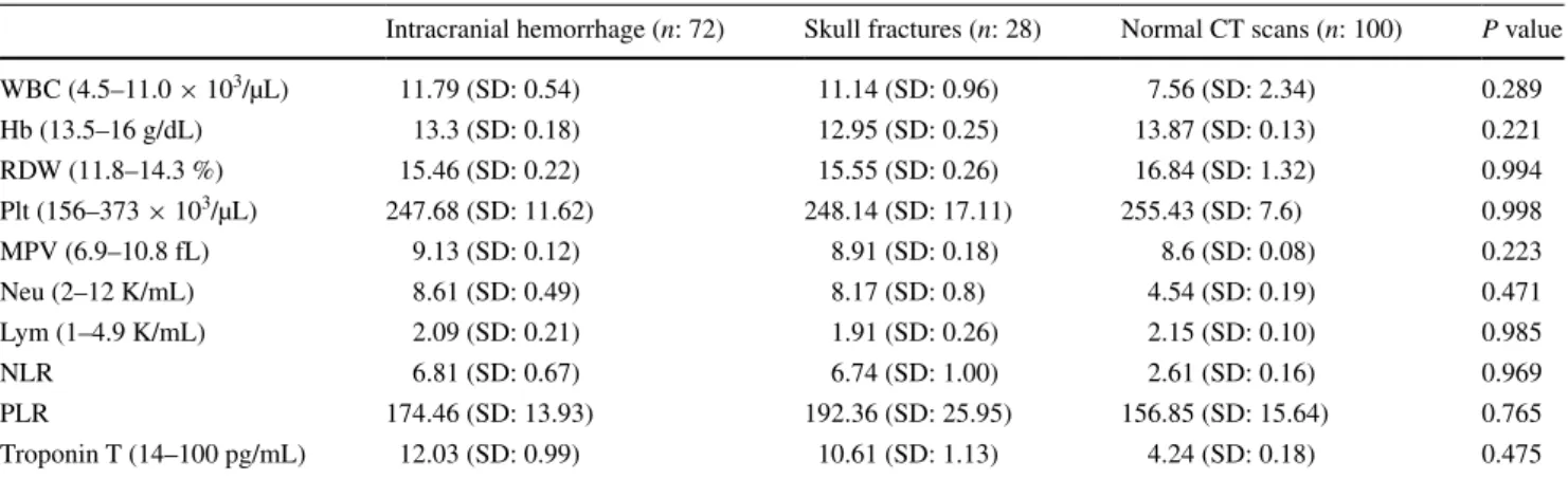
The evaluation of the patients in terms of type of pathol-ogy observed in the CT scan showed that 72 patients had intracranial hemorrhage, 28 patients had skull fractures and 100 patients had normal CT scans. The assessment of the hematological inflammatory parameters showed that there were no significant differences between patients with intracranial hemorrhage and patients with skull fractures (p ≥ 0.05) (Table 4). However, there were significant differ-ences between patients with normal CT scans and patients with intracranial hemorrhage in terms of WBC, Hb, RDW, MPV, Neu, lym, NLR, PLR and troponin T (p ≤ 0.05). Moreover, significant differences between patients with normal CT scans and patients with skull fractures in terms of WBC, Hb, neu, lym, troponin T and NLR were also observed (p ≤ 0.05).
Discussion
In this study, we found that parameters such as WBC, RDW, MPV, neu, troponin T and NLR were significantly different between patients with the presence and absence of brain pathologies on the CT scans presenting to the emer-gency department with isolated minor head trauma and a GCS of 15.
There is a common protocol in place for patients with minor head trauma concerning the requirement for CT scan in patients. However, some factors such as exagger-ated patient complaints and overcrowded emergency room may force us when evaluating patients [6]. The study by Boran et al. [3] aimed to determine the clinical symptoms that could indicate intracranial pathologies and serve as indications for CT scan requirement in patients that pre-sented to the emergency room with head trauma and had a GCS of 15. They found that the occurrence of pathologies in the CT scan were significantly higher in patients older
Fig. 1 The remaining areas under the ROC curve of hematological markers
Table 3 Specificity and sensitivity values of hematological markers Parameter Cut-off value Specificity (%) Sensitivity (%)
Troponin T 3.46 43 90 Troponin T 6.16 90 73 NLR 1.2 26 90 NLR 4.29 90 58 MPV 7.85 24 89 MPV 9.61 89 28
Table 4 The relationship between the hematological markers and intracranial hemorrhage, skull fractures or normal CT scans
Intracranial hemorrhage (n: 72) Skull fractures (n: 28) Normal CT scans (n: 100) P value
WBC (4.5–11.0 × 10³/µL) 11.79 (SD: 0.54) 11.14 (SD: 0.96) 7.56 (SD: 2.34) 0.289 Hb (13.5–16 g/dL) 13.3 (SD: 0.18) 12.95 (SD: 0.25) 13.87 (SD: 0.13) 0.221 RDW (11.8–14.3 %) 15.46 (SD: 0.22) 15.55 (SD: 0.26) 16.84 (SD: 1.32) 0.994 Plt (156–373 × 103/µL) 247.68 (SD: 11.62) 248.14 (SD: 17.11) 255.43 (SD: 7.6) 0.998 MPV (6.9–10.8 fL) 9.13 (SD: 0.12) 8.91 (SD: 0.18) 8.6 (SD: 0.08) 0.223 Neu (2–12 K/mL) 8.61 (SD: 0.49) 8.17 (SD: 0.8) 4.54 (SD: 0.19) 0.471 Lym (1–4.9 K/mL) 2.09 (SD: 0.21) 1.91 (SD: 0.26) 2.15 (SD: 0.10) 0.985 NLR 6.81 (SD: 0.67) 6.74 (SD: 1.00) 2.61 (SD: 0.16) 0.969 PLR 174.46 (SD: 13.93) 192.36 (SD: 25.95) 156.85 (SD: 15.64) 0.765 Troponin T (14–100 pg/mL) 12.03 (SD: 0.99) 10.61 (SD: 1.13) 4.24 (SD: 0.18) 0.475
than 60 years, patients that had penetrating trauma, patients exposed to non-vehicle traffic accidents, and patients who experienced posttraumatic seizures. However, patients with minor head trauma in their study did not belong to the above described group but rather were under 60 years of age, had a GCS of 15, did not have focal neurological deficits, and did not experience either loss of consciousness or seizures after the trauma. Only two patients in their study group had pathologies on their CT scans, thus giving a pathol-ogy detection rate of 0.6 %. In another study, Probst et al. [8] evaluated factors that influenced indications for order-ing CT scans and concluded that factors such as easy access to CT, clinical culture, changes and expectations regarding the patient, physicians’ experience, educational status, error and malpractice fear, and fear of exposing the patient to material or radiation damage may influence the decision to order a CT scan in the emergency department. In our study, we found that hematological parameters and troponin T were significantly different based on the presence of brain pathologies on the CT scans. While there is not a universal consensus, there are two very well developed and validated decision rules, the Canadian and New Orleans head CT cri-teria, but we did not comply with these criteria due to some factors. Therefore, the use of these markers may be valuable for suspected cases. We think these results can be controlled or developed by more extensive studies.
The most important result of our study was that MPV, NLR, and troponin T were significantly elevated in patients that had pathologies present on their CT scans. We believe that these parameters could be used as objective indicators for CT scan requirement in borderline patients. MPV and NLR are known to indicate an inflammatory process [9, 10]. However, the role of the inflammatory process is not well defined in the pathophysiology of intracranial events in head traumas. The physical damage that occurs immedi-ately following head trauma is followed by secondary tis-sue damage. The posttraumatic inflammatory response is one of the links in the chain of events that leads to tissue damage. Continuation of the inflammatory process leads to damage of vascular structures, which in turn leads to blood–brain barrier disruption and brain edema [1]. MPV and NLR are markers that can be examined by complete blood count within minutes of patients’ admission to the emergency department. This method is easily accessible and effortless, which are the major advantages of this test. In addition, these parameters can serve as excellent inflam-matory markers and therefore have been used in many stud-ies. Lippi et al. [11] evaluated hematological markers of patients with isolated minor head trauma and healthy con-trols and determined that MPV was significantly different in patients with head trauma. The authors concluded that the higher prevalence of small and hyporeactive thrombo-cytes in minor head trauma might be the reason for their
observed difference. NLR has been studied in solid tumors and cardiac disease and has been reported to be related to prognosis. In fact, in many studies it has been suggested that inflammation might be responsible for the prognostic value of the NLR [12–14].
Many markers have been studied in minor head trauma. For example Wolf et al. [15] evaluated S100 protein and NSE of patients with isolated minor head trauma and healthy controls and determined that NSE and S100 protein were significantly different in patients with head trauma. In another study, Kavalci et al. [16] evaluated tau protein in patients with minor head trauma and determined that tau protein was not significantly different in patients with head trauma. In another study, Hensler et al. [17] evaluated inter-leukins 10 and 13 in patients with severe head trauma. They determined that interleukins 10 and 13 were significantly different in patients with severe head trauma, and they explained that this was due to inflammation. To the best of our knowledge, both MPV and NLR have never been reported to be used as markers in minor head trauma; there-fore we can claim that the present study is the first regard-ing this subject. The elevated levels of markers in our study can be explained by inflammation, which is also compat-ible with the literature. Our work is valuable in terms of being the first such study in the literature; however, further studies are needed to support our results.
Troponins, along with tropomyosins, are structural pro-teins that play a role in the regulation of skeletal and car-diac muscle contraction. Since the year 2000, they have been used as standard markers in acute coronary syndromes [18]. Moreover, these proteins have been reported to be elevated in chronic kidney failure, pulmonary embolism, and sepsis [19]. In recent years, some studies reported that troponin values increased after head trauma. Salim et al. [20] reported elevated troponin levels in patients with head trauma, and this increase was associated with the severity of the head trauma. Moreover, they noted that, although the reason for the increase in troponin levels following neuro-logic events is not fully understood, it might be related to increased catecholamine levels after brain injury. In addi-tion, Lippi et al. [21] also reported that cardiac troponin I levels were significantly higher in patients with minor head trauma and stated that it might be due to intracranial pathologies affecting the myocardium. In our study, the tro-ponin levels were also significantly elevated. Although we cannot fully explain the pathology, we also believe that it might be due catecholamines in the circulation and cranial pathologies affecting the myocardium.
In conclusion, we believe MPV, NLR, and troponin T can be used as parameters that indicate brain patholo-gies on CT scans of patients presenting to the emergency department with isolated minor head trauma and a GCS of 15 when the necessity of a CT scan is otherwise unclear.
Limitation
Firstly, the study had undergone planning to prospectively investigate as planned; we may also have a lot of data in our control. But this was a file screening study, which tried to determine the importance of the inflammatory markers in minor head trauma. Essentially as the most important marker of inflammation CRP, interleukins and the oth-ers were not observedly tested in file screening studies. If they were tested, then inflammatory process could be more clearly revealed. These deficiencies are the limitation of our study.
Compliance with ethical standards
Conflict of interest Ethem Acar, Ahmet Demir, Ömer Dogan Alatas, Halil Beydilli, Birdal Yıldırım, Ulviye Kırlı, D. Burcu Hazer, M.Rabia Kılınç, Ülkü Karagöz and Serhan Derin declare that they have no con-flict of interest.
Compliance with ethical requirements The study was conducted in the Mugla Universty Training and Education Hospital in Turkey. Ethical clearance was obtained from the ethics committee of Mugla University (no:2014/12).
References
1. Dalbayrak S, Gumustas S, Bal A, Akansel G. Early and delayed CT findings in patients with mild-to-moderate head trauma. Turk Neurosurg. 2011;21(4):591–8.
2. Gentry LR. Imaging of closed head injury. Radiology. 1994;1:1–17.
3. Boran BO, Barut N, Akgün C, Çelikoğlu E, Bozbuğa M. Indica-tions for computed tomography in patients with mild head inju-ries. Ulus Travma Acil Cerrahi Derg. 2005;3:218–23.
4. Stiell IG, Wells GA, Vandemheen K, Clement C, Lesiuk H, Lau-pacis A, et al. The Canadian CT head rule for patients with minor head injury. Lancet. 2001;357:1391–6.
5. Kavalci C, Aksel G, Salt O, Yilmaz MS, Demir A, Kavalci G, et al. Comparison of the Canadian CT head rule and the New Orleans criteria in patients with minor head injury. World J Emerg Surg. 2014;9:31.
6. Jeret SJ, Menachem M, Anziska B, Lipitz M, Vilceus AP, Ware JA, et al. Clinical predictors of abnormality disclosed by com-puted tomography after mild head trauma. Neurosurgery. 1993;32:9–15 (discussion 15–6).
7. MacLaren RE, Ghoorahoo HI, Kirby NG. Use of an accident and emergency department observation ward in the management of head injury. Br J Surg. 1993;80:215–7.
8. Probst MA, Kanzaria HK, Schriger DL. A conceptual model of emergency physician decision making for head computed tomog-raphy in mild head injury. Am J Emerg Med. 2014;32:645–50. 9. Peng W, Li C, Wen TF, Yan LN, Li B, Wang WT, et al.
Neutro-phil to lymphocyte ratio changes predict small hepatocellular carcinoma survival. J Surg Res. 2014;192:402–8.
10. Bhat T, Teli S, Rijal J, Bhat H, Raza M, Khoueiry G, et al. Neu-trophil to lymphocyte ratio and cardiovascular diseases: a review. Expert Rev Cardiovasc Ther. 2013;11:55–9.
11. Lippi G, Carbucicchio A, Benatti M, Cervellin G. The mean platelet volume is decreased in patients with mild head trauma and brain injury. Blood Coagul Fibrinolysis. 2013;24:780–3. 12. Templeton AJ, Pezero C, Omlin A, McNamara MG,
Leibow-itz-Amit R, Vera-Badillo FE, et al. Simple prognostic score for metastatic castration-resistant prostate cancer with incor-poration of neutrophil-to-lymphocyte ratio. Cancer Month. 2014;120:3346–52.
13. Soylu K, Yuksel S, Gulel O, Erbay AR, Meric M, Zengin H, Museyibov M, et al. The relationship of coronary flow to neutro-phil/lymphocyte ratio in patients undergoing primary percutane-ous coronary intervention. J Thorac Dis. 2013;5:258–64. 14. Shen XH, Chen Q, Shi Y, Li HW. Association of
neutrophil/lym-phocyte ratio with long-term mortality after ST elevation myo-cardial infarction treated with primary percutaneous coronary intervention. Chin Med J (Engl). 2010;123:3438–43.
15. Wolf H, Frantal S, Pajenda GS, Salameh O, Widhalm H, Hajdu S, et al. Predictive value of neuromarkers supported by a set of clinical criteria in patients with mild traumatic brain injury: S100B protein and neuron-specific enolase on trial: clinical arti-cle. J Neurosurg. 2013;118:1298–303.
16. Kavalci C, Pekdemir M, Durukan P, Ilhan N, Yildiz M, Serhatli-oglu S, et al. The value of serum tau protein for the diagnosis of intracranial injury in minor head trauma. Am J Emerg Med. 2007;25:391–5.
17. Hensler T, Sauerland S, Riess P, Hess S, Helling HJ, Andermahr J, et al. The effect of additional brain injury on systemic inter-leukin (IL)-10 and IL-13 levels in trauma patients. Inflamm Res. 2000;49:524–8.
18. Sheehan P, Vasikaran SD. The evolving clinical role of car-diac troponins and new acute myokardial infarction guidelines: implications for the clinical laboratory. Clin Bichemist Rev. 2001;23:52–65.
19. Hays A, Diringer MN. Elevated troponin levels are associated with higher mortality following intracerebral hemorrhage. Neu-rology. 2006;66:1330–4.
20. Salim A, Hadjizacharia P, Brown C, Inaba K, Teixeira PG, Chan L, et al. Significance of troponin elevation after severe traumatic brain injury. J Trauma. 2008;64:46–52.
21. Lippi G, Dipalo M, Carbucicchio A, Aloe R, Benatti M, Cer-vellin G. The concentration of highly-sensitive troponin I is increased in patients with brain injury after mild head trauma. Int J Cardiol. 2013;168:1617–8.