https://doi.org/10.1007/s11033-020-05833-5
ORIGINAL ARTICLE
Antioxidant activity, phytochemical composition of Andricus
tomentosus and its antiproliferative effect on Mia-Paca2 cell line
Özge Kılınçarslan Aksoy1 · Ramazan Mammadov2 · Mücahit Seçme3Received: 17 June 2020 / Accepted: 7 September 2020 / Published online: 28 September 2020 © Springer Nature B.V. 2020
Abstract
Plant derived products are widely used in cancer treatment. Gall species has been preferred for treatment various diseases in folk medicine, but there are few studies on the anticancer effects of gall species. The present study reports the antioxidant activity and total secondary metabolites of extracts of A. tomentosus galls. The antioxidant potency of galls was carried out using different in-vitro model systems. Their cytotoxic efficacy on Mia-Paca cell line was also explored. Gall extract was found to contain a large amount of phenolic acids. The extract potently scavenged free radicals including DPPH (IC50
9.56 ± 1.08 µg/mL), ABTS (IC50 18.51 ± 0.25 µg/mL). It can be seen as a potential source of antioxidants according to β-carotene/linoleic acid method (%92.58 ± 0.92) and Phosphomolybdenum assays (104.36 ± 4.95 mgAE/g). Gall extract also posses ability of metal chelating (%40.07 ± 2.30) and Fe3+ (184.01 ± 2.83 mgTE/g) and Cu2+ (89.81 ± 0.96 mgTE/g)
reducing activity. According to total secondary metabolites tests, gall extract showed high total phenolic, total flavonoid and total tannin amount. HPLC analysis of the extract suggested it to contain caffeic acid (424.068.479 µg/g), ellagic acid (187.696.132 µg/g). XTT assay revealed gall extract to enhance percent survival of Mia-Paca2 cell line exposed A.
tomento-sus extracts. The best cytotoxic effect was determined in acetone extracts (IC50: 124.7 µM). Expression of some genes (Bax,
Bcl-2, FAS, BID, caspase-3, caspase-8, caspase-9, caspase-10, FADD, TRADD) in the apoptosis pathway was determined
to invastigate apoptosis inducing activity. These results indicate that A. tomentosus galls possess potent antioxidant activity, when tested both in chemical as well as biological models.
Keywords Andricus tomentosus · Galls · Antioxidant · XTT · HPLC
Introduction
In recent years, the use of herbs for the prevention and treat-ment of cancer has gained more attention, due to various phytochemical components and fewer side effects. Cytotoxic potentials of four different medicinal plants against six dif-ferent cancer cell lines (A-549, CCC-222, DU-145, MCF-7, K-562 and PC-3) and one normal cell line (Beas-2B) were investigated [1]. In another study was revealed that three
different plant extracts have cytotoxic effect on MCF-7 cell line [2].
In one of the studies, phenolic compounds found in
Oci-mum basilicum and Thymus algeriensis essential oils were
analyzed by High Pressure Liquid Chromatography (HPLC) and a rich flavonoid content was revealed. According to this research, the essential oil analysis of Ocimum basilicum and
Thymus algeriensis shows that they respectively contains 26
and 29 different components. These two phytochemically rich species were found to have moderate antioxidant activ-ity, a weak antimicrobial effect and cytotoxic effects on dif-ferent cell lines [3].
Coriander (Coriandrum sativum L.) is a plant from the Apiaceae family and has been used for both medicinal and nutritional properties for centuries. In a study investigat-ing the effects of C. sativum extract on gene expression, viability, colony formation, migration and invasion of human prostate cancer cell line (PC-3) and Lymph Node Carcinoma
* Özge Kılınçarslan Aksoy [email protected]
1 Department of Biology, Science & Art Faculty, Pamukkale University, Denizli, Turkey
2 Department of Molecular Biology an Genetic, Muğla Sıtkı Koçman University, Mugla, Turkey
3 Department of Medical Biology, Medicine Faculty, TurkeyPamukkale University, Denizli, Turkey
of the Prostate cancer cell lines (LNCaP), the effects of C.
sativum extracts on prostate cancer are presented [4]. Pancreatic cancer is one of the leading causes of cancer-related death in World [5]. The incidence rate of pancreatic cancer worldwide was shared as 2.5 in the data of Globo-can in 2018. Pancreas Globo-cancer is the 9th common type of cancer in Turkey. Mortality rate was recorded at %99.1 in Turkey and %94.2 in the world [6]. It has been suggested that traditional chemotherapy is not sufficient in the treatment of pancreatic cancer and some studies that investigate the effects of natural products on treatments attract attention [7]. Some studies investigating the anticancer properties of plant derived natural components show that phenolic components such as quercetin, myricetin, apigenin, naringenin, epigal-locatechin-3-gallate have antipancreatic cancer [8]. Tannic acid (TA) is an another polyphenolic compound obtained from plant origin. Previous studies have shown multiple human health benefits of TA including the anti-cancer abil-ity [9, 10]. In a research have been reported that efficient pancreatic cancer therapy was developed with tannic
acid-pectine nano-complexes [11]. Some galls of Quercus
spe-cies possess hydrolyzable tannin is an antioxidant compound
and is produced by gall wasp [12]. The main component
of oak gall is tannin (50–70%) especially the galicine acid that is formed of tannin acid, gallic acid, ellagic acid etc. [13]. Quercus galls are widely used in folkmedicine due to their multiple therapeutic properties [14]. In some traditional medicine, gall samples were used for treatment of some disease and pharmacological studies were shown that oak galls possess some biological activities such as antibacterial, antioxidant, local anaesthetic, anti-inflammatory, anticancer, analgesic, Angiotensin converting enzyme (ACE) inhibitory and antivenom activities [15, 16].
In the present study, we purposed to revealed the anti-oxidant capacity, phytochemical ingredient and cytotoxic activity of extracts prepared from Andricus tomentosus is an oak gall.
Materials and methods
Plant materialThe galls result from the tumors provoked by the bite of female cynipid wasps in the buds of this plant [17]. A.
tomentosus (Trotter, 1901) was collected from Seydikemer,
Muğla (September 2018, May 2019), is an asexual oak gall.
A. tomentosus prefers Q. cerris, Q. frainetto, Q. infectoria, Q. lusitanica, Q. petraea, Q. pubescens, Q. robur [18, 19] such as host. We collected from Q. infectoria Oliver in this study.
A. tomentosus has a single chamber. Its best
distinguish-ing feature is that it is covered with a velvety cover. It is
usually in the form of a cone. The mature gall is 12–18 mm tall and the base is 10–15 mm in diameter. This gall develops in late summer and matures in autumn [20]. The collected gall samples were identified by Prof.Dr. Yusuf Katılmış in Entomology Laboratory, Pamukkale University, Turkey.
Preparation of extracts
The pulverised gall (30 g) were separately subjected to sol-vent extraction in a Water Bath with acetone, ethanol, meth-anol and water (50 °C, 6 h). Then solvents were evaporated with IKA RV 10 Digital Rotary Evaporator, samples were lyophilised (Labconco Freezone 6). Lyophilized samples were stored at − 20 °C. The dried extracts were weighed to determine the percent of yield [21].
The percentage yield was obtained using this formula W1/W0 × 100. Where W1 is the final weight of the extract and W0 is the initial weight of sample.
Determination of total antioxidant activity
β-carotene-linoleic acid assay
This method is based on the monitoring of the color open-ing of β-carotene by alkyl peroxides formed by free radical chain reaction by heat and air oxidation of linoleic acid. β-carotene-linoleic acid assay was evaluated by Amin et al. [22]. The results are calculated with the formula using the initial and final absorbances of samples and control group. where AC and ACº are absorbance values initial and final measurement of contol group ; As and ASº are absorbance
values of samples or standards, respectively.
Phosphomolybdenum assay
This assay is based on the reduction of Mo (IV) to Mo (V) with antioxidant agents. The green color resulting from the reduction is measured at 695 nm. Phosphomolydenum assay of gall extracts carried out Prieto et al. [23]. Results are given as equivalent to the ascorbic acid (AE) standard.
Determination of radical scavenging activity
DPPH radical scavenging activity
The free radical removal activities of the extracts were deter-mined using 1,1-diphenyl-2-picrilhydrazil (DPPH) free radi-cal. DPPH analysis of plant extracts prepared at different concentrations (0.005–0.025 mg/ml) was performed accord-ing to Wu et al. [24]. %50 effect concentrations (IC50) values
[1 − (AC− As∕A◦ C− A
◦
were also evaluated after the results were calculated accord-ing to the followaccord-ing equation. Butylated Hydroxytoluene (BHT) was used as positive control.
ABTS radical cation activity
2,2’-azino-bis(3-ethylbenzothiazoline-6-sulfonic acid) (ABTS) radical removal activity assays according to the Re et al. [25] method. It is based on the discoloration of the blue solution prepared with ABTS with potassium persulfite with antioxidants. IC50 values were given after % inhibitions were calculated according to the above formula. Positive control is BHT in this assay.
Reducing power activity
Cupric reducing antioxidant capacity (CUPRAC)
In this assay, Cu (II)-neokuproine complex becomes Cu (I)-neocuproine as a result of redox reaction with samples. Apak et al. [26]’ s spectrophotometric method was based and absorbances were measured 450 nm. Trolox (TE) was used as standard.
Ferric reducing antioxidant power (FRAP)
The principle of this method is based on the reduction of a Fe(III)-tripyridyltriazine (TPTZ) complex to Fe(II)-TPTZ in the presence of antioxidants. The results measured at 593 nm are given as equivalent to trolox. This assay was carried out according to Apak et al. [27].
Metal chealating activity
The ferrous chealating capacity of gall extracts was deter-mined with Dinis et al. [28]’ s method with slight modifi-cation. According to this method, extracs inhibit ferrozine complexing with Fe2+ and color expansion is observed
spec-trophotometrically (562 nm). Ethylenediaminetetraacetic acid (EDTA) was used as standard and results were given equivalent to it.
Determination of total secondary metabolites amount
Total phenolic content
Total phenolic content of extracts was determined as equiva-lent to gallic acid using Folin-Ciocalteu Reagent [29]. The [Acontrol − Asample∕Acontrol] × 100.
formation of the colored mixture is determined spectropho-tometrically at 760 nm.
Total flavonoid content
The total amount of flavonoids is determined by using the colorimetric method of Aluminum chloride (AlCl3) based
on the formation of stable complexes of flavon and flavonols with C-4 keto group, C-3 or C-5 hydroxyl groups and alu-minum chloride in acid. Results are presented as equivalent to quercetine [30].
Total tannin content
The vanillin-sulfuric acid method is widely used for the quantitative determination of the amount of condensed tan-nin in plants. The basic principle of this method is based on spectrophotometric measurement of red color formed by reacting sulfuric acid oxidized tannins with vanillin in the range of 500 nm. Total tannin amount of gall extracts were determined to Bekir et al. [31]’s method and equvelant to catechin (CE).
Phenolic compound characterization by HPLC
High-Performance Liquid Chromatography with Diode-Array Detection (HPLC-DAD) analysis of phenolic com-pounds was carried out at Mehmet Akif Ersoy University Scientific and Technology Application and Research Center. For the determination of phenolic compounds by HPLC, the method of Caponio et al. [32] was used with some modifica-tion. Gallic acid, 3,4-dihydroxybenzoic acid, 4-hydroxyben-zoic acid, caffeic acid, cinnamic acid, 2,5-dihydroxy ben4-hydroxyben-zoic acid, chlorogenic acid, vanillic acid, p-coumaric acid, feru-lic acid, epicatechin, naringin, rutin, quercetin, ellagic acid were used as standard.
Determination of cytotoxic activity and anti‑apopototic activity
Cell culture and XTT assay
Pancreatic cancer cell line, MiaPaca-2 (ATCC Cat# CRL-1420, RRID:CVCL_0428) were used in this study. The cells were grown in Dulbecco’s modified Eagle’s medium (DMEM) supplemented with %10 fetal bovine serum ((FBS) 04-121-18, Biological Industries), 0.1 mM Aminoacid solu-tion (01-340-1B, Biological Industries), 100U/0.1 mg peni-cillin/streptomycin (03-031-1C, Biological Industries) in a humidified incubator with %5 CO2 at 37 °C. Briefly, MIA PaCa-2 cells were seeded at a density of 2 × 10⁴ cells/well
in 96-well plates and incubated for 24 h (37 C and 5% CO2) without extract. After incubation, the cells were treated with
a mixture of Dimethyl Sulfoxide (DMSO) (at a concentra-tion of 0.5%) and the cell culture medium or with different concentrations of Andricus tomentosus extracts (5, 10, 20, 30, 40, 50, 75, 100, 250, 500 µg/mL) extracts during in the mixture of DMSO (at 0.5%) and the cell culture medium 24, 48 and 72 h, considering a time and dose dependent manner.
Cell viability and IC50 doses of A. tomentosus extracts
in Mia-Paca2 were performed by using trypan blue dye exclusion test and sodium 3,3′-[1(phenylamino)carbonyl]-3,4-tetrazolium]-3is(4-methoxy-6-nitro) benzene sulfonic acid hydrate (XTT) assay was carried out with Cell Pro-liferation Assay with XTT Reagent—Cell ProPro-liferation Kit (Cat # 30007-1000-BIOTIUM) according to manufacturers’ instruction. Viability was calculated using as follows:
RNA isolation and real-time PCR
Total RNA was isolated from the cells after exposed to IC50 dose of A. tomentosus extracts and control cells with Tri-zol reagent (Invitrogen, USA) according to manufacturer instructions. Complementary DNA (cDNA) synthesis was performed on the total RNAs using cDNA Synthesis Kit (WIZBIO- Cat. No W2211-1, USA).
% Viability = (Absorbance value of experiment well∕Absorbance value of control well) × 100
Bax, Bcl-2, FAS, Bid, Caspase-3, Caspase-8, Cas-pase-9, Caspase-10, FADD, TRADD gene expression was
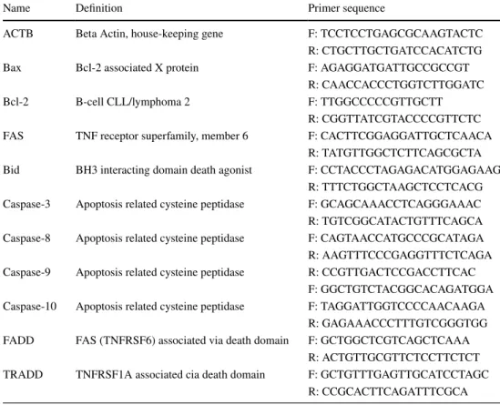
performed by Real Time Online RT-PCR according to the WizPure™ qPCR Master (SYBR) Mix protocol (WIZBIO- Cat.No. W1711-5, USA). The expression results were pro-portioned to the Beta- actine gene (housekeeping gene) expressions to calculate relative expression rations. Primer sequences are given in Table 1 [33].
Statistical analysis
Data were analyzed by using ∆∆CT method and quantified by computer program. VolcanoPlot analyses were used in
the web-based “RT² Profiler ™ PCR Array Data Analysis“ program. The aim of the method is based on the compari-son of two expression results with ± 3SD. Thus, in cases where mRNA expression was compared, expression values of mRNAs in the groups were relatively determined. Com-parison of groups was statistically evaluated by ANOVA and Tukey analysis in SPSS Analysis program.
Table 1 Primer sequences of
the genes used in this study Name Definition Primer sequence
ACTB Beta Actin, house-keeping gene F: TCC TCC TGA GCG CAA GTA CTC R: CTG CTT GCT GAT CCA CAT CTG
Bax Bcl-2 associated X protein F: AGA GGA TGA TTG CCG CCG T
R: CAA CCA CCC TGG TCT TGG ATC
Bcl-2 B-cell CLL/lymphoma 2 F: TTG GCC CCC GTT GCTT
R: CGG TTA TCG TAC CCC GTT CTC FAS TNF receptor superfamily, member 6 F: CAC TTC GGA GGA TTG CTC AACA
R: TAT GTT GGC TCT TCA GCG CTA Bid BH3 interacting domain death agonist F: CCT ACC CTA GAG ACA TGG AGAAG
R: TTT CTG GCT AAG CTC CTC ACG Caspase-3 Apoptosis related cysteine peptidase F: GCA GCA AAC CTC AGG GAA AC R: TGT CGG CAT ACT GTT TCA GCA Caspase-8 Apoptosis related cysteine peptidase F: CAG TAA CCA TGC CCG CAT AGA R: AAG TTT CCC GAG GTT TCT CAGA Caspase-9 Apoptosis related cysteine peptidase R: CCG TTG ACT CCG ACC TTC AC
F: GGC TGT CTA CGG CAC AGA TGGA Caspase-10 Apoptosis related cysteine peptidase F: TAG GAT TGG TCC CCA ACA AGA
R: GAG AAA CCC TTT GTC GGG TGG FADD FAS (TNFRSF6) associated via death domain F: GCT GGC TCG TCA GCT CAA A
R: ACT GTT GCG TTC TCC TTC TCT TRADD TNFRSF1A associated cia death domain F: GCT GTT TGA GTT GCA TCC TAGC
Results
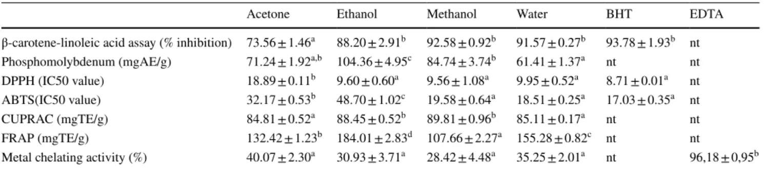
Antioxidant activity
To determined antioxidant activity of A. tomentosus gall extracts, we preferred seven different methods to com-pare the results with each other and provide more reliable data (Table 2). β-carotene-linoleic acid and phosphomo-lybdenum assay were used to evaluated total antioxidant capacity of extracts. Methanol extracts (%92.58 ± 0.92) have highest antioxidant activity according to β-carotene-linoleic acid assay, but ethanol extracts (104.36 ± 4.95 mgAE/g) posses most effective activity in phosphomo-lybdenum assay.
DPPH and ABTS radical scavenging assays are com-monly carried out for fast evaluation of antioxidant activity because of them stability in the radical form and simplic-ity of the assay. In both DPPH (IC50: 9.56 ± 1.08 µg/mL) and ABTS (IC50: 18.51 ± 0.25 µg/mL) tests, water extracts showed the highest radical scavenging activity. FRAP and CUPRAC assays were carried out to determine the reduc-tion power of the extracts. Results which are equivalent to trolox standard, show that extracts have high reducing power activity (CUPRAC methanol: 89.81 ± 0.96 mgTE/g;
FRAPethanol: 184.01 ± 2.83 mgTE/g). And also, ferrious metal chelating capacity of extracts were evaluated and
results were given in comparison with EDTA standard. According to this test, acetone extract had highest chelat-ing capacity (%40.07 ± 2.30).
Extract yield and total secondary metabolites amount
The efficiency of extracts prepared with solvents with differ-ent polarities was calculated. As seen in Table 3, the highest extract amount obtained from water extract (%24,138). This result can be related to that water have the highest polarity.
Total phenolic content of extracts was calculated equiv-alent to gallic acid and the highest content was observed in ethanol extract (297.47 ± 2.52 mgGAE/g). Our results showed that water extract have the highest total flavonoid amount (46.88 ± 0.21 mgQE/g), while acetone extract have the highest total tannin amount (48.22 ± 1.09 mgCE/g).
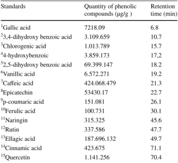
Phenolic compound characterisation by HPLC
Phenolic compounds of extracted gall samples were analysed by HPLC method. The results have been listed in Table 4. According to this results, caffeic acid (424.068.479 µg/g), ellagic acid (187.696.132 µg/g) and 2,5-dihydroxy ben-zoic acid (69.399.147 µg/g) are most common phenolic
Table 2 Antioxidant activity of A.tomentosus Gall extracts
Data represent mean values ± standard error (n = 3). In the same extract, data marked with different letters indicate significant difference (p < 0.05)
AEs ascorbic acid equivalents, TEs trolox equivalents, nd not detected
Acetone Ethanol Methanol Water BHT EDTA
β-carotene-linoleic acid assay (% inhibition) 73.56 ± 1.46a 88.20 ± 2.91b 92.58 ± 0.92b 91.57 ± 0.27b 93.78 ± 1.93b nt Phosphomolybdenum (mgAE/g) 71.24 ± 1.92a,b 104.36 ± 4.95c 84.74 ± 3.74b 61.41 ± 1.37a nt nt DPPH (IC50 value) 18.89 ± 0.11b 9.60 ± 0.60a 9.56 ± 1.08a 9.95 ± 0.52a 8.71 ± 0.01a nt ABTS(IC50 value) 32.17 ± 0.53b 48.70 ± 1.02c 19.58 ± 0.64a 18.51 ± 0.25a 17.03 ± 0.35a nt
CUPRAC (mgTE/g) 84.81 ± 0.52a 88.45 ± 0.52b 89.81 ± 0.96b 85.11 ± 0.17a nt nt
FRAP (mgTE/g) 132.42 ± 1.23b 184.01 ± 2.83d 107.66 ± 2.27a 155.28 ± 0.82c nt nt
Metal chelating activity (%) 40.07 ± 2.30a 30.93 ± 3.71a 28.42 ± 4.48a 35.25 ± 2.01a nt 96,18 ± 0,95b
Table 3 Extract yield and total secondary metabolites amount of A. tomentous according to different solvent
Data represent mean values ± standard error (n = 3). In the same extract, data marked with different letters indicate significant difference (p < 0.05)
GAEs gallic acid equivalents, QEs quercetin equivalents, CEs catechine equivalents
Acetone Ethanol Methanol Water
Extraction yield (%) 8547 9,0856 17,867 24,138
Total phenolic amount (mgGAE/g) 280.60 ± 1.38a 297.47 ± 2.52a 284.14 ± 1.18a 287.89 ± 0.64a Total flavonoid amount (mgQE/g) 31.12 ± 0.67a 29.97 ± 0.59a 30.51 ± 0.46a 46.88 ± 0.21b Total tannin amount (mgCE/g) 48.22 ± 1.09d 23.00 ± 0.15c 14.72 ± 0.14b 6.63 ± 0.15a
compounds of methanol extract of A. tomentosus. It is seen that the other 12 components are also detected in high amounts.
Cytotoxic and anti‑apoptic activity
The dose (5–500 µM) and time (24, 48 and 72 h) depend-ent cytotoxic effects of the extracts were investigated with XTT analysis. For determining the lowest and most effec-tive dose rate 24, 48 and 72 h changes were also observed. A decrease in cell viability was detected at each of the
24, 48 and 72 h, but the lowest doses were obtained at the 24th h. This study showed that in the presence of acetone, ethanol, methanol and water extracts of A. tomentosus,
50% growth inhibition concentrations (IC50) occur in
Mia-Paca2 cell line after 24 h at concentrations 124.7 µg/ mL, 158.3 µg/mL, 187.4 µg/mL and 169.8 µg/mL respec-tively (Table 5). Results showed that, cellular proliferation decreased by concentration dependent manner.
We performed the expression of selected genes (Bax,
Bcl-2, FAS, Bid, Caspase-3, Caspase-8, Capspase-9, Caspase-10, FADD and TRADD) in cells exposed to the
extract using RT- PCR method. According to this method, mRNA levels were analyzed in extract-treated cells and untreated control group cells. In the expression level changes of the cell cycle and apoptosis related genes in dose groups comparing to control group, which are sta-tistically significant and absent, are shown in the Table 5. Expression results of acetonic extract showed that Bax,
caspase-10, FADD and TRADD gene expression levels in
Mia-Paca2 cells were increased in dose group cells com-pared to the control cells in a time-dependent manner. While Bax, Bid, caspase-3, caspase-8 and TRADD gene expression was significantly increased in the ethanolic dose group, the expression changes were not detected in other genes compared with the control cells. In methanol extract treated dose group, while expression of Bid and
caspase-3 were significantly increased, expression of cas-pase-10 was decreased. All gene expression without Bax
and Bid, were decreased in water extract dose group. The reason why the expression levels of genes have different effects in different solvents may be related to the fact that each solvent dissolves different components in the extract in different amounts. Since this affects cell signaling path-ways differently, it can change the outcome of apoptosis.
Table 4 Phenolic compounds characterization of methanolic gall extract by HPLC
Standards Quantity of phenolic
compounds (µg/g ) Retention time (min)
1Gallic acid 7218.09 6.8
23,4-dihydroxy benzoic acid 3.109.659 10.7
3Chlorogenic acid 1.013.789 15.7
44-hydroxybenzoic 3.859.173 17,2
52,5-dihydroxy benzoic acid 69.399.147 18.2
6Vanillic acid 6.572.271 19.2 7Caffeic acid 424.068.479 21.3 8Epicatechin 53430.17 22.7 9p-coumaric acid 151.081 26.1 10Ferulic acid 100.731 30.1 11Naringin 315.325 45.6 12Rutin 337.586 47.7 13Ellagic acid 187.696.132 49.7 14Cinnamic acid 423.675 71.1 15Quercetin 1.141.256 70.4
Table 5 Fold regulation of genes comparing to control group
*P < 0.05 values was marked with * Acetone (IC50: 124.7 µM–24 h) Ethanol (IC50: 158.3 µM–24 h) Methanol (IC50: 187.4 µM–24 h) Water (IC50: 169.8 µM–24 h)
Gene FC Pvalue FC Pvalue FC Pvalue FC Pvalue
Bax 25.05 0.001058* 26.08 0.000016* 8.13 0.150856 6.93 0.000001* Bcl-2 − 2.6 0.030786* − 1.44 0.820964 1.01 0.869223 − 12.75 0.008912* FAS − 11.96 0.1093 1.03 0.687722 − 1.07 0.674186 − 3.07 0.272017 Bid 10 0.009956* 6.4 0.011776* 4.05 0.003156* 1.42 0.127859 Caspase-3 − 5.67 0.085642 5.54 0.040855* 3.32 0.0001* − 1.79 0.023235* Caspase-8 − 1.8 0.000584* 1.78 0.007222* 3.17 0.179143 − 2.74 0.010501* Caspase-9 − 4.32 0.116836 − 1.82 0.250015 − 1.16 0.535897 − 1.85 0.27676 Caspase-10 1.17 0.016329* − 1.27 0.662722 − 1.71 0.00751* − 5.44 0.000378* FADD 5.73 0.02677* 5.46 0.058487 2.65 0.402193 − 3.17 0.270822 TRADD 3.91 0.000003* 1.56 0.020745* − 1.11 0.255985 − 9.83 0.000138*
Discussion
Quercus species are distributed in Iran, Iraq, and Turkey
and then spread to Asia Minor, Europe, and Northern Africa [15, 34]. Quercus infectoria (Family: Fagaceae) commonly known as gall oak, mazu or Manjakani. Phyto-chemical analysis of galls investigated the presence of sap-onins, alkaloids, tannins, glycosides, triterpenes, sterols,
phenolic compounds, carbohydrates, and flavonoids [35,
36]. It is known that the gall extracts have some biological activity such as antimicrobial, analgesic, anticarcinogenic, antioxidant and antioxidant activity thanks to their chemi-cal composition [37].
In present study, some phytochemical analysis and bio-logical activity of A. tomentosus extracts was determined. Our studies were revealed that A. tomentosus gall extracts possess the high total antioxidant capacity, radical scav-enging activity, metal reducing and chealating power. In our previous study, DPPH (8.67 ± 0.58 µg/mL), ABTS (IC50: 44.97 ± 2.56 µg/mL) radical scavenging activity of Andricus quercustozae asexual gall extract was deter-mined. And also, we were examined β–carotene/linoleic acid (%87.49 ± 1.27), Phosphomolybdenum (78.20 ± 1.63 mgAE/g) antioxidant capacity, CUPRAC (245.82 ± 1.06 mgTE/g) reducing power activity and the high total sec-ondary metabolites of A. quercustozae extracts. Another studies showed that oak galls have the high antioxidant activity of using DPPH and ABTS method [38, 39]. With some research, reducing power activity of some gall
spe-cies detected by CUPRAC and FRAP assays [40, 41].
Sukkor et al. [42] was carried out extraction of phenolic acids from oak galls and they were observed that gallic acid (497.34 mg/g), tannic acid (2430.48 mg/g) was the maximum amount phenolic acids. It was found that the most common phenolic acid in the A. quercustozae extract is caffeic acid, which has antioxidant, anti-aging properties [15]. Plant extracts with components such as caffeic acid, 2,5-dihydroxy benzoic acid and ellagic acid are used for pharmaceutical purposes because they have antioxidant, anticarcinogenic and anti-inflammatory properties [43].
Previous study was carried out to determine the poten-tial of galls of Q.infectoria as an antiproliferative agent towards cervical cancer cells (HeLa, IC50: 2.82 ± 0.21 µg/
mL) and ovarian cancer cells (Caov-3, IC50:6.50 ± 0.24 µg/
mL). In their study, it was stated that galls can be recom-mended as an anticancer agent [44]. In another research showed that Galla Chinensis that is an oak gall, has impor-tant cytotoxic activity with IC50 = 4.339 µg/mL [45]. It was showed that oak gall had high reduction to mice mam-mary carcino cell line 2003 AMN3 cancer cell line with 2 µg/mL IC50 value [46]. The results of a study investigat-ing the anticancer effect of extract of Quercus infectoria
in colon cancer HT29 cell line show that aqueous extract increases the expression of the Bax and Bcl-2 genes
com-pared to GAPDH reference gene [47].
The antioxidant and anticancer properties of A.
tomento-sus extracts are strong because of their high content of
caf-feic acid (424.068.479), ellagic acid (187.696.132, 2,5-dihy-droxy benzoic acid (69.399.147) and This is due to the fact that it contains phenolic, which has a lot of OH groups. It is possible to say that antioxidants may have an anticancer effect by inactivating certain transcription factors that oxida-tive stress can activate [48].
Acknowledgements The authors are grateful for the financial support from the Pamukkale University Scientific Research Projects Coordina-tion Unit (Project No: 2018FEBE062).
Compliance with ethical standards
Conflict of interest All authors declare that they have no conflict of interest.
References
1 Ugur D, Gunes H, Gunes F, Mammadov R (2017) Cytotoxic activities of certain medicinal plants on different cancer. Iran J Pharm Res 13:299
2 Tokgun O, Akca H, Mammadov R, Aykurt C, Deniz G (2012) Convolvulus galaticus, Crocus antalyensis, and Lilium candi-dum extracts show their antitumor activity through induction of p53-mediated apoptosis on human breast cancer cell line MCF-7 cells. J Med Food 15(11):1000–1005. https ://doi.org/10.1089/ jmf.2012.0050
3 Rezzoug M, Bakchiche B, Gherib A, Roberta A, Kilinçarslan Ö, Mammadov R, Bardaweel SK (2019) Chemical composition and bioactivity of essential oils and ethanolic extracts of Oci-mum basilicum L. and Thymus algeriensis Boiss. & Reut. from the Algerian Saharan Atlas. BMC Complement Altern Med 19(1):146. https ://doi.org/10.1186/s1290 6-019-2556-y
4 Elmas L, Secme M, Mammadov R, Fahrioglu U, Dodurga Y (2019) The determination of the potential anticancer effects of Coriandrum sativum in PC-3 and LNCaP prostate cancer cell lines. J Cell Biochem 120(3):3506–3513. https ://doi.org/10.1002/ jcb.27625
5 Ferlay J, Soerjomataram I, Dikshit R, Eser S, Mathers C, Rebelo M, Parkin DM, Forman D, Bray F (2015) Cancer incidence and mortality worldwide: sources, methods and major patterns in GLOBOCAN 2012. Int J Cancer 136(5):E359–E386. https ://doi. org/10.1002/ijc.29210
6 Bray F, Ferlay J, Soerjomataram I, Siegel RL, Torre LA, Jemal A (2018) Global cancer statistics 2018: GLOBOCAN estimates of incidence and mortality worldwide for 36 cancers in 185 coun-tries. CA Cancer J Clin 68(6):394–424. https ://doi.org/10.3322/ caac.21492
7. Han Y, Ma L, Zhao L, Feng W, Zheng X (2019) Rosmarinic inhib-its cell proliferation, invasion and migration via up-regulating miR-506 and suppressing MMP2/16 expression in pancreatic can-cer. Biomed Pharmacother 115:108878. https ://doi.org/10.1016/j. bioph a.2019.10887 8
8 Bhuyan DJ, Vuong QV, Chalmers AC, Bowyer MC, Scarlett CJ (2018) An array of bioactive compounds from Australian
eucalypts and their relevance in pancreatic cancer therapeutics. Pancreas 47(6):690–707. https ://doi.org/10.1097/MPA.00000 00000 00107 4
9 Chung KT, Wong TY, Wei CI, Huang YW, Lin Y (1998) Tannins and human health: a review. Crit Rev Food Sci Nutr 38:421– 464. https ://doi.org/10.1080/10408 69989 12742 73
10 Cai Y, Zhang J, Chen NG, Shi Z, Qiu J, He C, Chen M (2017) Recent advances in anticancer activities and drug delivery systems of Tannins. Med Res Rev 37:665–701. https ://doi. org/10.1002/med.21422
11 Chauhan SS, Shetty AB, Hatami E, Chowdhury P, Yallapu MM (2020) Pectin-tannic acid nano-complexes promote the delivery and bioactivity of drugs in pancreatic cancer cells. Pharmaceu-tics 12(3):285. https ://doi.org/10.3390/pharm aceut ics12 03028 5
12 Türkan F, Taslimi P, Saltan FZ (2019) Tannic acid as a natural antioxidant compound: discovery of a potent metabolic enzyme inhibitor for a new therapeutic approach in diabetes and Alzhei-mer’s disease. J Biochem Mol Toxicol 33(8):e22340. https ://doi. org/10.1002/jbt.22340
13 Claus EP (1962) Pharmacognosy. Acad Med 37(1):79
14 Sukor N, Jusoh R, Rahim SA, Kamarudin N (2018) Ultrasound assisted methods for enhanced extraction of phenolic acids from Quercus infectoria galls. Mater Today 5(10):21990–21999. https ://doi.org/10.1016/j.matpr .2018.07.060
15 Azmaz M, Kılınçarslan Aksoy Ö, Katılmış Y, Mammadov M (2020) Investigation of the antioxidant activity and phenolic com-pounds of Andricus quercustozae gall and host plant (Quercus infectoria). Int J Secondary Metab 7(2):77–87. https ://doi. org/10.21448 /ijsm.67493 0
16. Akbar S (2020) Quercus infectoria G. Olivier (Fagaceae). In: Handbook of 200 medicinal plants. pp. 1505–1511. https ://doi. org/10.1007/978-3-030-16807 -0_155
17. Tavakoli M, Melika G, Sadeghi SE, Pénzes Z, Assareh MA, Atkinson R et al (2008) New species of oak gallwasps from Iran (Hymenoptera: Cynipidae: Cynipini). Zootaxa 1699(1):1–64 18. Azmaz M, Katılmış Y (2017) Cynipidae (Insecta: Hymenoptera)
fauna of Istanbul. Mun Ent Zool 12(1):151
19. Ionescu MA (1957) Insecta: Cynipinae. Editura Academiei Republicii Populare România
20 Melika G (2006) Gall wasps of Ukraine. Cynipidae Vestnik zoologii 21(1–2):301–644
21 Kılınçarslan O, Mammadov R (2018) HPLC analysis and antioxi-dant, antibacterial and cytotoxicity activities of various solvent extracts of Erysimum kotschyanum Gay.(Brassicaceae). J Chem Soc Pak 40(04):707
22. Amin I, Zamaliah MM, Chin WF (2004) Total antioxidant activity and phenolic content in selected vegetables. Food Chem 87:581– 586. https ://doi.org/10.1016/j.foodc hem.2004.01.010
23 Prieto P, Pineda M, Aguilar M (1999) Spectrophotometric quanti-tation of antioxidant capacity through the formation of a phospho-molybdenum complex: specific application to the determination of vitamin E. Anal Biochem 269(29):337–41
24. Wu C, Chen F, Wang X, Kim HJ, He GQ, Haley-Zitlin V, Huang G (2006) Antioxidant constituents in feverfew (Tanace-tum parthenium) extract and their chromatographic quantifica-tion. Food Chem 96(2):220–227. https ://doi.org/10.1016/j.foodc hem.2005.02.024
25. Re R, Pellegrini N, Proteggente A, Pannala A, Yang M, Rice-Evans C (1999) Antioxidant activity applying an improved ABTS radical cation decolorization assay. Free Radical Biol Med 26:1231–1237. https ://doi.org/10.1016/S0891 -5849(98)00315 -3 26. Apak R, Guclu K, Ozyurek M, Karademir SE, Ercag E (2006) The
cupric ion reducing antioxidant capacity and polyphenolic content of some herbal teas. Int J Food Sci Nutr 57:292–304. https ://doi. org/10.1080/09637 48060 07981 32
27. Apak R, Özyürek M, Güçlü K, Çapanoğlu E (2016) Antioxidant activity/capacity measurement. 1. Classification, physicochemical principles, mechanisms, and electron transfer (ET)-based assays. J Agric Food Chem 64(5):997–1027. https ://doi.org/10.1021/acs. jafc.5b047 39
28 Dinis TCP, Madeira VMC, Almeida LM (1994) Action of phe-nolic derivates (acetoaminophen, salicylate and 5-aminosalycilate) as inhibitors of membrane lipid peroxidation and as peroxyl radi-cal scavengers. Arch Biochem Biophys 315:161–169. https ://doi. org/10.1006/abbi.1994.1485
29 Slinkard K, Vernon L, Singleton VL (1977) Total phenol analyses: automation and comparison with manual methods. Am J Enol Viticult 28:49–55
30 Arvouet-Grand A, Vennat B, Pourrat A, Legret P (1994) Stand-ardization of propolis extract and identification of principal con-stituents. J Pharm Belg 49:462–468
31. Bekir J, Mars M, Souchard JP, Bouajila J (2013) Assessment of antioxidant, anti inflammatory, anti-cholinesterase and cytotoxic activities of pomegranate (Punica granatum) leaves. Food Chem Toxicol 55:470–475. https ://doi.org/10.1016/j.fct.2013.01.036 32 Caponio F, Alloggio V, Gomesb T (1999) Phenolic compounds of
virgin olive oil; influence of paste preparation techniques. Food Chem 64:203–209. https ://doi.org/10.1016/S0308 -8146(98)00146 -0
33. Fahrioğlu U, Dodurga Y, Elmas L, Seçme M (2016) Ferulic acid decreases cell viability and colony formation while inhibiting migration of MIA PaCa-2 human pancreatic cancer cells in vitro. Gene 576(1):476–482. https ://doi.org/10.1016/j.gene.2015.10.061 34 Ahmad W (2016) Ethnopharmacology of Quercus infectoria
olivier galls: a review. Hippocratic J Unani Med 11:105–18 35. Shrestha S, Kaushik VS, Eshwarappa RS, Subaramaihha SR,
Ramanna LM, Lakkappa DB et al (2014) Pharmacognostic stud-ies of insect gall of Quercus infectoria Olivier (Fagaceae). Asian Pac J Trop Biomed 4(1):35–39. https ://doi.org/10.1016/S2221 -1691(14)60205 -7
36 Zachariah SM, Kumar NM, Darsana K, Gopal D, Thomas N, Ramkumar M et al (2014) Phytochemical screening, formulation and evaluation of dried galls of Quercus infectoria Olivier. Int J Pharm Sci Rev Res 26:125–30
37 Fathabada AE, Schariatifar N, Mardania K, Pourfard MI (2015) Study on antibacterial and antioxidant activiy of Oak gall Quercus infectoria (extracts from Iran). Int J Curr Scı 14:44–50
38 Arina MI, Harisun Y (2019) Effect of extraction temperatures on tannin content and antioxidant activity of Quercus infectoria (Manjakani). Biocatal Agric Biotechnol 19:101104. https ://doi. org/10.1016/j.bcab.2019.10110 4
39. Kaur G, Athar M, Alam MS (2008) Quercus infectoria galls pos-sess antioxidant activity and abrogates oxidative stress-induced functional alterations in murine macrophages. Chem Biol Interact 171:272–282. https ://doi.org/10.1016/j.cbi.2007.10.002 40. Doğan Abdioğlu M (2019) Bazı meşe gallerinin kolinesteraz,
tirozinaz ve üreaz enzim inhibisyonu ile antioksidan aktivitesinin belirlenmesi. Master’s thesis, Batman Üniversitesi Fen Bilimleri Enstitüsü, Türkiye
41 Shahin S, Ahmad N (2014) Antioxidant properties and total phe-nolic content of herbs used in post partum diet therapy in Patna (Bihar), India. J Pharm Biol Sci 9:17–20
42 Sukor NF, Jusoh R, Kamarudin NS, Halim NA, Sulaiman AZ, Abdullah SB (2020) Synergistic effect of probe sonication and ionic liquid for extraction of phenolic acids from oak galls. Ultrason Sonochem 62:104876. https ://doi.org/10.1016/j.ultso nch.2019.10487 6
43 Aydin Ç, Rakhimzhanova A, Kilinçarslan Ö, Mammadov R (2020) Antioxidant and phenolic characterization with HPLC of vari-ous extract of Verbascum glomeratum Linneus. J Chem Soc Pak 42(2):222
44 Hasmah A, Nurazila Z, Chow CY, Rina R, Rafiquzzaman M (2010) Cytotoxic effects of Quercus infectoria extracts towards cervical (Hela) and Ovarian (Caov-3) cancer cell lines. Health Environ J 1(2):17–23
45 Gao J, Yang X, Yin W, Li M (2018) Gallnuts: a potential treasure in anticancer drug discovery. Evid Based Complement Altern Med 2018:4930371
46 Jalill RDA (2018) Chemical analysis and anticancer effects of Juniperus polycarpos and oak gall plants extracts. Res J Pharm Technol 11(6):2372–2387
47 Abdalan S, Baghbani-Arani F, Sadat Shandiz SA (2018) Evalu-ation of anticancer effect of aqueous and hydroalcoholic extracts
of Quercusin fectoria leaf against colon cancer HT29 cell line. J Arak Univ Med Sci 21(4):48–57
48 Reuter S, Gupta SC, Chaturvedi MM, Aggarwal BB (2010) Oxida-tive stress, inflammation, and cancer: how are they linked? Free Radical Biol Med 49(11):1603–1616. https ://doi.org/10.1016/j. freer adbio med.2010.09.006
Publisher’s Note Springer Nature remains neutral with regard to jurisdictional claims in published maps and institutional affiliations.