Address for correspondence: Dr. Taner Ulus, Eskişehir Osmangazi Üniversitesi Tıp Fakültesi, Kardiyoloji Anabilim Dalı, Meşelik Kampüsü, 26480, Odunpazarı, 26200 Eskişehir-Türkiye
Phone: +90 222 239 29 79 Dahili: 3700 E-mail: [email protected] Accepted Date: 11.01.2019 Available Online Date: 24.01.2019
©Copyright 2018 by Turkish Society of Cardiology - Available online at www.anatoljcardiol.com DOI:10.14744/AnatolJCardiol.2019.60687
AUTHORS: Taner Ulus
1, Kaan Okyay
2, Hasan Kutsi Kabul
3, Emin Evren Özcan
4, Özcan Özeke
5, Hakan Altay
6REVIEWERS: Bülent Görenek
1, Aylin Yıldırır
2, Sercan Okutucu
7, Abdullah Tekin
81Department of Cardiology, Faculty of Medicine, Eskişehir Osmangazi University; Eskişehir-Turkey 2Department of Cardiology, Faculty of Medicine, Başkent University; Ankara-Turkey 3Department of Cardiology, Gülhane Training and Research Hospital; Ankara-Turkey 4Department of Cardiology, Faculty of Medicine, Dokuz Eylül University; İzmir-Turkey
5Department of Cardiology, Health Sciences University, Türkiye Yüksek İhtisas Training and Research Hospital; Ankara-Turkey 6Department of Cardiology, Faculty of Medicine, Başkent University; İstanbul-Turkey
7Department of Cardiology, Memorial Ankara Hospital; Ankara-Turkey 8Department of Cardiology, Faculty of Medicine, Başkent University; Adana-Turkey
Turkish Society of Cardiology consensus paper on management of
arrhythmia-induced cardiomyopathy
Background
Heart failure (HF) is one of the major causes of mortality and morbidity. The identification of causes of left ventricular (LV) sys-tolic dysfunction is important in terms of initiating causal treat-ment and improving prognosis. Arrhythmia-induced cardiomy-opathy (AIC) is a potentially reversible form of cardiomycardiomy-opathy (CMP) in which LV dysfunction results from atrial or ventricular arrhythmias (1). It can be resolved by eliminating or effectively treating responsible arrhythmia (2).
Aim of the document
Early recognition of the relationship between responsible ar-rhythmia and CMP is of great importance in terms of the improve-ment of symptoms, LV systolic dysfunction, and functional status with effective treatment. However, in the clinical practice, AIC is often overlooked, and arrhythmias are generally seen as the result of HF. Again, there is a lack of information about the patho-physiology of AIC and the course of the disease after effective treatment of the responsible arrhythmia. This document is writ-ten to give clear messages for further recognition and treatment of AIC based on the current literature.
Definition
AIC is defined as LV systolic dysfunction due to supraven-tricular or vensupraven-tricular arrhythmia that can be either sustained
or paroxysmal or is characterized by highly frequent ectopic activity (3). AIC can be divided into two categories. Type 1 AIC (arrhythmia-induced): arrhythmia is accepted as the absolute reason of ventricular dysfunction that returns to normal after successful treatment of arrhythmia. Type 2 AIC (arrhythmia-me-diated): arrhythmia exacerbates the LV dysfunction in patients with concomitant heart disease, and treatment of the arrhythmia provides partial improvement (4).
Epidemiology
The prevalence of HF is increasing worldwide due to better treatment of acute cardiac events, improvements in medical and surgical treatment methods, and aging of the population. Approx-imately 1%–2% of the general population, and >10% of over 70 years old are affected with HF (5). Cardiac arrhythmias generally occur during the natural course of HF, but sometimes they are the sole etiology of the unexplained systolic HF or dilated CMP. Reli-able epidemiological data regarding the AIC are lacking, and the prevalence in general is underestimated, given that arrhythmia is often considered to be a result of rather than a possible cause of CMP.
Although age is the major determinant of incidence and prevalence of overall HF, AIC appears to occur at any age. How-ever, the common types of arrhythmias causing AIC differ among age groups. Focal atrial tachycardia (FAT) (59%) and permanent
causes of AIC in children in the largest pediatric series of AIC, whereas ventricular arrhythmias are rare (4). The incidence of AIC was 9%–34% in adult patients with frequent premature ven-tricular complexes (PVC) and/or nonsustained venven-tricular tachy-cardia (VT) referred for electrophysiological evaluation (6).
The most common cause of AIC in adults is atrial fibrillation (AF). Most common arrhythmia coexisting with HF is also AF. The LV systolic dysfunction is found in 20%–30% of all patients with AF, and 10%–50% of patients with HF have AF (7). In the Framing-ham study, those with AF had a higher risk of developing HF [haz-ard ratio of 2.22 (CI 1.47–3.34) p<0.0001] (8). Both AF and HF can directly lead to the other, so it is not easy to assess the causal link between AF and systolic dysfunction. The definite diagnosis of AIC in this context can only be made if systolic dysfunction is reversible after restoration of sinus rhythm. Recent ablation stud-ies have revealed that approximately one-third of patients with
was detected in 58%–88% of these cases (9, 10). Pathophysiology and mechanisms
The main three mechanisms that appear to be responsible for the AIC development are tachycardia, irregular rhythm, and dyssynchrony. There is significant overlap among these mecha-nisms (11).
In animal models, rapid stimulation has been shown to re-sult in LV dysfunction within weeks after tachycardia begins (4). Three phases have been defined in this situation (Fig. 1). In the compensatory phase (the first 3–7 days of rapid pacing), the LV pump function is normal, and there is an increased neurohor-monal activation with early changes in the extracellular matrix. In the LV dysfunction phase (about 1–3 weeks after the onset of rapid pacing), there is cellular remodeling, contractile dysfunc-tion with LV systolic dysfuncdysfunc-tion, and dilatadysfunc-tion. Continued neu-Consensus statements
References
Main clinical scenarios that increase the suspicion of arrhythmia-induced or arrhythmia-mediated 19 cardiomyopathy (CMP) (AIC) can be listed as follows:
• Simultaneous presentation of a tachyarrhythmia or frequent ectopy and systolic dysfunction in a patient with no preexisting heart disease.
• Asymptomatic CMP in the setting of a persistent arrhythmia or frequent ectopy.
• A patient with known structural heart disease now presenting with worsening left ventricular (LV) dysfunction and heart failure (HF) secondary to an arrhythmia.
Treatment of AIC should be primarily aimed at eliminating or controlling the arrhythmia using either 1, 4, 12, 31, 50 pharmacological or ablative techniques with the goal of improving symptoms and reversing systolic
dysfunction. The exact approach should be selected depending on the underlying arrhythmia. Curative ablation is the preferred method of choice in appropriate patients.
Following the normalization of LV function, continuation of HF medication is recommended. 1, 4 Close follow-up is recommended given that recurrent arrhythmia can result in rapid decline in 1, 4 LV function with development of HF.
The following laboratory findings can help distinguish AIC from other CMPs: 22-28 • A smaller LV end diastolic diameter and mass index
• A more profound reduction in apical longitudinal strain compared to the mid and basal segments • Absence of late gadolinium enhancement and early right ventricular systolic dysfunction on cardiac magnetic resonance imaging
• Significant smaller percentage of the LV endocardium with abnormal unipolar voltage • Rapid decline of the N-terminal pro-brain natriuretic peptide levels at one week following control of arrhythmia
Implantable cardioverter defibrillator implantation for primary prevention should be delayed for a reasonable 4 period to see the response to the optimal medical treatment in AIC.
rohormonal activation and upregulation of the renin angiotensin system are observed. The LV failure phase (>3 weeks) is char-acterized by severe LV pump failure, severe LV dilatation, signifi-cant neurohormonal activation, and defects in Ca+2 handling (4).
Myocardial energy store depletion, increased oxidative stress, blunted response to beta adrenergic stimulation (12, 13), reduced myocardial blood flow (14), and abnormal calcium han-dling (15-17) have all been implicated in the pathogenesis of AIC. The proposed mechanisms described above are seen in multiple forms of chronic HF, and they may be related, at least in part, to the effects of elevated filling pressures and decreased cardiac output. Changes seen early after initiation of responsible arrhythmia are more likely to be related to elevated heart rates, whereas later changes are more likely to be due to a combina-tion of the arrhythmia as well as the downstream effects of the HF syndrome (12).
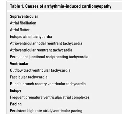
Causes of arrhythmia-induced cardiomyopathy
There is a wide range of arrhythmias and clinical conditions related with development of AIC. They are listed in Table 1.
Clinical features
AIC has varied presentations. In fetal life, AIC might be pre-sented with hydrops fetalis (18). In children and adults, clinical picture of AIC can vary from asymptomatic arrhythmia to end-stage HF. Many patients are asymptomatic from arrhythmias or describe subtle or challenging symptoms making early diagnosis more difficult. In this point, AIC could not be detected until mani-fest HF develops. When symptoms of HF exist, without aggressive treatment of HF and arrhythmia, worsening of HF is inevitable (19). Signs and/or symptoms are related to the tachyarrhythmia (e.g., palpitations, dyspnea, chest discomfort), HF (e.g., dyspnea,
edema, weight gain, orthopnea), or both. However, shortness of breath was reported as the primary complaint (20). It can be argued that patients with relatively higher heart rates often present earlier with symptoms related to the tachycardia, and they can be detected earlier and treated without development of CMP. Main clinical scenarios that increase the suspicion of arrhythmia-induced or arrhythmia-mediated CMP can be listed as follows (19).
1. Simultaneous presentation of a tachyarrhythmia or frequent ectopy and systolic dysfunction in a patient with no preexist-ing heart disease.
2. Asymptomatic CMP in the setting of a persistent arrhythmia or frequent ectopy.
3. A patient with known structural heart disease now present-ing with worsenpresent-ing LV dysfunction and HF secondary to an arrhythmia.
Diagnosis
The key diagnostic criterion of AIC is the detection of a path-ological tachycardia or persistent arrhythmia in the presence of an otherwise unexplained LV systolic dysfunction (4). The proof of AIC is to document LV systolic dysfunction in serial measure-ments after pathological tachycardia or resistant arrhythmia in a patient with normal LV functions prior to onset of the arrhythmia (21). However, this proof is rarely seen in clinical practice. A sin-gle 12-lead electrocardiogram (ECG) may not diagnose respon-sible arrhythmia. Continuous ambulatory ECG (Holter) monitoring is able to identify recurrent tachycardia, mean ventricular rate in AF, and the frequency of PVC (1).
Some laboratory tests can help distinguish AIC from other CMPs. Patients with AIC have a smaller LV end diastolic diameter and mass index compared those with preexisting dilated CMP
Responsible arrhythmia
Within one week
1-3 weeks
3 weeks Compensatory phase
Normal LV pump function Sympathetic system activation
LV dysfunction phase
LV pump dysfunction and dilatation Myocardial contractile dysfunction Neurohormonal activation
Heart failure phase Severe LV pump dysfunction Significant neurohormonal activation Systemic hemodynamic compromise Pulmonary/systemic edema
Figure 1. Schematic presentation of the pathophysiology of AIC in animal models
AIC - arrhythmia-induced cardiomyopathy, LV - left ventricle
Table 1. Causes of arrhythmia-induced cardiomyopathy
Supraventricular Atrial fibrillation Atrial flutter
Ectopic atrial tachycardia
Atrioventricular nodal reentrant tachycardia Atrioventricular reentrant tachycardia
Permanent junctional reciprocating tachycardia Ventricular
Outflow tract ventricular tachycardia Fascicular tachycardia
Bundle branch reentry ventricular tachycardia Ectopy
Frequent premature ventricular/atrial complexes Pacing
AIC had a more profound reduction in apical longitudinal strain compared to that in the mid and basal segments, while patients with other forms of CMP after arrhythmia correction had a pre-dominant reduction of longitudinal strain in the basal segments (24). Absence of late gadolinium enhancement and early right ventricular systolic dysfunction on cardiac magnetic resonance imaging (MRI) might help to differentiate AIC from other heart dis-eases (25, 26). Unipolar electroanatomic mapping can be helpful in differentiating AIC from patients with irreversible CMP. Campos et al. (27) have demonstrated that patients with irreversible CMP had a significant larger percentage of the LV endocardium with abnormal unipolar voltage. In addition, a study showed that rapid decline of the N-terminal pro-brain natriuretic peptide levels at one week following control of arrhythmia was associated with AIC (28).
Endomyocardial biopsy specimens from patients with AIC exhibit features distinct from those of other types of CMP, includ-ing scant or absent myocardial fibrosis, increased expression of major histocompatibility complex class II molecules, CD68+ mac-rophage infiltration, and an enrichment of mitochondria in close proximity to intercalated disks (29). However, the potential role of the myocardial biopsy in the diagnosis of AIC requires additional investigations.
When AIC cannot be differentiated from dilated CMP with consequent tachycardia, treatments for both problems are nec-essary (3). The correct diagnosis can only be established after demonstration of the improvement of LV function within a few weeks or months after successful treatment of arrhythmia (21).
Table 2 shows diagnostic tests, which can help differentiate AIC from other forms of nonischemic dilated CMP.
Management
a) Principles of management
Treatment of AIC should be primarily aimed at eliminating or controlling the arrhythmia using either pharmacological or ablative techniques with the goal of improving symptoms and reversing systolic dysfunction (Fig. 2). The exact approach should be selected depending on the underlying arrhythmia.
priate patients. Along with arrhythmia control, HF medication is recommended as well (30).
b) Arrhythmia-specific diagnostic tests and treatment Atrial fibrillation
AF is the most common cause of AIC in adults (4, 31). AF and CMP often coexist and precipitate one another (31). Figuring out whether LV systolic dysfunction is due to underlying structural heart disease or arrhythmia itself is challenging. Rapid and ir-regular heart rate and loss of atrial contraction are the proposed pathophysiologic mechanisms for the development of AIC in pa-tients with AF (32, 33).
It was thought that LV systolic function improvement could be achieved with any number of AF treatment strategies, whether that is rate control or pacing and ablation procedure or rhythm control with catheter ablation or antiarrhythmic drugs. In AF-CHF trial, no mortality advantage was observed between patients with
Table 2. Diagnostic tests that distinguish AIC from other forms of nonischemic dilated CMP
Tests AIC Dilated CMP
LV end diastolic diameter A smaller LV end diastolic diameter A larger LV end diastolic diameter
RV systolic dysfunction Early Late
Strain distribution Decreased longitudinal strain in the Decreased longitudinal strain in the apical segments basal segments
Late gadolinium enhancement on cardiac MRI No Yes
NT-proBNP following control of arrhythmia Fast reduction Slow or limited reduction
AIC - arrhythmia-induced cardiomyopathy, CMP - cardiomyopathy, LV - left ventricle, MRI - magnetic resonance imaging, NT-proBNP - N-terminal pro-brain natriuretic peptide, RV - right ventricle
Figure 2. Flow diagram of diagnosis and treatment approaches in AIC
AIC - arrhythmia-induced cardiomyopathy, HF - heart failure, LV - left ventricle *: This condition may be related to more extensive myocardial damage produced by longer periods of tachycardia and/or contribution of underlying heart disease
Persistent tachyarrhythmia or frequent ectopy
Neurohormonal activation Systolic contractility ↓ LV systolic dysfunction LV dilatation Cardiac output ↓
Termination or suppression of arrhythmia (Successful rhythm or effective rate control)
HF therapy HF resolution and LV function recovery
Confirms diagnosis of AIC No improvement in LV functionNot AIC or may be AIC* Continuation of HF therapy Follow-up:
Maintain sinus rhythm/strict rate control Close follow-up to avoid arrhythmia recurrence Continue neurohormonal antagonists
AF and HF who were randomized to either rate control or rhythm control (amiodarone with cardioversion) (34). However, AIC can occur in AF and normal ventricular rates given the fact that irreg-ular ventricirreg-ular contraction is associated with LV dyssynchrony in the long term (35, 36).
In the AATAC-AF trial, 203 patients with persistent AF and HF with LV ejection fraction (LVEF) <40% were randomized to cathe-ter ablation arm or amiodarone arm. Arrhythmia recurrences and hospitalization rates were significantly lower with improved qual-ity of life in the ablation arm. The LVEF improved by 9.6%±7.4% in the ablation arm compared to 4.2%±6.2% in the amiodarone arm (37). Another systematic review of 19 studies (914 patients) also demonstrated the superiority of catheter ablation to restore sinus rhythm with LVEF improvement by 13.3% (38).
CAMERA-MRI (9) and CASTLE-AF 2 (39) trials also demon-strated the significance of restoring sinus rhythm with catheter ablation with improvements in systolic function and quality of life. In the CAMERA-MRI trial, the primary end point of LVEF improve-ment was significantly higher in catheter ablation group com-pared to that in medical rate control group at a median follow-up of 6 months (18.3% vs. 4.4%, p<0.0001). Similarly, in CASTLE-AF 2 trial, LVEF improved by 8% in ablation arm compared with 0% in the medical arm at 60 months.
Briefly, in patients with AIC secondary to AF, restoration of sinus rhythm should be the primary goal. In terms of rhythm control, multiple studies proved the superiority of catheter ab-lation compared to pharmacological treatment (40-43). Despite primarily successful catheter ablation, recurrence of AF is high, and repeat ablation(s) is/are generally needed. In patients with treatment-refractory AF with a high ventricular rate, atrioven-tricular node ablation and bivenatrioven-tricular pacing seems a ratio-nale option (44, 45).
Atrial flutter
Atrial flutter is one of the common causes of AIC. In one study, LV systolic dysfunction was observed in 25% of patients present-ing with atrial flutter (46). Another study evaluatpresent-ing >1000 patients with atrial flutter found the incidence of AIC nearly 8% (47). Ven-tricular rate control is difficult in atrial flutter; therefore, rhythm control is often necessary. Although electrical cardioversion is effective in restoring sinus rhythm, recurrences may occur in follow-up period. Given the high success and low complication rate, catheter ablation approach should be primarily considered (48). The LV systolic function improvement was seen in >50% of patients with atrial flutter undergoing ablation (47).
Supraventricular tachycardias
The AIC has been associated with essentially any frequent and persistent supraventricular tachycardia (SVT). The common reentrant SVTs such as atrioventricular nodal reentrant tachy-cardia and atrioventricular reciprocating tachytachy-cardia are most commonly paroxysmal, but rarely can be incessant. Therefore, they are rarely associated with AIC. The most classic incessant
SVT-mediated AICs in adults are FAT, atrial flutter, dual atrioven-tricular nodal nonreentrant tachycardia, and PJRT (49, 50). The FAT and atrial flutter are often refractory to pharmacological sup-pression (50). The radiofrequency ablation of SVTs is effective in over 95% of cases, and it should be recommended as a first-line therapy for SVT-mediated AIC in adults (4).
Frequent premature ventricular contractions and VT
Idiopathic ventricular arrhythmias in the absence of structural heart disease are considered a benign entity. In a normal healthy population, PVCs have been observed in up to 75 % of subjects on 48-hour Holter monitoring (51). However, there is clear associa-tion between frequent PVCs and CMP (20, 52, 53).
Underlying pathophysiological mechanism is not entirely clear. Various cellular and clinical mechanisms have been sug-gested. The excitation-contraction coupling is impaired because of the decreased Ca+2 release from the sarcoplasmic reticulum
that results in contractile dysfunction (54). Ventricular dyssyn-chrony is another suspected mechanism that may also lead to LV impairment.
The predisposition to the development of CMP is another co-nundrum. PVC burden is the well-accepted risk factor. Burdens above 15%–25% of the total cardiac beats are associated with CMP (20, 52, 53). On the other hand, the vast majority of patients with frequent PVCs will not develop CMP. Although many other risk factors such as lack of palpitations, nonsustained VT, a retro-grade P-wave after the PVCs, interpolated PVCs, PVC QRS dura-tion (e.g., ≥150 ms), and epicardial origin are suggested, current data are not sufficient for accurate risk prediction (55, 56). Novel imaging modalities such as real-time three-dimensional speckle tracking echocardiography seems promising to detect subtle LV dysfunction.
Regardless of whether PVCs are the cause or the result of CMP, catheter ablation is the preferred treatment option. Suc-cessful ablation results in significant improvement of cardiac functions. PVCs burden before and after ablation are the main predictors of LVEF recovery (57). Since frequent PVCs worsen CMP, presence of structural heart disease does not diminish the benefit of ablation.
For patients who require arrhythmia suppression for symp-toms or declining ventricular function suspected to be due to fre-quent PVCs (generally >15% of beats and predominately of one morphology) and for whom antiarrhythmic medications are inef-fective, not tolerated, or not the patient’s preference, catheter ablation is recommended in the 2017 AHA/ACC/HRS Ventricular Arrhythmias Guideline (Class I recommendation) (56). Pharmaco-logic treatment has a class IIa recommendation. Beta-blockers and amiodarone are usually preferred. There is a general res-ervation against other antiarrhythmics because of the increased mortality observed in patients with LV dysfunction and history of myocardial infarction (58, 59). In selected patients suspected of having PVC-induced CMP, Class IC antiarrhythmic drugs effec-tively suppressed PVCs, leading to LVEF recovery in the
major-cohort (60).
Implantable cardioverter defibrillator (ICD) implantation in PVC-induced CMP is not a rare scenario. In these patients, it should be evaluated whether LV function improves after an ef-fective treatment such as ablation.
AIC in adults with congenital heart disease
Cardiac arrhythmias are a major source of morbidity and mor-tality in adults with congenital heart disease (ACHD). In patients with ACHD, symptomatic tachyarrhythmias may be seen after surgery, or they may be seen in the absence of any intervention, as in Ebstein’s anomaly (patients with this anomaly may have ac-cessory connections) (61, 62). Intra-atrial reentrant tachycardia is the most common form of SVT in the population with ACHD (63). Ventricular tachyarrhythmias are well-known late sequelae after surgical repair of a variety of forms of the disease (61). The possible mechanisms for the development of AIC in this group of patients are similar to those in other patients (such as high heart rates, ventricular dyssynchrony) (64-66).
In patients with ACHD with AIC, it is reasonable to apply the principles of rate and rhythm control as in other patient groups (62, 63). Digitalis is less effective in younger, active patients (63). Additional ventricular scarring or systemic ventricular dysfunc-tion/hypertrophy often limits the choice of an antiarrhythmic drug in patients with ACHD. Amiodarone is often the only option for pa-tients with ACHD. However, long-term therapy with amiodarone has severe side effects, particularly in young adults (64). Cath-eter ablation has additional difficulties due to unusual conduc-tion anatomy and difficulty accessing vessels and intracardiac chambers in such patients (63).
AIC in children
The most common arrhythmias associated with pediatric AIC include FAT and PJRT (67-69). The junctional ectopic tachycardia (JET) is usually seen after congenital heart surgery (“postopera-tive JET”) (58). Atrial flutter, AF or incessant VT may also lead to pediatric AIC although these arrhythmias are typically seen in adults. Beta-blockers are the most common first-line agent in most pediatric AIC (67, 69). There is also a trend toward in-creased use of flecainide in pediatric CMPs (70). In addition to first-line beta-blockers (67, 69, 71), the combination of sotalol and propafenone has been used in effectively controlling FAT (72). Since the spontaneous resolution is common for young children with FAT (69), the catheter ablation is only recommended in in-fants and young children if pharmacological treatment is not fea-sible or unsuccessful (73). However, the FAT is unlikely to resolve spontaneously, and antiarrhythmics are frequently ineffective in children aged ≥3 years (74). The spontaneous resolution of PJRT is almost unlikely with 12% of reported rate (68), and the antiar-rhythmics result in complete control only in few patients. There-fore, the catheter ablation is the primary treatment for PJRT with reported success rates of 90% (68).
Following effective and timely treatment of arrhythmia with either elimination or adequate suppression, LV systolic function completely or partially recovers in case of AIC. However, such recovery may be totally absent. This may be related to more ex-tensive myocardial damage produced by longer periods of tachy-cardia and/or contribution of underlying heart disease (21, 75). Researchers reported that patients with HF with recovered EF have better clinical courses including lower mortality than pa-tients with permanently reduced or permanently preserved EF (76). It may take weeks to months for the recovery of systolic function after treatment of the arrhythmia. This represents the importance of timely interventions before an adverse burden of remodeling goes too far. One study showed that even after years of complete recovery of systolic function, mild LV dilatation and ultrastructural myocardial lesions may be seen (77). This may ex-plain the rare incidences of sudden cardiac death in patients with AIC who had already recovered systolic function (31).
It is also evident that recurrent tachyarrhythmias following initial recovery can cause relapses of AIC (31). Although CMP in response to an arrhythmia may take months to years to develop, recurrent arrhythmia can result in rapid decline in ventricular function with development of HF, suggesting residual ultrastruc-tural abnormalities in the so-called HF with recovered EF. Very close patient follow-up with ambulatory Holter electrocardiogram and imaging studies and aggressive treatment if needed could prevent the devastating consequences of recurrent arrhythmias.
AF-induced CMP is also associated with a more benign prog-nosis compared to new-onset AF in a patient with established HF because in the latter form, AF is a marker of more advanced HF and associated with a worse outcome (78). Distinguishing AIC in case of AF and HF is very important because aggressive at-tempts for restoration of sinus rhythm in case of AIC could lead to complete recovery of systolic function and favorable prognosis. Both CAMERA-MRI and CASTLE HF trials have demonstrated that catheter ablation in patients with AF and systolic dysfunction re-sulted in improvements in HF symptoms, LVEF with reductions in hospitalizations, and total mortality (9, 39). Part of these positive effects of catheter ablation in these studies, although inconclu-sive, may be attributed to the substantial number of AIC patients presented in these two studies.
Although the decisive treatment of AIC is the control of arrhyth-mia, treatment with disease-modifying drugs (angiotensin convert-ing enzyme inhibitor, beta-blockers, mineralocorticoid receptor antagonists) still play an important role. The continuation of these medical treatments following recovery of LV systolic function is controversial. It is advisable to continue these medications for HF after recovery of systolic function considering that the persistence of subtle negative remodeling in such patients (31, 77).
Summary
Patients with AIC are the relatively favorable subgroup of the patients with CMP. Correcting the underlying arrhythmia can
pro-vide a dramatic improvement in the LV functions. On the other hand, it may not always be easy to determine whether the ar-rhythmia is the result or the cause of the CMP. Even though it re-minds the chicken and egg conundrum, both groups benefit from appropriate arrhythmia treatment.
Patients with AIC will undoubtedly have the chance to be cured. Regardless of the cause, frequent arrhythmias worsen the preexisting CMP, in which case arrhythmia treatment may lead to partial but important recovery of the LV dysfunction. These pa-tients are those who mostly need sinus rhythm. Catheter ablation is the effective treatment to restore sinus rhythm. The efficacy of drugs that can be used in HF is limited, and the incidence of side effect is high.
Unfortunately, treatment of arrhythmias and restoring sinus rhythm may not always be possible. In this case, efficient rate control is crucial to prevent the development of CMP. Atrioven-tricular node ablation and pacemaker implantation may be an un-pleasant but mandatory solution in patients with atrial arrhythmia progressing to CMP where speed control is insufficient.
It should be kept in mind that many arrhythmias are curable in the ablation era, and LV systolic functions usually improve with an effective arrhythmia treatment. Therefore, the risk assessment of sudden cardiac death and the ICD implantation for primary pvention should be delayed for a reasonable period to see the re-sponse to the optimal medical treatment.
Acknowledgments: The authors thank the members of the Turkish Society of Cardiology Guidelines Committee (Aylin Yıldırır, Bülent Görenek, Gökhan Kahveci, Mustafa Çetin, Taner Ulus, Regayip Zehir, Ali Baturak, Abdullah Tekin, Beste Özben Sadıç, Asife Şahinarslan, Kaan Okyay).
Conflict of interest: None declared.
Peer-review: Externally and internally peer-reviewed.
Authorship contributions: Concept – T.U.; Design – T.U.; Supervision – T.U.; Data collection &/or processing – T.U., K.O., H.K.K., E.E.Ö., Ö.Ö., H.A.; Analysis &/or interpretation – T.U., K.O., H.K.K., E.E.Ö., Ö.Ö., H.A.; Literature search – T.U., K.O., H.K.K., E.E.Ö., Ö.Ö., H.A.; Critical review – B.G., A.Y., S.O., A.T.
References
1. Sossalla S, Vollmann D. Arrhythmia-Induced Cardiomyopathy. Dtsch Arztebl Int 2018; 115: 335-41. [CrossRef]
2. Shinbane JS, Wood MA, Jensen DN, Ellenbogen KA, Fitzpatrick AP, Scheinman MM. Tachycardia-induced cardiomyopathy: a review of animal models and clinical studies. J Am Coll Cardiol 1997; 29: 709-15. 3. Gopinathannair R, Sullivan R, Olshansky B. Tachycardia-mediated
cardiomyopathy: recognition and management. Curr Heart Fail Rep 2009; 6: 257-64. [CrossRef]
4. Gopinathannair R, Etheridge SP, Marchlinski FE, Spinale FG, Lak-kireddy D, Olshansky B. Arrhythmia-Induced Cardiomyopathies: Mechanisms, Recognition, and Management. J Am Coll Cardiol 2015; 66: 1714-28. [CrossRef]
5. Redfield MM, Jacobsen SJ, Burnett JC Jr, Mahoney DW, Bailey KR, Rodeheffer RJ. Burden of systolic and diastolic ventricular dysfunc-tion in the community: appreciating the scope of the heart failure epidemic. JAMA 2003; 289: 194-202. [CrossRef]
6. Yokokawa M, Good E, Crawford T, Chugh A, Pelosi F Jr, Latchamset-ty R, et al. Recovery from left ventricular dysfunction after ablation of frequent premature ventricular complexes. Heart Rhythm 2013; 10: 172-5. [CrossRef]
7. Maisel WH, Stevenson LW. Atrial fibrillation in heart failure: epide-miology, pathophysiology, and rationale for therapy. Am J Cardiol 2003; 91: 2D-8D. [CrossRef]
8. Santhanakrishnan R, Wang N, Larson MG, Magnani JW, McManus DD, Lubitz SA, et al. Atrial Fibrillation Begets Heart Failure and Vice Versa: Temporal Associations and Differences in Preserved Versus Reduced Ejection Fraction. Circulation 2016; 133: 484-92. [CrossRef]
9. Prabhu S, Taylor AJ, Costello BT, Kaye DM, McLellan AJA, Vosko-boinik A, et al. Catheter Ablation Versus Medical Rate Control in Atrial Fibrillation and Systolic Dysfunction: The CAMERA-MRI Study. J Am Coll Cardiol 2017; 70: 1949-61. [CrossRef]
10. Hsu LF, Jais P, Sanders P, Garrigue S, Hocini M, Sacher F, et al. Cath-eter ablation for atrial fibrillation in congestive heart failure. N Engl J Med 2004; 351: 2373-83. [CrossRef]
11. Sugumar H, Prabhu S, Voskoboinik A, Kistler PM. Arrhythmia in-duced cardiomyopathy. J Arrhythm 2018; 34: 376-83. [CrossRef]
12. Ellis ER, Josephson ME. What About Tachycardia-induced Cardio-myopathy? Arrhythm Electrophysiol Rev 2013; 2: 82-90. [CrossRef]
13. Gupta S, Figueredo VM. Tachycardia mediated cardiomyopathy: Pathophysiology, mechanisms, clinical features and management. Int J Cardiol 2014; 172: 40-6. [CrossRef]
14. Spinale FG, Tanaka R, Crawford FA, Zile MR. Changes in myocardial blood flow during development of and recovery from tachycardia induced cardiomyopathy. Circulation 1992; 85: 717-29. [CrossRef]
15. O'Brien PJ, Ianuzzo CD, Moe GW, Stopps TP, Armstrong PW. Rapid ventricular pacing of dogs to heart failure: biochemical and physi-ological studies. Can J Physiol Pharmacol 1990; 68: 34-9. [CrossRef]
16. Perreault CL, Shannon RP, Komamura K, Vatner SF, Morgan JP. Ab-normalities in intracellular calcium regulation and contractile func-tion in myocardium from dogs with pacing-induced heart failure. J Clin Invest 1992; 89: 932-8. [CrossRef]
17. Balijepalli RC, Lokuta AJ, Maertz NA, Buck JM, Haworth RA, Valdiv-ia HH, et al. Depletion of T-tubules and specific subcellular changes in sarcolemmal proteins in tachycardia-induced heart failure. Car-diovasc Res 2003; 59: 67-77. [CrossRef]
18. Krapp M, Gembruch U, Baumann P. Venous blood flow pattern sug-gesting tachycardia-induced 'cardiomyopathy' in the fetus. Ultra-sound Obstet Gynecol 1997; 10: 32-40. [CrossRef]
19. Dhawan R, Gopinathannair R. Arrhythmia-Induced Cardiomyopa-thy: Prevalent, Under-recognized, Reversible. J Atr Fibrillation 2017; 10: 1776. [CrossRef]
20. Hasdemir C, Ulucan C, Yavuzgil O, Yuksel A, Kartal Y, Simsek E, et al. Tachycardia-induced cardiomyopathy in patients with idiopathic ventricular arrhythmias: the incidence, clinical and electrophysi-ologic characteristics, and the predictors. J Cardiovasc Electro-physiol 2011; 22: 663-8. [CrossRef]
21. Fenelon G, Wijns W, Andries E, Brugada P. Tachycardiomyopathy: mechanisms and clinical implications. Pacing Clin Electrophysiol 1996; 19: 95-106. [CrossRef]
22. Jeong YH, Choi KJ, Song JM, Hwang ES, Park KM, Nam GB, et al. Di-agnostic approach and treatment strategy in tachycardia-induced cardiomyopathy. Clin Cardiol 2008; 31: 172-8. [CrossRef]
et al. Characteristics of congestive heart failure accompanied by atrial fibrillation with special reference to tachycardia-induced cardiomyopathy. Circ J 2007; 71: 936-40. [CrossRef]
24. Okada A, Nakajima I, Morita Y, Inoue YY, Kamakura T, Wada M, et al. Diagnostic Value of Right Ventricular Dysfunction in Tachycardia-Induced Cardiomyopathy Using Cardiac Magnetic Resonance Im-aging. Circ J 2016; 80: 2141-8. [CrossRef]
25. Hasdemir C, Yuksel A, Camli D, Kartal Y, Simsek E, Musayev O, et al. Late gadolinium enhancement CMR in patients with tachycardia-induced cardiomyopathy caused by idiopathic ventricular arrhyth-mias. Pacing Clin Electrophysiol 2012; 35: 465-70. [CrossRef]
26. Kusunose K, Torii Y, Yamada H, Nishio S, Hirata Y, Seno H, et al. Clini-cal Utility of Longitudinal Strain to Predict Functional Recovery in Patients with Tachyarrhythmia and Reduced LVEF. JACC Cardiovasc Imaging 2017; 10: 118-26. [CrossRef]
27. Campos B, Jauregui ME, Park KM, Mountantonakis SE, Gerstenfeld EP, Haqqani H, et al. New unipolar electrogram criteria to identify irreversibility of nonischemic left ventricular cardiomyopathy. J Am Coll Cardiol 2012; 60: 2194-204. [CrossRef]
28. Nia AM, Gassanov N, Dahlem KM, Caglayan E, Hellmich M, Erd-mann E, et al. Diagnostic accuracy of NT-proBNP ratio (BNP-R) for early diagnosis of tachycardia-mediated cardiomyopathy: a pilot study. Clin Res Cardiol 2011; 100: 887-96. [CrossRef]
29. Mueller KAL, Heinzmann D, Klingel K, Fallier-Becker P, Kandolf R, Kilias A, et al. Histopathological and immunological characteristics of tachycardia-induced cardiomyopathy. J Am Coll Cardiol 2017; 69: 2160-72. [CrossRef]
30. Ponikowski P, Voors AA, Anker SD, Bueno H, Cleland JG, Coats AJ, et al.; Authors/Task Force Members; Document Reviewers. 2016 ESC Guidelines for the diagnosis and treatment of acute and chron-ic heart failure: The Task Force for the diagnosis and treatment of acute and chronic heart failure of the European Society of Cardiol-ogy (ESC). Developed with the special contribution of the Heart Fail-ure Association (HFA) of the ESC. Eur J Heart Fail 2016; 18: 891-975. 31. Nerheim P, Birger-Botkin S, Piracha L, Olshansky B. Heart failure
and sudden death in patients with tachycardia-induced cardiomy-opathy and recurrent tachycardia. Circulation 2004; 110: 247-52. 32. Ferreira JP, Santos M. Heart failure and atrial fibrillation: from basic
science to clinical practice. Int J Mol Sci 2015; 16: 3133-47. [CrossRef]
33. Nedios S, Sommer P, Dagres N, Kosiuk J, Arya A, Richter S, et al. Long-term follow-up after atrial fibrillation ablation in patients with impaired left ventricular systolic function: the importance of rhythm and rate control. Heart Rhythm 2014; 11: 344-51. [CrossRef]
34. Roy D, Talajic M, Nattel S, Wyse DG, Dorian P, Lee KL, et al.; Atrial Fibrillation and Congestive Heart Failure Investigators. Rhythm con-trol versus rate concon-trol for atrial fibrillation and heart failure. N Engl J Med 2008; 358: 2667-77. [CrossRef]
35. Clark DM, Plumb VJ, Epstein AE, Kay GN. Hemodynamic effects of an irregular sequence of ventricular cycle lengths during atrial fi-brillation. J Am Coll Cardiol 1997; 30: 1039-45. [CrossRef]
36. Simantirakis EN, Prassopoulos VK, Chrysostomakis SI, Kochiadakis GE, Koukouraki SI, Lekakis JP, et al. Effects of asynchronous ven-tricular activation on myocardial adrenergic innervation in patients with permanent dual-chamber pacemakers; an I(123)-metaiodo-benzylguanidine cardiac scintigraphic study. Eur Heart J 2001; 22: 323-32. [CrossRef]
37. Di Biase L, Mohanty P, Mohanty S, Santangeli P, Trivedi C, Lakkireddy D, et al. Ablation Versus Amiodarone for Treatment of Persistent Atrial Fibrillation in Patients with Congestive Heart Failure and an
Trial. Circulation 2016; 133: 1637-44. [CrossRef]
38. Ganesan AN, Nandal S, Luker J, Pathak RK, Mahajan R, Twomey D, et al. Catheter ablation of atrial fibrillation in patients with concomi-tant left ventricular impairment: a systematic review of efficacy and effect on ejection fraction. Heart Lung Circ 2015; 24: 270-80. [CrossRef]
39. Marrouche NF, Brachmann J, Andresen D, Siebels J, Boersma L, Jordaens L, et al. Catheter Ablation for Atrial Fibrillation with Heart Failure. N Engl J Med 2018; 378: 417-27. [CrossRef]
40. Hunter RJ, Berriman TJ, Diab I, Kamdar R, Richmond L, Baker V, et al. A randomized controlled trial of catheter ablation versus medical treatment of atrial fibrillation in heart failure (the CAMTAF trial). Circ Arrhythm Electrophysiol 2014; 7: 31-8. [CrossRef]
41. Khan MN, Jais P, Cummings J, Di Biase L, Sanders P, Martin DO, et al.; PABA-CHF Investigators. Pulmonary-vein isolation for atrial fibrilla-tion in patients with heart failure. N Engl J Med 2008; 359: 1778-85. 42. Hsu LF, Jais P, Keane D, Wharton JM, Deisenhofer I, Hocini M, et al.
Atrial fibrillation originating from persistent left superior vena cava. Circulation 2004; 109: 828-32. [CrossRef]
43. MacDonald MR, Connelly DT, Hawkins NM, Steedman T, Payne J, Shaw M, et al. Radiofrequency ablation for persistent atrial fibril-lation in patients with advanced heart failure and severe left ven-tricular systolic dysfunction: a randomised controlled trial. Heart 2011; 97: 740-7. [CrossRef]
44. Ozcan C, Jahangir A, Friedman PA, Munger TM, Packer DL, Hodge DO, et al. Significant effects of atrioventricular node ablation and pacemaker implantation on left ventricular function and long-term survival in patients with atrial fibrillation and left ventricular dys-function. Am J Cardiol 2003; 92: 33-7. [CrossRef]
45. Chatterjee NA, Upadhyay GA, Ellenbogen KA, McAlister FA, Choudhry NK, Singh JP. Atrioventricular nodal ablation in atrial fi-brillation: a meta-analysis and systematic review. Circ Arrhythm Electrophysiol 2012; 5: 68-76. [CrossRef]
46. Pizzale S, Lemery R, Green MS, Gollob MH, Tang AS, Birnie DH. Fre-quency and predictors of tachycardia-induced cardiomyopathy in patients with persistent atrial flutter. Can J Cardiol 2009; 25: 469-72. 47. Brembilla-Perrot B, Ferreira JP, Manenti V, Sellal JM, Olivier A, Vil-lemin T, et al. Predictors and prognostic significance of tachycar-diomyopathy: insights from a cohort of 1269 patients undergoing atrial flutter ablation. Eur J Heart Fail 2016; 18: 394-401. [CrossRef]
48. Katritsis DGC, Boriani G, Cosio FG, Hindricks G, Jaïs P, Josephson ME, et al. European Heart Rhythm Association (EHRA) consensus document on the management of supraventricular arrhythmias, en-dorsed by Heart Rhythm Society (HRS), Asia-Pacific Heart Rhythm Society (APHRS), and Sociedad Latinoamericana de Estimulacion Cardiaca y Electrofisiologia (SOLAECE). Europace 2017; 19: 465-511. 49. Wang NC. Dual atrioventricular nodal nonreentrant tachycardia: a
systematic review. Pacing Clin Electrophysiol 2011; 34: 1671-81. 50. Medi C, Kalman JM, Haqqani H, Vohra JK, Morton JB, Sparks PB, et
al. Tachycardia-mediated cardiomyopathy secondary to focal atrial tachycardia: long-term outcome after catheter ablation. J Am Coll Cardiol 2009; 53: 1791-7. [CrossRef]
51. Gaita F, Giustetto C, Di Donna P, Richiardi E, Libero L, Brusin MC, et al. Long-term follow-up of right ventricular monomorphic extrasys-toles. J Am Coll Cardiol 2001; 38: 364-70. [CrossRef]
52. Ban JE, Park HC, Park JS, Nagamoto Y, Choi JI, Lim HE, et al. Elec-trocardio- graphic and electrophysiological characteristics of pre-mature ventricular com- plexes associated with left ventricular dysfunction in patients without structural heart disease. Europace 2013; 15: 735-41. [CrossRef]
53. Baman TS, Lange DC, Ilg KJ, Gupta SK, Liu TY, Alguire C, et al. Rela-tionship between burden of premature ventricular complexes and left ventricular function. Heart Rhythm 2010; 7: 865-9. [CrossRef]
54. Wang Y, Eltit JM, Kaszala K, Tan A, Jiang M, Zhang M, et al. Cellular mechanism of premature ventricular contraction-induced cardio-myopathy. Heart Rhythm 2014; 11: 2064-72. [CrossRef]
55. Sadron Blaye-Felice M, Hamon D, Sacher F, Pascale P, Rollin A, Du-parc A, et al. Premature ventricular contraction-induced cardiomy-opathy: Related clinical and electrophysiologic parameters. Heart Rhythm 2016; 13: 103-10. [CrossRef]
56. Al-Khatib SM, Stevenson WG, Ackerman MJ, Bryant WJ, Callans DJ, Curtis AB, et al. 2017 AHA/ACC/HRS Guideline for Management of Patients with Ventricular Arrhythmias and the Prevention of Sud-den Cardiac Death: A Report of the American College of Cardiology/ American Heart Association Task Force on Clinical Practice Guide-lines and the Heart Rhythm Society. J Am Coll Cardiol 2018; 72: e91-e220. [CrossRef]
57. Latchamsetty R, Yokokawa M, Morady F, Kim HM, Mathew S, Tilz R, et al. Multicenter Outcomes for Catheter Ablation of Idiopathic Premature Ventricular Complexes. JACC Clin Electrophysiol 2015; 1: 116-23. [CrossRef]
58. Echt DS, Liebson PR, Mitchell LB, Peters RW, Obias-Manno D, Bark-er AH, et al. Mortality and morbidity in patients receiving encainide, flecainide, or placebo. The cardiac arrhythmia suppression trial. N Engl J Med 1991; 324: 781-8. [CrossRef]
59. Waldo AL, Camm AJ, deRuyter H, Friedman PL, MacNeil DJ, Pauls JF, et al. Effect of d-sotalol on mortality in patients with left ventricu-lar dysfunction after recent and remote myocardial infarction. The SWORD Investigators. Survival with Oral d-Sotalol. Lancet 1996; 348: 7-12. [CrossRef]
60. Hyman MC, Mustin D, Supple G, Schaller RD, Santangeli P, Arkles J, et al. Class IC antiarrhythmic drugs for suspected premature ven-tricular contraction-induced cardiomyopathy. Heart Rhythm 2018; 15: 159-63. [CrossRef]
61. Philip Saul J, Kanter RJ; WRITING COMMITTEE, Abrams D, Asir-vatham S, Bar-Cohen Y, et al. PACES/HRS expert consensus state-ment on the use of catheter ablation in children and patients with congenital heart disease: Developed in partnership with the Pediat-ric and Congenital Electrophysiology Society (PACES) and the Heart Rhythm Society (HRS). Endorsed by the governing bodies of PACES, HRS, the American Academy of Pediatrics (AAP), the American Heart Association (AHA), and the Association for European Pediat-ric and Congenital Cardiology (AEPC). Heart Rhythm 2016; 13: e251-89. [CrossRef]
62. Epstein MR, Saul JP, Weindling SN, Triedman JK, Walsh EP. Atrio-ventricular reciprocating tachycardia involving twin atrioventricu-lar nodes in patients with complex congenital heart disease. J Car-diovasc Electrophysiol 2001; 12: 671-9. [CrossRef]
63. Wasmer K, Eckardt L. Management of supraventricular arrhythmias in adults with congenital heart disease. Heart 2016; 102: 1614-9. 64. Khairy P, Van Hare GF, Balaji S, Berul CI, Cecchin F, Cohen MI, et al.
PACES/HRS Expert Consensus Statement on the Recognition and Management of Arrhythmias in Adult Congenital Heart Disease: developed in partnership between the Pediatric and Congenital Electrophysiology Society (PACES) and the Heart Rhythm Society (HRS). Endorsed by the governing bodies of PACES, HRS, the Ameri-can College of Cardiology (ACC), the AmeriAmeri-can Heart Association (AHA), the European Heart Rhythm Association (EHRA), the Cana-dian Heart Rhythm Society (CHRS), and the International Society
for Adult Congenital Heart Disease (ISACHD). Heart Rhythm 2014; 11: e102-65. [CrossRef]
65. Khairy P, Fernandes SM, Mayer JE Jr, Triedman JK, Walsh EP, Lock JE, et al. Long-term survival, modes of death, and predictors of mortality in patients with Fontan surgery. Circulation 2008; 117: 85-92. [CrossRef]
66. Khairy P, Harris L, Landzberg MJ, Fernandes SM, Barlow A, Mer-cier LA, et al. Sudden death and defibrillators in transposition of the great arteries with intra-atrial baffles: a multicenter study. Circ Arrhythm Electrophysiol 2008; 1: 250-7. [CrossRef]
67. Moore JP, Patel PA, Shannon KM, Albers EL, Salerno JC, Stein MA, et al. Predictors of myocardial recovery in pediatric tachycardia-induced cardiomyopathy. Heart Rhythm 2014; 11: 1163-9. [CrossRef]
68. Kang KT, Potts JE, Radbill AE, La Page MJ, Papagiannis J, Garnreiter JM, et al. Permanent junctional reciprocating tachycardia in chil-dren: a multicenter experience. Heart Rhythm 2014; 11: 1426-32. 69. Kang KT, Etheridge SP, Kantoch MJ, Tisma-Dupanovic S, Bradley
DJ, Balaji S, et al. Current management of focal atrial tachycardia in children: a multicenter experience. Circ Arrhythm Electrophysiol 2014; 7: 664-70. [CrossRef]
70. Moffett BS, Valdes SO, Lupo PJ, delaUz C, Miyake C, Krenek M, et al. Flecainide use in children with cardiomyopathy or structural heart disease. Pediatr Cardiol 2015; 36: 146-50. [CrossRef]
71. Collins KK, Van Hare GF, Kertesz NJ, Law IH, Bar-Cohen Y, Dubin AM, et al. Pediatric nonpost-operative junctional ectopic tachycar-dia medical management and interventional therapies. J Am Coll Cardiol 2009; 53: 690-7. [CrossRef]
72. Ge H, Li X, Liu H, Jiang H. Predictors of Pharmacological Therapy of Ectopic Atrial Tachycardia in Children. Pediatr Cardiol 2017; 38: 289-95. [CrossRef]
73. Philip Saul J, Kanter RJ; WRITING COMMITTEE, Abrams D, Asir-vatham S, Bar-Cohen Y, et al. PACES/HRS expert consensus state-ment on the use of catheter ablation in children and patients with congenital heart disease: Developed in partnership with the Pediat-ric and Congenital Electrophysiology Society (PACES) and the Heart Rhythm Society (HRS). Endorsed by the governing bodies of PACES, HRS, the American Academy of Pediatrics (AAP), the American Heart Association (AHA), and the Association for European Pediat-ric and Congenital Cardiology (AEPC). Heart Rhythm 2016; 13: e251-89. [CrossRef]
74. Salerno JC, Kertesz NJ, Friedman RA, Fenrich AL Jr. Clinical course of atrial ectopic tachycardia is age-dependent: results and treat-ment in children < 3 or > or =3 years of age. J Am Coll Cardiol 2004; 43: 438-44. [CrossRef]
75. O'Neill B, Klein C, Cuiraudon G, Yee R, Fujimura O, Boahene A, et al. Results of operative therapy in the permanent form of junctional reciprocating tachycardia. Am J Cardiol 1989; 63: 1074-9. [CrossRef]
76. Kalogeropoulos AP, Fonarow GC, Georgiopoulou V, Burkman G, Si-wamogsatham S, Patel A, et al. Characteristics and Outcomes of Adult Outpatients with Heart Failure and Improved or Recovered Ejection Fraction. JAMA Cardiol 2016; 1: 510-8. [CrossRef]
77. Ling LH, Kalman JM, Ellims AH, Iles LM, Medi C, Sherratt C, et al. Diffuse ventricular fibrosis is a late outcome of tachycardia-medi-ated cardiomyopathy after successful ablation. Circ Arrhythm Elec-trophysiol 2013; 6: 697-704. [CrossRef]
78. Smit MD, Moes ML, Maass AH, Achekar ID, Van Geel PP, Hillege HL, et al. The importance of whether atrial fibrillation or heart failure develops first. Eur J Heart Fail 2012; 14: 1030-40. [CrossRef]