Yazışma Adresi/Address for Correspondence: Dr. Özkan Oğuz, Cukurova University Faculty of Medicine, Department ARAŞTIRMA / RESEARCH
Anatomical assessment of chest radiographs
Göğüs radyografilerinin anatomik olarak değerlendirilmesi
Esin Özşahin1, Neslihan Boyan2, Emine Kızılkanat2, Şenay Demir3, Can Pelin4, Roger W. Soames5, Özkan Oğuz2
1Baskent University Adana Research and Teaching Hospital, Dept. Anatomy, 3Dept. Radiology, Adana, Turkey; 2Cukurova University Faculty of Medicine, Department of Anatomy, Adana, Turkey;
4Baskent University Faculty of Medicine, Department of Anatomy, , Ankara, Turkey;
5Centre for Anatomy and Human Identification College of Life Sciences University of Dundee, UK.
Cukurova Medical Journal 2018;43(Suppl 1):75-79
Abstract Öz
Purpose: The aim of this study was to determine the
frequency of anatomical parameters and their measurement as seen on normal posteroanterior (PA) chest radiographs in a Turkish population.
Materials and Methods: We evaluated fifty normal PA
chest radiographs of Turkish population adults. The frequency of anatomical parameters and their measurement with respect to the diaphragm was evaluated.
Results: The frequency of anatomical parameters and
their measurement with respect to the diaphragm is as follows: level (right side higher in 98%, left and right sides same level 2%), lobulation (88% absent, 12% present), eventration (98% absent, 2% present) and contour (90% smooth, 10% not smooth); level of hilum (right and left sides same level 52%, left side higher 36%, right side higher 12%); number of pairs of ribs (twelve 96%, unable to determine 4%); number of ribs superposing the lung parenchyma (seven 2%, eight 4%, nine 24%, ten 70%); distance from the lateral margin of the vertebral body to the aorta (16.44 ± 4.35 mm); angle between the vertebral body and the aorticopulmonary line (16.04 ± 3.110); the carina angle (58.46 ± 11.130) and the cardiothoracic ratio (38.75 ± 4.27).
Conclusion: The data presented in this study may be
useful in understanding normal thoracic structures. A knowledge of the normal anatomy as well as variations are significant for physicians in the assessment of chest radiographs.
Amaç: Bu çalışmanın amacı bir Türk populasyonunda
normal posteroanterior (PA) göğüs radiyografisinde normal anatomik bulguların frekansı ve ölçülerinin tespit edilmesidir.
Gereç ve Yöntem: Elli sağlıklı Türk erişkinin normal PA
radyografileri değerlendirilmiştir. Anatomik parametrelerin sıklıkları ve diyaframa göre ölçümleri de değerlendirilmiştir.
Bulgular: Anatomik parametrelerin ve ölçümlerinin
diaframa göre frekansları şöyleydi: Seviye (sağ taraf daha yüksek %98, eşit %2), lobulasyon (%88 yok, %12 var), evantrasyon (%98 yok, %2 var), kontür (%90 düzenli, %10 düzensiz), hilum seviyesi (eşit %52, sol yüksek %36, sağ yüksek %12), Kosta çifti sayıları (12; %96, tespit edilemeyen %4), akciğer parankimine süperpoze olan Kosta sayısı (yedi %2, sekiz %4, dokuz %24, on %70), vertebral cismin lateral kenarının aortaya olan uzaklığı (16,44±4.35 mm) vertebra cismi ve ve aortikopulmoner hat arası açı (16.04±3.110), karina açısı(58.46±11.130) ve kardiyotorasik oran (38.75 ± 4.27).
Sonuç: Bu çalışmada sunulan veriler normal torasik
yapıların anlaşılmasında katkı sağlayacaktır. Normal anatominin yanı sıra varyasyonlarının da bilinmesi hekimlerin göğüs radyografilerini değerlendirmeleri açısından önemlidir..
Key words: Chest radiograph, carina angle, cardiothoracic
76 INTRODUCTION
Initial radiologic examination for many patients is the Thorax x-ray film1. Thorax x-ray film is the most commonly ordered radiologic routine which is inexpensive and can be performed in most health care settings. It is useful in medical decisions for countless medical conditions. It helps to discover abnormalities and diseases of the airways, blood vessels, bones, heart and lungs. Therefore it is fundemental that physicians have an accurate knowledge in interpreting chest x ray films to detect the variations manifested in these films1,2,3. A systematic assessment and experience is of top priority to extinguish reviewer induced mistakes for Thorax graphy. An accurate assessment of chest x-ray films nessecitates experience with normal anatomy and variations of the region2.
Diaphragma is an anatomic structure made of muscle and aponeurosis which seperates abdomen and thorax from each other. On a normal Thorax X-ray, right diaphragm is higher than the left diaphragma. Evantration is defined as the abnormal elevation and contour of the dome of diaphragma to due absence and deficiency of muscle of diaphragma or incomplete anterior muscularisation4,5,6. Depending on ones strength diaphragmatic deep breathing level (inspiration) changes.
This study aims to determine the presence and size of anatomical parameters as found on normal posteroanterior chest x-rays in an adult population.This will supply basic approach understandings for systematic review of chest x-ray
films and the frequency of common thoracic x-ray images on random graphies.
MATERIALS AND METHODS
We evaluated fifty normal PA adult chest radiographs. The cases were between 20-63 years of age (mean 42 years) with 21 men and 29 women. The height and weight of the cases are unknown. The frequency of anatomical parameters and their measurement with respect to the diaphragm was evaluated.
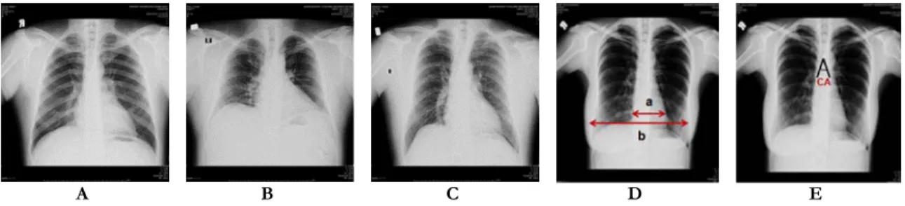
The level (left or right side higher, left and right sides same level), lobulation (absent or present) (Fig. 1A), eventration (absent or present) (Fig. 1B) and contour (smooth or not smooth) (Fig.1C) of diaphragm; level of hilum (right and left sides same level, left side higher or right side higher); number of pairs of ribs; number of ribs superposing the lung parenchyma were assessed. The distance from the lateral margin of the vertebral body to the aorta; the angle between the vertebral body and the aorticopulmonary line; the cardiothoracic ratio (maximum transverse diameter of the heart shadow/maximum transverse diameter of the thorax X 100) (Fig 1D) and the carina bifurcation angle were measured (Fig.1E).
Statistical analysis
Statistical analysis was performed using SPSS software (Version 17.0, SPSS Inc., Chicago, IL, USA). Descriptive analysis (mean, standard deviation, freguencies) are evaluated and reported.
A B C D E
Figure 1. The lobulation (A), eventration (B) and contour (not smooth) (C) of diaphragm were shown with arrows. The cardiothoracic ratio was estimated with following formula: a/bX100 (a: maximum transverse diameter of the heart shadow, b: maximum transverse diameter of the thorax) (D). The carina bifurcation angle (CA) were measured (E).
RESULTS
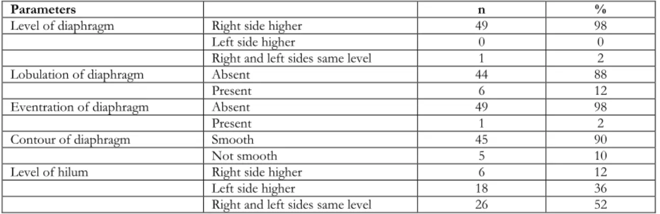
The frequency of anatomical parameters and their measurement with respect to the diaphragm is as follows: level (right side higher in 98%, left and right sides same level 2%), lobulation (88% absent, 12% present), eventration (98% absent, 2% present) and contour (90% smooth, 10% not smooth); level of hilum (right and left sides same level 52%, left side higher 36%,
right side higher 12%) (Table 1); number of pairs of ribs (twelve 96%, unable to determine 4%); number of ribs superposing the lung parenchyma (seven 2%, eight 4%, nine 24%, ten 70%) (Table 2); distance from the lateral margin of the vertebral body to the aorta (16.44 ± 4.35 mm); angle between the vertebral body and the aorticopulmonary line (16.04 ± 3.110); the carina bifurcation angle (58.46 ± 11.130) and the cardiothoracic ratio (38.75 ± 4.27) (Table 3).
Table 1. The frequency of anatomical parameters of diaphragm.
Parameters n %
Level of diaphragm Right side higher 49 98
Left side higher 0 0
Right and left sides same level 1 2
Lobulation of diaphragm Absent 44 88
Present 6 12
Eventration of diaphragm Absent 49 98
Present 1 2
Contour of diaphragm Smooth 45 90
Not smooth 5 10
Level of hilum Right side higher 6 12
Left side higher 18 36
Right and left sides same level 26 52
Table 2. The frequency of anatomical parameters of ribs
Parameters n %
Number of pairs of ribs Twelve 48 96
Unable to determine 2 4
Number of ribs superposing the
lung parenchyma Seven 1 2
Eight 2 4
Nine 12 24
Ten 35 70
Table 3. The measurements of thorax
Parameters Range Mean±SD
VB-A (mm) 5-27.71 16.44±4.35
AVB-APL (0) 9-22 16.04±3.11
CTR 30.77-49.26 38.75±4.27
CA (0) 40-80 58.46±11.13
VB-A: The distance from the lateral margin of the vertebral body to the aorta; AVB-APL: the angle between the vertebral body and the aorticopulmonary line; CTR: the cardiothoracic ratio; CA: the carina bifurcation angle.
DISCUSSION
Most of the studies on this particular topic reflect results from European and American population7. There have been so few reports describing the normal radiographic anatomy and variations of the
we assessed normal PA chest radiographs and analysed the radiographic anatomy in detail among Turkish population. The results represented here show adult thorax graphies of our cases to be in line with literature displaying expected measurements. On a normal Thorax X-ray, visible structures are;
78
ribs, scapulae, breasts and bowel gas. Important obscured and invisible structures are: sternum, esophagus, spine, pleura, fissures and aorta1,2. Bilateral diaphragmatic evantration usually displays poor prognosis. It may be congenital in nature and with a defect of cervical myotomes migrating with intact nerve supply posteriorly. It maybe primary or a result of phrenic nerve damage. These cases are generally asymptomatic but if serious evantration is present acute lung, heart and gastrointestinal consequences may occur especially in children8. The surface of diaphragma is smooth. However due to diaphragmatic hernias and abnormal enlargement of the liver and some other abdominal content it may be irregular or lobular. Lobulation of the diaphragma may point to a serious problem in the prescence of a large hernia2.Hila consists of vessels, lymph nodes and bronchi. The left hilum is usually higher than the right. Abnormalities of these structures are presented by a change in position or enlargement of hiler size or elevated density. Asymmetric hilar enlargement may be due to malignancy2. The ribs are important points in assessment ofthorax X-rays. The anterior edge of 5-7 ribs and posterior edge of 8-10 ribs should be visible to indicate complete breath. Visualization of more than 7 ribs from the anterior suggest lung hyperextansion. The image must be demonstrating the sixth rib on the anterior and the ninth rib on the posterior over the diaphragma dome for an accurate Thorax X-rays9,10. Cardiothoracic ratio helps to evaluate the size of the heart. It is the ratio of the image of the cardiac diameter to thoracic diameter. In normal subjects this ratio must be >0.5. Increased cardiothoracic ratio refers to increased heart size and warrants search for associating pathologies11,12,13.Carina is a part of the trachea found at the base and the carina bifurcation angle is formed when the main bronchi is divided into right and left branches. It usually sits in the T4/5 plane. The absolute mean value of the tracheal bifurcation angle was found to be 79.7 degrees and the range 37-105 degrees14,15. An increased tracheal carinal bifurcation angle is shown to indicate left atrial enlargement. If it is greater than 100 degrees it can be concluded that left atrial enlargement is present16,17.
The assessment of thorax X-ray of our cases displayed the usual primary anatomic structures to be in line with textbook and literature data. The relatively small sample size may weaken the power
of the study but that, these graphies are obtained from healthy subjects will help better evaluate the variables of thoracic x-ray films. The data presented in this study may be useful in understanding normal thoracic structures. A knowledge of the normal anatomy as well as variations are significant for physicians in the assessment of chest radiographs.
Acknowledgement
This manuscript was presented as a poster presentation at the IVth International Symposium of Clinical and Applied Anatomy (ISCAA), Ankara/TURKEY, June 28- July 01, 2012.
REFERENCES
1. Abiru H, Ashizawa K, Hashmi R, Hayashi K. Normal radiographic anatomy of thoracic structures: analysis of 1000 chest radiographs in Japanese population. Br J Radiol. 2005;78:398-404.
2. Oğur T, Uçar E. Systematic interpretation of adult thorax radiography. TAF Prev Med Bull. 2009;8:427-36.
3. Gao N, Kwan BC, Chow KM, Chung KY, Leung CB, Lİ PK et al. Measurements on the routine chest radiograph as prognostic markers in Chinese peritoneal dialysis patients. Clin Nephrol. 2011;76:16-22.
4. Christensen P. Eventration of the diaphragm. Thorax. 1959;14:311-9.
5. Groth SS, Andrade RS. Diaphramatic eventration. Thorac Surg Clin. 2009;19:511-9.
6. Boyan N, Uzum AK, Demir S. Asymptomatic diaphragm eventration caused by a case. CU Saglık Bil Derg. 2006;22:21-5.
7. Nakashima K, Ashizawa K, Ochi M, Hashmi R, Hayashi K, Gotoh S, Honda S, Igarashi A, Komaki T. Interpretation of normal anatomic structures on chest radiography: comparison of Fuji computed radiography (FCR) 5501D with FCR 5000 and screen-film system. J Appl Clin Med Phys. 2003;4:85-90.
8. Shah Mirany J, Schmitz GL, Watson RR. Eventration of the diaphragm: physiologic and surgical significance. Arch Surg. 1968;96:844-50.
9. Yamaguchi I, Itoh H. Radiologic-anatomic correlation of thoracic vertebrae and rib shadows in chest digital radiograph. Nihon Hoshasen Gijutsu Gakkai Zasshi. 2007;20;63:1145-51.
10. Kurihara Y, Yakushiji YK, Matsumoto J, Ishikawa T, Hirata K. The ribs: anatomic and radiologic considerations. RadioGraphics. 1999;19:105-19. 11. Gao N, Kwan BC, Chow KM, Chung KY, Leung
CB, Li PK et al. Longitudinal changes of cardiothoracic ratio and vascular pedicle width as predictors of volume status during one year in
chinese peritoneal dialysis patients. Kidney Blood Press Res. 2009;32:45-50.
12. Zhu Y, Xu H, Zhu X, Wei Y, Yang G, Xu Y and Tang L. Association between cardiothoracic ratio, left ventricular size and systolic function in patients undergoing computed tomography coronary angiography. Exp Ther Med. 2014;8:1757-63. 13. Hemingway H, Shipley M, Christie D, Marmot M. Is
cardiothoracic ratio in healthy middle aged men an independent predictor of coronary heart disease mortality? Whitehall study 25 year follow up. BMJ. 1998;316:1353-4.
14. Coppola V, Vallone G, Coscioni E, Coppola M, Maraziti G, Alfinito M et al. Normal value of the tracheal bifurcation angle and correlation with left atrial volume. Radiol Med. 1998;95:461-5.
15. Kamel SK, Lau G, Stringer MD. In vivo and in vitro morphometry of the human trachea. Clin Anat. 2009;22:571-9.
16. Dennie CJ, Coblentz CL. The trachea: normal anatomic features, imaging and causes of displacement. Can Assoc Radiol J. 1993;44:81-9. 17. Taskin V, Bates MC, Chillag SA. Tracheal carinal
angle and left atrial size. Arch Intern Med. 1991;151:307-8.