Ankara Üniv Vet Fak Derg, 63, 329-331, 2016
Short Communication / Kısa Bilimsel Çalışma
The first report of ehrlichiosis in a cat in Turkey
Metin Koray ALBAY
1, Necmettin Sarp SEVGİSUNAR
1, Sima ŞAHİNDURAN
1, Özlem ÖZMEN
2Mehmet Akif Ersoy University, Faculty of Veterinary Medicine, 1Departments of Internal Medicine, 2Departments of Pathology, Burdur, Turkey.
Summary: An 11-year old cat presented to the Veterinary Medical Teaching Hospital in Mehmet Akif Ersoy University from a farm in Burdur province. She presented fever, lethargy, icteric mucous membrans, abdominal bloating and anorexia. An Ehrlichia
canis infection was confirmed by blood smear and indirect immunofluorescence assay (IFAT) method. At the examination of blood
smear marked anemia and anisocytosis were observed in erythrocytes. Ehrlichia spp. morulae stage were observed in cytoplasm of neutrophils. Blood samples were taken from cat for haematological and biochemical analysis. The cat was treated with doxycycline and recovered rapidly.
Keywords: Cat, ehrlichia, treatment.
Türkiye'de bir kedide ilk ehrlichiosis olgusu
Özet: 11 yaşında dişi bir kedi ateş, letarji, mukozalarda ikter, karında şişkinlik ve iştahsızlık şikayetleri ile Mehmet Akif Ersoy Üniversitesi Veteriner Fakültesi Eğitim Hastanesine getirildi. Ehrlichia canis enfeksiyonu, sürme froti hazırlanarak ve ayrıca indirek immunofloresan (IFAT) yöntemiyle teşhis edildi. Sürme frotiler incelendiğinde belirgin anemi ve eritrositlerde anizositoz gözlendi. Nötrofillerin sitoplazmasında Ehrlichia spp morulası mevcuttu. Hematoloji ve biyokimya analizleri için kan örnekleri alındı. Tedavide doxycycline kullanılarak iyileşme sağlandı.
Anahtar sözcükler: Ehrlichia, kedi, tedavi.
Ehrlichiosis is a worldwide distributed disease caused by microorganisms of Ehrlichia genus that is transmitted by arthropod vectors (9). Rhipicephalus sanguineus and Dermacentor variabilis are the known vectors (6). Ehrlichia spp. belongs to the Rickettsiales order and the Anaplasmataceae family (15). The first case of feline monocytic ehrlichiosis was described in France by Charpentier and Groulade (1986). Since then, several cases of E. canis infection have been reported in cats (7, 12, 14).
In this study, we found Ehrlichia canis agent in a cat in the south-west of Turkey by demonstrating inclusions of the agent on blood smear.
The Ehrlichia species that naturally infect cats have not yet been fully determined, although monocyte and lymphocyte inclusions and, more consistently, E. canis DNA have been detected in cats (6, 13), as well as some granulocytic inclusions related to feline granulocytic ehrlichiosis caused by Anaplasma phagocytophilium (1, 16). The clinical signs and abnormal laboratory findings relating to ehrlichiosis are similar in felids and canids (2). The role of cats in the epidemiology of ehrlichiosis is unknown. It has been claimed that cats are more resistant to infection than are dogs (11).
An 11-year old female cat were brought to the Mehmet Akif Ersoy University Veterinary Medical Teaching Hospital with complain of progressive anorexia. According to the anamnesis, the cat presented severe anorexia and icterus in oral mucous, lost weight in one month. In physical examination fever, lethargy, icteric mucous membranes, dehydration, abdominal bloating were detected. The cat had been living indoors with access to the outdoors. No ticks and external parasites were found on the her body.
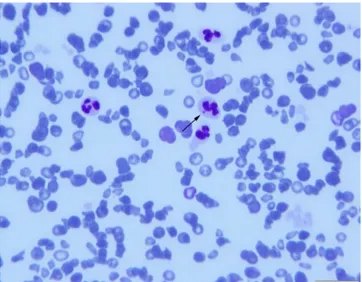
For diagnostic purposes, blood smear was prepared and examined under immersion oil at light microscope. Ehrlichia spp. were diagnosed as granular bodies in the cytoplasm of neutrophils. In addition, marked anemia and anisosytosis were observed in erithrocytes (Figure 1). Diagnoses of ehrlichiosis were supported by using IFAT with a 1:40 antibody titre as cut-off (FLUO EHRLICHIA CANIS, Kit fort he detection of anti-Ehrlichia canis, Agrolabo, Biopronix). This test is originally developed for use in dog ehrlichiosis, but it can be used in serological diagnosis for cats (10, 12). Rapid diagnostic kit were used for feline immunodeficiency virus and feline leukaemia virus all tests were negative.
Metin Koray Albay - Necmettin Sarp Sevgisunar - Sima Şahinduran - Özlem Özmen 330
Figure 1: An Ehrilichia spp. morulae (arrow) in a neutrophil leukocyte and anisocytic erythrocytes in blood smear.
Şekil1: Giemsa morulası boyalı preparatta nötrofil lökositlerde
Ehrilichia spp. morulası ve eritrositlerde anizositoz.
Table 1: Haematological results before and after treatment. Tablo 1: Tedaviden önce ve sonraki hematoloji sonuçları.
Parameters Before treatment One week after treatment Three weeks after treatment Total leukocyte (×109 /l) 35.87 24.95 20.12 Lymphocyte (×109/l) 1.73 4.33 7.52 Granulocyte (×109/l) 30.43 17.59 14.58 Erythrocyte (x1012/l) 4.31 6.25 6.45 Haemoglobin (g/l) 71 89 102 Haematocrite (%) 48 43.82 39.02 Trombocyte (×109/l) 57 170 376
Table 2: Serum biochemical results before and after treatment. Tablo 2: Tedaviden önce ve sonraki serum biyokimyasal sonuçları. Parameters Before treatment One week after treatment Three weeks after treatment Glucose (mg/dl) 160 113 109 Albumin (g/dl) 1.77 2.18 2.09 Total protein(mg/dl) 4.8 6.1 6.3 Creatinine (mg/dl) 0.95 1.01 1.07 Urea (mg/dl) 26 23 29 ALT (U/l) 94 95 95 AST (U/l) 69.8 36.4 36.1 ALP (U/l) 130 106 49 GGT (U/l) 7 6 7 Direct bilirubin (mg/dl) 1.12 0.39 0.18
Blood sample was taken from the cephalic vein for haematological and biochemical examinations. MS9 blood counting equipment and IDEXX Vet-Test equipment and reagents were used for hematological and biochemical analysis. The serum was analyzed for glucose, alanine aminotransferase (ALT), aspartate aminotransferase (AST), alkaline phosphatase (ALP), gamma glutamyl transferase (GGT), creatinine, urea, albumin, total protein, and direct bilirubin. In haematological analysis white blood cell and granulocyte values increased, but trombocyte value was decreased. Although AST and ALP and direct bilirubin values showed a rise, ALT, albumin, total protein, and creatinine values had fallen. Treatment was started with doxycycline orally in 10 mg/kg body weight for 21 days once a day. Cell blood counting and serum biochemical analyzes were repeated after end of treatment (Table 1 and 2).
Research about the presence of Ehrlichia spp. in domestic cats has been conducted in some countries, such as Spain (1), Sweden (4), France (3), and United State (5). First diagnosis of ehrlichiosis case as clinically was reported from a dog in Aydin province in 2007 (18). But there is no report about present of Ehrlichia spp. in domestic cat before. As our knowledge it is the first report of ehrlichiosis in a cat in Turkey. Although described, disease due to tick-borne agent in cats is not well understood and appear rare compared with that in dogs. Clinical signs and laboratory findings in feline ehrlichiosis are similar to including anorexia, lethargy, weight loss, joint pain, dyspnes, lymphadenomegaly, and anemia (17). While Ebani and Bertelloni (10), reported that seroprevalence is higher in elderly cats, the cat was 11 years old in this study. According to authors it can be thought that older cats are exposed to vectors more frequently compared to the younger cats. The same author (10), reported that for determination of ehrlichiosis, haematological examinations revealing decreased thrombocyte values should be observed. In our study before treatment, (first results) leukocytosis, lymphopenia, granulocytosis, erythrocytopenia, and thrombocytopenia were determined as haematological findings. These results are in agreement with previous studies (4, 10). In this study, haemoglobin value was abnormal. Although Bjoersdorff et al. (4), reported that levels of liver enzymes were, in this study, between physiological limits, in this study direct bilirubin, AST and ALP values were significantly increased. Whereas levels of creatinine, urea, ALT and GGT concentrations were normal. The cat was treated with doxycycline at a dosage of 10 mg/kg orally for 3 weeks. After 1 and 3 weeks in treatment priod with doxycycline blood analysis was repeated 2 times again (second and third results). Haematological and biochemical analysis results
Ankara Üniv Vet Fak Derg, 63, 2016 331
gradually returned to reference values and during the 6 months of follow-up the cat did not show any secondary ehrlichial manifestation.
This study is demonstrated first time Ehrlichia canis have been reported in domestic cat in Turkey.
References
1. Aguirre E, Tesouro MA, Amusategui I, et al. (2004):
Assessment of feline ehrlichiosis in central Spain using serology and a polymerase chain reaction technique. Ann
NY Acad Sci, 1026, 103-105.
2. Almosny NRP, Almeida LE, Moreira NM, et al. (1998):
Erliquiose clinica em gato (Felis catus). Rev Bras Cienc
Vrt , 5, 82-83.
3. Beaufils JP, Marin-Granel J, Jumelle P (1995):
Ehrlichia infection in cats: a review of three cases. Part
Med Chir Anim Com, 30, 397-402.
4. Bjoersdorff A, Svendenius L, Owenst JH, et al. (1999):
Feline granulocytic ehrlichiosis- a report of a new clinical entity and characterisation of the infectious agent. J Small
Anim Pract, 40, 20-24.
5. Bouloy RP, Lappin MR, Holland CH, et al. (1994):
Clinical ehrlichiosis in a cat. J Am Vet Med Assoc, 204,
1475-1478.
6. Braga MSCO, Andre MR, Freschi CR, et al. (2012):
Molecular and serological detection of Ehelichia spp. in cats on Sao Luis Island, Maranhao, Brazil. Rev Bras
Parasitol Vet, 21, 37-41.
7. Breitschwerdt E.B, Abrams-Ogg ACG, Lappin MR, et al. (2002): Molecular evidence supporting Ehrlichia
canis-like infection in cats. J Vet Int Med, 16, 642-649.
8. Charpentier F, Groulade P (1986): Report of one case of
probable feline ehrlichiosis. Bull Acad Vet, 59, 287-290.
9. Couto CG (1998): Doenças rickettsiais. In: Birchard, S.J., Sherding, R.G. (eds) Manual Saunders: Clínica de Pequenos Animais. Roca, São Paulo, pp 139-142. 10. Ebani VV, Bertelloni F (2014): Serological evidence of
exposure to Ehrlichia canis and Anaplasma
phagocytophilum in Central Italian healthy domestic cats.
Ticks Tick Borne Dis, 5, 668-671.
11. Lappin MR, Griffin B, Brunt J, et al. (2006): Prevalence
of Bartonellaspeciesi haemoplasma speciesi Ehrlichia species, Anaplasma phagocytophilium, and Neorickettsia risticii DNA in the blood of cats and their fleas in the Unites States. J Feline Med Surg, 8, 85-90.
12. Little SF (2010): Ehrlichiosis and anaplasmosis in dogs
and cats. Vet Clin Small Anim, 40, 1121-1140.
13. Oliveria LS, Mourao LC, Oliveria KA, et al. (2009):
Molecular detection of Ehrlichia canis in cats in Brazil.
Clin Microbiol Infect,15, 53-54.
14. Ortuno A, Gauss CBL, Garcia F, et al. (2005):
Serological evidence of Ehrlichia spp. exposure in cats from northeastern Spain. J Vet Med, B, 52, 246-248.
15. Paddock CD, Childs JE (2003): Ehrlichia chaffeensis: A
prototypical emerging pathogen. Clin Microbiol Rev, 16,
37-64.
16. Shaw SE, Day MJ, Birtles RJ, et al. (2001): Tick-born
infectious diseases of dogs. Trends Parasitol, 17, 74-80.
17. Stubbs CJ, Holland CJ, Reif JS, et al. (2000): Feline
ehrlichiosis. Comp Cont Ed Pract Vet, 22, 307-318.
18. Ulutas B, Bayramlı G, Karagenc T (2007): First case of
Anaplasma (Ehrlichia) platys infection in a dog in Turkey.
Turk J Vet Animal Sci, 31, 279-282.
Geliş tarihi: 29.04.2015 / Kabul tarihi: 12.10.2015
Address for correspondence:
Dr. Sima Sahinduran
Mehmet Akif Ersoy University, Faculty of Veterinary Medicine. Department of Internal Medicine, İstiklal Yerleskesi, Burdur-Turkey. e-mail: sahinduran@mehmetakif.edu.tr
