donkeys, horses and native geese in Turkey
Article in Revue de médecine vétérinaire · March 2018 CITATIONS
0
READS
83
5 authors, including:
Some of the authors of this publication are also working on these related projects:
Serological investigation of West Nile Virus (WNV) infection in cats and dogs in the Burdur districtView project
ArbovirusesView project Yakup Yildirim
Mehmet Akif Ersoy University
46PUBLICATIONS 234CITATIONS SEE PROFILE Aykut Ozkul Ankara University 302PUBLICATIONS 2,705CITATIONS SEE PROFILE
Introduction
West Nile virus (WNV) is an enveloped, single-stranded positive polarity-bearing RNA virus, which belongs to the genus Flavivirus in the family Flaviviridae. The agent is also part of the JE-serocomplex that contains the Japanese encephalitis virus (JEV), Saint Louis encephalitis virus (SLEV), Murray Valley encephalitis virus (MVEV) and Kunjin virus (KUNV). It causes infections characterized by mild inflammatory diseases, meningitis, encephalitis or deaths in various animals such as humans, horses and birds [3, 4, 28, 31].
The natural transmission cycle of WNV infection occurs between wild and domestic birds and mosquitoes, especially of the Culex species [23]. In this respect, presenting in this
study the status of the infection in native species of free-range geese raised in small family-owned businesses, which share joint basins with wild birds [17] is important for the region. Migratory birds play in the epizootiology of WNV infection.
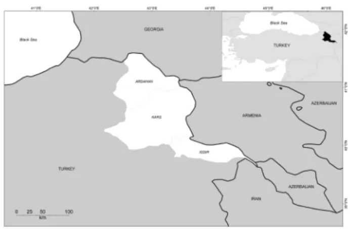
The study was performed in Kars, Ardahan and Iğdır provinces (43.05° E and 40. 36° N), which is undergoing ecological and socioeconomical changes and also has a rich mosquito fauna. It is located in the Northern Anatolian. Vector-borne viral diseases such as Bluetongue (BT) and Akabane (AKA) are endemic in this region, which is the most important livestock production area in Turkey.
In order to differentiate the agent from other flaviviruses and prevent cross-reactivity, specific tests such as the plaque reduction neutralization test (PRNT), enzyme linked SUMMARY
West Nile virus (WNV), an arthropod-borne viral pathogen of global importance, is considered to be the most widespread flavivirus in the world. Here we present a serological and virological study on WNV in horses, donkeys and Turkish native geese in the North-eastern Anatolian province. Blood sera were collected randomly from 118 horses, 70 donkeys and 378 geese, and tested for antibodies against WNV using a commercial competitive enzyme-linked immunosorbent assay (C-ELISA). The overall results revealed that 0.8% (1/118) of the horses, 20% (14/70) of the donkeys and 1.1% (4/378) of the geese were WNV seropositive. To determine the presence of WNV nucleic acid, positive blood sera were tested by the reverse transcription polymerase chain reaction (RT-PCR) technique. WNV nucleic acid was not found in horse and goose samples, while it was demonstrated in four donkey samples. The results suggest that the infection was spreading in private small-scale production units. This study is the first molecular and serological study to determine virus prevalence and seroprevalence of WNV infection in horses, donkeys and Turkish native geese in the North-eastern Anatolian province of Turkey. It is also the first to be conducted on Turkish native goose in Turkey.
Keywords: Donkey, Horse, Turkish native goose, West Nile virus, C-ELISA, RT-PCR, seroprevalence
RÉSUMÉ
Investigation moléculaire et sérologique de l’infection par le virus de West Nile chez les ânes, les chevaux et les oies indigènes en Turquie Le virus de West Nile (VWN) est considéré comme le flavivirus le plus répandu dans le monde entier. Nous présentons ici une étude sérologique et virologique réalisée chez les chevaux, ânes et oies turques indigènes dans le nord-est de l’Anatolie en Turquie. Des sérums ont été prélevés au hasard sur 118 chevaux, 70 ânes et 378 oies, et testé pour les anticorps contre le VWN à l’aide d’un dosage immuno-enzymatique compétitif (C-ELISA). Les résultats globaux ont révélé que 0,8 % (1/118) des chevaux, 20 % (14/70) des ânes et 1,1 % (4/378) des oies étaient séropositifs. Les sérums positifs ont été testés par RT-PCR. L’acide nucléique du VWN n’a pas été trouvé dans des échantillons de chevaux et d’oies, alors qu’il était présent dans quatre échantillons obtenus sur les ânes. Les résultats suggèrent que l’infection se propageait dans les petites unités de production privées. Cette étude est la première étude moléculaire et sérologique visant à déterminer la prévalence du virus et la séroprévalence de l’infection VWN chez les chevaux, les ânes et les oies indigènes turques dans la province d’Anatolie du nord-est de la Turquie.
Mots-clés : Ane, Cheval, Oie, Turquie, Virus de West Nile, C-ELISA, RT-PCR, séroprévalence
Molecular and serological investigation of West Nile
virus (WNV) infection in donkeys, horses and native
geese in Turkey
Y. YILDIRIM1*, V. YILMAZ2, K. YAZICI3, C. ÖZİC4, A. OZKUL5
1Department of Virology, Faculty of Veterinary Medicine, Mehmet Akif Ersoy University, Burdur, Turkey 2Department of Virology, Faculty of Veterinary Medicine, Kafkas University, Kars, Turkey
3Vocational School of Technical Sciences, Ardahan University, Ardahan, Turkey
4Department of Bioengineering, Faculty of Engineering-Architecture, Kafkas University, Kars, Turkey 5Department of Virology, Faculty of Veterinary Medicine, Ankara University, Ankara, Turkey
immunosorbent assay (ELISA), indirect fluorescent antibody test (IFAT), and especially PCR, in which WNV-specific RNA sequences are used [2, 5, 19, 22, 27, 32]. Serological tests detect the host immune response to WNV infection. Although viremia is detectable earlier than the immune response, serologic (IgG and IgM) assays are typically more sensitive for detecting active and convalescent WNV infection. IgM is typically detectable at the time of initial presentation. The IgM antibody capture enzyme-linked immunosorbent assay (MAC–ELISA) is the most conclusive laboratory method for diagnosis of WNV infection of the CNS [3, 32]. The method is high sensitivity and specificity >95% [14]. RT-PCR has proven to be an effective method for detection of WNV nucleic acid from a variety of sample types [18]. WNV viremia peaks at about the time of symptom onset and rapidly fades to undetectable levels. Thus, RT-PCR assays can detect WNV RNA in clinical samples as early as several days before symptom onset, prior to seroconversion. Although this assay has excellent analytical sensitivity, it lack the clinical sensitivity of antibody tests and is typically not used alone for screening or diagnosis [3].
The 2010 human cases (n=47) of WNV infections in Turkey were observed in several provinces mostly in the western part. Ten of the patients died. [16]. The WNV infections were included in the national notifiable diseases list as of April 2011.
This study aims to detect the WNV-seroprevalence in horses, donkeys and free-range Turkish native geese raised in three provinces (Kars, Ardahan and Igdir) located in the Northeast Turkey and make suggestions to help prevent economic losses. In addition, this survey is important in terms of acquiring initial data for the aforementioned infection in horses, donkeys and Turkish native geese and forming a basis for studies to be conducted in the future. Unlike previous studies, it is studied that in the region of Turkey which includes the migratory birds route. Considering the epidemiology of infection (the animals which use common water sources with migratory birds) are investigated the role of migratory birds for WNV infection in the transmission.
Materials and methods
THE STUDY AREA AND ANIMALS SAMPLED
The study was carried out on small family-owned businesses located between February 2014 to January 2015 in the Northeast Anatolia in Turkey (Figure 1). In the region climate differences can be seen due to elevation. Some of the most important migration routes of the Western Palearctic region pass through Turkey. There are three migration routes in Turkey. These are the Istanbul Bosphorus, Belen Passage (Antakya) and Çoruh Valley.
One of the important territorial features of the region is the wide use of odd-toed animals (e.g. horses and donkeys) for haulage, transportation and in agriculture, and thus, one or two horses are raised on every farm in the study area. The equines used in this study comprised healthy appearing horses or donkeys older than 1 year of age and unvaccinated against the aforementioned infection. Likewise, materials were obtained from regional native geese older than 3 months of age and from these family-owned small-scale businesses, which raise 20-30 geese on average for both food-supply and source of income purposes. In accordance with this, distribution of a total of 566 randomly selected animals in terms of province of origin and species were shown in Table I. All sampling were performed after the approval of local
Location Species Number of samples C-ELISA Positive
Ardahan Province Goose 129 1 (0.8%)
Horse 38 0 (0%)
Donkey 24 2 (8.3%)
Igdir Province Goose 124 2 (1.6%)
Horse 40 1 (2.5%)
Donkey 25 9 (36%)
Kars province Goose 125 1 (0.8%)
Horse 40 0 (0%)
Donkey 21 3 (14.3%)
Total 566 19 (3.4%)
Figure 1: Geographical positioning of the Turkish provinces in which the study was performed.
ethical committee for animal studies (Approval Number: ERÜ-HADYEK-2013/13-103). This research was conducted after the approval of Erciyes University Animal Testing Local Ethics Council.
SERUM SAMPLES
Blood serum samples were collected by puncture of jugular (horse and donkey) and brachial veins (goose) into vacuum tubes with clot activator from randomly selected animals. After clotting at room temperature for 15-30 minutes and centrifugation at 3000 g, at 4°C for 10 minutes, serum samples were carefully harvested, heat-inactivated and stored at –20°C until analysis.
REVERSE TRANSCRIPTION POLYMERASE CHAIN REACTION (RT-PCR)
Total RNA was isolated from serum samples using TRIzol reagent (Sigma). Total RNA was treated with RQ1 DNAse I (Promega). To obtain cDNA from total RNA was used Fermentas Revert Aid First Strand cDNA synthesis Kit (#K1622) and applied to kit protocol. Reverse transcription (RT) was performed according to the manufacturer’s directions (Fermentas) using 1 unit of MMLV reverse transcriptase with 5 μg of total RNA and oligo dT22 primer. The 25μl PCR mixture contained were performed using 1 μL of cDNA template, 0.25 μL (5u/ μL) of Taq polymerase, 2.5 μL (100 ng) of primers, 1.5 μL (25mM) dNTP, 2.5 μL 10× PCR buffer and 1.5 μL MgCl2. The RT-PCR assay was used to detect WNV RNA. WNV specific primers were designed as follows: WN233 (5’-TTGTGTTGGCTCTCTTGGCGTTCTT-3’) and WN640c (5’-CAGCCGACAGCACTGGACATTCATA-3’) which are specific for WNV and sizes are 408 bp [10, 19].
COMPETITIVE ENZYME LINKED IMMUNOSORBENT ASSAY (C-ELISA)
West Nile Competition Multi-species ELISA kit (ID Screen®, Productc code: West Nile Competition Multi-species (WNC), IDvet, Grabels, France) were used to detect anti-pr-E antibodies against the WNV. The ELISA is high sensitivity and specificity >95% [14].
Tests were performed according to the manufacturer’s directions. The OD of each well was read using an ELISA reader at a wavelength of 450 nm.
STATISTICAL ANALYSIS
Statistical analysis was carried out via the Statistical Package for Social Sciences software (IBM SPSS Statistics 20.0, SPSS, Inc., Chicago, IL, USA) [15]. The main differences between species and regions were evaluated using the Chi-Square (χ2) test. At the end of the study, the data from which the value of P<0.05 was derived was accepted as significant.
Results
WNV nucleic acid was not found in horse and geese samples by RT-PCR. In contrast, WNV nucleic acid originated amplicon (408 bp in length) was detected in 4 donkey seropositive samples.
In this study, 566 samples were examined in terms of the presence of WNV-specific IgG using the C-ELISA method. WNV-specific antibodies were detected in 20% (14/70) of tested donkey sera, 0.8% (1/118) of horse sera, and 1.1% (4/378) of goose sera (Table I).
According to the statistical analyses, the difference in seropositivity rate confirmed in donkeys statistically compared with the seropositivity rate confirmed in horses and Turkish native geese was determined to be very significant (p<0.001).
Distribution of seroprevalence ratios according to the provinces in which the study was carried out is as follows: 1.6% (3/191) in Ardahan, 6.3% (12/189) in Igdir, and 2.2% (4/186) in Kars (Table I). According to the statistical analyses, the antibody positivity rate in the Igdir region statistically compared with the seropositivity rate in the Kars and Ardahan regions was noted to be significant (p<0.05).
Discussion
Arthropod-borne viruses are transmitted by blood-sucking arthropods such as mosquitos, ticks and sand flies. Because the actual cycle of the West Nile virus is between birds and mosquitoes, many studies have been done to research the link between virus transmission and migratory birds [9, 11, 13, 21]. Two of the most important migratory routes enter Turkey from the northeast and northwest, meet in Antakya, and continue down through continental Africa. Millions of birds flying to the Middle East and Africa from Russia and the Caucasus in autumn and returning in the spring travel over the Northeast Anatolia region of Turkey from end to end. The serological identification of the existence of WNV infection in domestic geese sharing wetlands used by migratory birds to take breaks along their migratory routes, as well as other equid species, is important for this study in terms of the role of migratory birds play in the epizootiology of WNV infection.
The study was carried out in the regions of Igdir Valley, Kars Plateau and Ardahan Plateau, which are located in the Northeast Anatolia and have different climatic and geographical features. In addition, the area chosen for the study provides suitable habitats for mosquito larvae with a variety of wetlands, and because of the extensive amount of animal-farming done in the area, there is a variety of host types on which mature insects are able to feed. When all the area’s conditions are considered, it can be said that it is a potentially risky area for WNV infection.
Wernery et al. [30] detected WNV seropositivity in the blood serum samples they took from equid species to be 20% in the study they conducted in the United Arab Emirates.
When Turkey’s geographic location and the geographical distribution of the infection are considered, the country is susceptible to many arboviral infections. The first data regarding WNV infection in Turkey dates back to 1970. According to a hemagglutination inhibition assay (HAI) done on human and sheep serum collected from the West Anatolia region, seropositivity in humans was detected to be 6%, and 1-5% in sheep [1, 29]. Five years later, human serum samples collected from the Southeast Anatolia region using HAI were checked in terms of the existence of WNV antibodies, and infection seroprevalence was found to be 40% in human serum samples [24]. According to a plaque reduction neutralization test (PRNT) done on 764 blood serum samples belonging to mules, cats, cattle, dogs, horses, humans and sheep from various provinces in Turkey (Hatay, Muğla, Şanlıurfa, Izmir, Adana, Bursa, Ankara, Antalya), WNV seroprevalence was reported to be 2-5% in mules, 4% in cattle, 7-37% in dogs, 5-13% in horses, 4-20% in humans, and 1% in sheep, however, seropositivity was not found in cats [26]. In another study, mosquito samples (Culex pipiens,
Ochlerotatus caspius and Aedes species) and human blood
serum samples were collected in the province of Şanlıurfa. While RT-PCR, VecTest and virus isolation attempts on Vero cell culture done on mosquito samples produced no positive results, an indirect immunofluorescence assay (IFA) done one 181 human blood serum samples detected WNV seropositivity to be 16% [25]. Ergunay et al. [8] reported that seroprevalance for WNV infection was 9.9% in duck, 12.5% in horse, 1.9% in sheep and 12.1% in human using PRNT to identify WNV antibodies in blood serum samples collected from 423 duck, 389 horse, 102 sheep and 266 human in 15 provinces in Turkey. Same researchers tested plasma samples from 256 horse and 266 human in Mersin, Adana and Mugla provinces for the presence of WNV RNA using nested and rRT-PCR and WNV RNA was detected in a total of 31 samples in the study. In another study of Ergunay et al. [7] found close relationships to WNV lineage 1 strain ArB310/67 from the Central African Republic, distinct from other WNVs circulating in the Mediterranean Basin, the Middle East, and Eastern Europe.
In this study, 566 samples were examined in terms of the presence of WNV-specific antibodies using the C-ELISA method. In 20% (14/70) of tested donkey serums, 0.8% (1/118) of horse serums, and 1.1% (4/378) of goose serums WNV-specific antibodies were detected. Distribution of seropositivity according to the regions in which the study was carried out is as follows: 1.6% (3/191) in Ardahan, 6.3% (12/189) in Igdir, and 2.2% (4/186) in Kars.
In the molecular part of the study, WNV nucleic acid presence in serum samples was examined using the reverse transcriptase polymerase chain reaction method. WNV
nucleic acid was only detected in four of the positive blood serum samples belonging to donkeys.
In parallel with the study performed by Ledermann et al. [20], detection of WN nucleic acid presence by RT-PCR method in serum samples detected as antibody positive by ELISA technique set us thinking that donkeys whose serum we collected were subclinically infected and samplings might have been done at the end of viremia, that is at the stage where the animals became seroconversion. Because detecting nucleic acid presence in serum samples is a prominent evidence of WN infection existence [12, 20].
It is thought that the reason seropositivity was high in donkeys is due to the fact that because the donkeys in the village and plateaus where the study was conducted are used especially for carrying water, they have a higher risk of being exposed to vectors carrying infectious agents in wetland areas. It was decided that the reason seropositivity was higher in the Igdir region compared to the other two regions is due to the fact that it has a hotter climate and vector population could be more widespread.
In conclusion, this study has shown that WNV infection is found at different rates in horses, donkeys and domestically-bred geese. In order to avoid the aforementioned disease, reducing the contact between humans and vector mosquitoes and ticks is a helpful way to drop the rate of mortality, morbidity and infection, and this can be achieved through control activities of mosquitoes, ticks, and winged insects or vectors. In addition, the importance of vaccination should not be forgotten and more effort than ever should be made in this area. Also, the necessity of informing animal breeders about infectious diseases and the preparation of animal shelters in modern conditions gains importance.
Acknowledgement
This project was supported by the Commission for the Scientific Research Projects of Kafkas University (Project name: Serological investigation of West Nile virus infections in horses, donkeys and Turkish native geese in Kars, Ardahan and Igdir provinces. Project No.: 2013-VF-98).
References
1. - ARI A.: Studies on activity and ecology of arboviruses in Turkey. Türk Hij. Tecr. Biyol. Derg., 1972, 32, 134-143. 2. - BLITVICH B.J., BOWEN R.A., MARLENEE N.L., HALL
R.A., BUNNING M.L., BEATY B.J.: Epitope-blocking enzyme-linked immunosorbent assays for detection of west nile virus antibodies in domestic mammals. J. Clin.
Microbiol., 2003, 41, 2676-2679.
3. - BUNNING M.L., BOWEN R.A., CROPP C.B., SULLIVAN K.G., DAVIS B.: Experimental infection of horses with West Nile virus. Emerg. Infect. Dis., 2002, 380-386.
4. - CDCP (Centers for disease control and prevention). Fact sheet: West Nile virus (WNV) infection: information forclinicians. Available at http://www.cdc.gov/ncidod/ dvbid/westnile/resources/fact_sheet_clinician.htm 5. - DIAMOND S.M.: Evasion of innate and adaptive
immunite by flavivirus. Immunol. Cell. Biol., 2003, 81, 196-206.
6. - DURAND B., CHEVALIER V., POUILLOT R., LABIE J.: West Nile virus outbreak in horses, southern France, 2000: results of a serosurvey. Emerg. Infect. Dis., 2002, 8, 777-782.
7. - ERGUNAY K., BAKONYI T., NOWOTNY N., OZKUL A.: Close Relationship between West Nile Virus from Turkey and Lineage 1 Strain from Central African Republic. Emerg. Infect. Dis., 2015, 21, 352-355.
8. - ERGUNAY K., GUNAY F., ERISOZ KASAP O., OTER K., GARGARI S., KARAOGLU T., TEZCAN S., CABALAR M., YILDIRIM Y., EMEKDAS G., ALTEN B., OZKUL A.: Serological, Molecular and Entomological Surveillance Demonstrates Widespread Circulation of West Nile Virus in Turkey. Neglected Tropical Diseases, 2014, 8, e3028.
9. - ESTEVES A., ALMEIDA A.P.G., GALÃO R.P., PARREIRA R.: West Nile virus in southern Portugal.
Vector Borne Zoonot. Dis., 2005, 5, 410-413.
10. - FARFAN-ALE J.A., LOROÑO-PINO M.A., GARCIA-REJON J.E., HOVAV E., POWERS A.M., LIN M., DORMAN K.S., PLATT K.B., BARTHOLOMAY L.C., SOTO V., BEATY B.J., LANCIOTTI R.S., BLITVICH B.J.: Detection of RNA from a Novel West Nile-like Virus and High Prevalence of an Insect-specific Flavivirus in Mosquitoes in the Yucatan Peninsula of Mexico. Am. J.
Trop. Med. Hyg., 2009, 80, 85-95.
11. - FIGUEROLA J., SORIGUER R., ROJO G., GÓMEZ-TEJEDOR C.: Seroconversion in wild birds and local circulation of West Nile virus, Spain. Emerg. Infec. Dis., 2007, 13, 1915-1917.
12. - GUBLER D.J., KUNO G., MARKOFF L.: Flaviviruses, West Nile virus. In: Knipe, D.M., Howley, P.M. (ed.) Fields Virology. 5nd ed. Wolters Kluwer/Lippincott W.W., Philadelphia, 2007, pp. 1153-1197.
13. - HUBÁLEK Z., HALOUZKA J., JUŘICOVÁ Z., ŠIKUTOVÁ S., RUDOLF I., HONZA M.: Serologic survey of birds for West Nile flavivirus in southern Moravia (Czech Republic). Vector Borne Zoonot. Dis., 2008, 8, 659-666.
14. - HUHN G.D., SEJVAR J.J., MONTGOMERY S.P., DWORKIN M.S.: West Nile virus in the United States: an update on an emerging infectious disease. Am. Fam.
Physician, 2003, 68, 653-660.
15. - IBM SPSS Statistics. SPSS for Windows Release 21.0, 2012, SPSS Inc. Chicago. IL. USA.
16. - KALAYCIOGLU H., KORUKLUOGLU G., OZKUL A., ONCUL O., TOSUN S., KARABAY O., GOZALAN A., UYAR Y., CAGLAYIK D.Y., ATASOYLU G., ALTAS A.B., YOLBAKAN S., OZDEN T.N., BAYRAKDAR F., SEZAK N., PELITLI T.S., KURTCEBE Z.O., AYDIN E., ERTEK M.: Emergence of West Nile virus infections in
humans in Turkey, 2010 to 2011. Euro. Surveill., 2012, 17, pii: 20182.
17. - KIRMIZIBAYRAK T., ÖNK K., YAZICI K.: Effects of Age and Sex on Slaughtering and Carcass Characteristics of Turkish Native Geese Reared in Free Range Production Conditions in Kars Province. Kafkas Univ. Vet. Fak.
Derg., 2011, 7, 41-45.
18. - LANCIOTTI R.S., KERST A.J.: Nucleic acid sequence-based amplification assays for rapid detection of West Nile and St. Louis encephalitis viruses. J. Clin. Microbiol., 2001, 39, 4506-4513.
19. - LANCIOTTI R.S., KERST A.J., NASCI R.S., GODSEY M.S., MITCHELL C.J., SAVAGE H.M., KOMAR N., PANELLA N.A., ALLEN B.C., VOLPE K.E., DAVIS B.S., ROEHRIG J.T.: Rapid detection of West Nile Virus from human clinical specimens, field-collected mosquitoes, and avian samples by a TaqMan reverse transcriptase-PCR assay. J. Clin. Microbiol., 2000, 38, 4066-4071. 20. - LEDERMANN P.J., LORONO-PINO M.A., ELLIS
C., SAXTON-SHAW K.D., BLITVICH B.J., BEATY B.J., BOWEN R.A., POWERS A.M.: Evaluation of Widely Used Diagnostic Tests to Detect West Nile Virus Infections in Horses Previously Infected with St. Louis Encephalitis Virus or Dengue Virus Type 2. Clin.
Vaccine Immunol., 2011, 18, 580-587.
21. - MALKINSON M., BANET C., WEISMAN Y., POKAMUNSKI S., KING R., DROUET M.T., DEUBEL V.: Introduction of West Nile virus in the Middle East by migrating white storks. Emerg. Infect. Dis., 2002, 8, 392-397.
22. - MARTIN D.A., MUTH A.D., BROWN T., JOHNSON J.A., KARABATSOS N., ROEHRIG T.J.: Standardization of immunoglobulin M capture enzyme linked immunosorbent assays for routine diagnosis of arboviral infections. J. Clin. Microbiol., 2000, 38, 1823-1826. 23. - MCMINN C.P.: The molecular basis of virulence of the
encephalitogenic flaviviruses. J. Gen. Virol., 1997, 78, 2711-22.
24. - MECO O.: Investigation of West Nile virus specific haemagglutination-inhibiting antibodies in southeastern Anatolian people. Mikrobiol. Bul., 1977, 11, 3-17.
25. - OZER N., ERGUNAY K., SIMSEK F., KAYNAS S., ALTEN B.: West Nile virus studies in the Sanliurfa Province of Turkey. J. Vector. Ecol., 2007, 32, 202-206. 26. - OZKUL A., YILDIRIM Y., PINAR D., AKCALI A.,
YILMAZ V., COLAK D.: Serologicalevidence of West Nile Virus (WNV) in mammalian species in Turkey.
Epidemiol. Infect., 2006, 134, 826-829.
27. - PADILLA J.A., RUBIO E.L., ROMERO E.E., CORDOBABA L., CUEVAS S., MEJIA F., CALDERON R., MILIAN F., ROSA A.T.D., WEAVER S.C., FRANCO J.G.E., SAIZ J.C.: The continous spread of west nile virus (WNV): seroprevalance in asymptomatic horses.
Epidemiol. Infectn., 2009, 137, 1163-1168.
28. - PETERSEN L.R., ROEHRING J.T.: West Nile virus: a reemerging global pathogen. Emerg. Infect. Dis., 2001, 7, 611-614.
29. - RADDA A.: Studies on the activity and ecology of arboviruses in Turkey. Zentralbl. Bakteriol., 1973, 225, 19-26.
30. - WERNERY U., KETTLE T., MOUSSA M., BABIKER H., WHITING CENTRAL J.: West Nile Fever in the United Arab Emirates. Wildlife Middle East News, 2007, 2, 1-2.
31. - YAZICI Z.: West Nile virus infection. Turk. J. Infect., 2005, 19, 139-143.
32. - ZIEGLER U., ANGENVOORT J., KLAUS C., NAGEL-KOHL U., SAUERWALD C., THALHEIM S., HORNER S., BRAUN B., KENKLIES S., TYCZKA J., KELLER M., GROSCHUP M.H.: Use of Competition ELISA for Monitoring of West Nile Virus Infections in Horses in Germany. Int. J. Environ. Res. Public Health, 2013, 10, 3112-3120.