P. Zorlutuna,1A. Tezcaner,2I. Kıyat,3A. Aydınlı,3V. Hasırcı1
1Department of Biological Sciences, Biotechnology Research Unit, Middle East Technical University,
Ankara 06531, Turkey
2Department of Engineering Sciences, Middle East Technical University, Ankara 06531, Turkey 3Department of Physics, Bilkent University, Ankara 06800, Turkey
Received 3 August 2005; revised 16 January 2006; accepted 9 February 2006
Published online 6 June 2006 in Wiley InterScience (www.interscience.wiley.com). DOI: 10.1002/jbm.a.30772 Abstract: In this study, biodegradable polyester based
car-riers were designed for tissue engineering of the epithelial and the stromal layers of the cornea, and the final construct was tested in vitro. In the construction of the epithelial layer, micropatterned films were prepared from blends of biode-gradable and biocompatible polyesters of natural (PHBV) and synthetic (P(L/DL)LA) origin, and these films were seeded with D407 (retinal pigment epithelial) cells. To im-prove cell adhesion and growth, the films were coated with fibronectin. To serve as the stromal layer of the cornea, highly porous foams of P(L/DL)LA-PHBV blends were seeded with 3T3 fibroblasts. Cell numbers on the polyester carriers were significantly higher than those on the tissue culture polystyrene control. The cells and the carriers were characterized scanning electron micrographs showed that the foam was highly porous and the pores were intercon-nected. 3T3 Fibroblasts were distributed quite
homoge-neously at the seeding site, but probably because of the high thickness of the carrier (⬃6 mm); they could not sufficiently populate the core (central parts of the foam) during the test duration. The D407 cells formed multilayers on the mi-cropatterned polyester film. Immunohistochemical studies showed that the cells retained their phenotype during cul-turing; D407 cells formed tight junctions characteristic of epithelial cells, and 3T3 cells deposited collagen type I into the foams. On the basis of these results, we concluded that the micropatterned films and the foams made of P(L/ DL)LA-PHBV blends have a serious potential as tissue en-gineering carriers for the reconstruction of the epithelial and stromal layers of the cornea. © 2006 Wiley Periodicals, Inc. J Biomed Mater Res 79A: 104 –113, 2006
Key words: cornea; tissue engineering; polyester; retinal pigment epithelial cells; fibroblasts
INTRODUCTION
Cornea is the outermost layer of the eye and corneal damages constitute⬃37% of all visual disabilities and 23% of medical visits for ocular problems in North America.1In this in vitro study, we aimed to construct
the top two layers of the cornea, the epithelial and stromal layers, using biodegradable polymeric carriers which were seeded with retinal pigment epithelial cells and fibroblasts.
Cornea is ⬃500 m in thickness.2It is transparent
even though the corneal tissue consists of a highly organized group of cells and extracellular matrix (ECM). The key factor in its transparency appears to
be the unique composition and organization of its ECM where the collagen fibers have a highly regular arrangement.3–5 They alternate as orthogonally ori-ented lamella especially in the central and posterior stroma. The proteoglycans (keratan sulfate, chon-droitin sulfate, and dermatan sulfate) of the ECM also participate in this regular arrangement.6
Damages to the cornea can be limited to the epithe-lium (such as in uncomplicated erosion) or could in-volve the epithelial basement membrane, the stromal lamella, and even the endothelium (the inner most layer). These damages can be caused by injuries due to foreign materials and chemical burns or due to viral or bacterial infections.1Despite currently available
treat-ments, corneal scar tissue formation occurs in many of these cases. This is a very severe problem that leads to loss of transparency of the cornea and eventually to blindness. In such cases, replacement of cornea is nec-essary. The current corneal replacements include do-nor and artificial corneas. Cornea is the most widely transplanted tissue and more than 50,000 corneal transplantations are performed each year in the
Correspondence to: V. Hasırcı; e-mail: [email protected] Contract grant sponsor: European Union FP6 STREP Project Cornea Engineering; contract grant number: 504017 Contract grant sponsor: Middle East Technical University Scientific Research Projects; contract grant numbers: BAP 2004 – 07-02– 00-16 and BAP 2005– 01-08 – 02
Contract grant sponsor: TUBITAK TBAG 2288
United States and Eastern Europe.7 Corneal
plants are considered to be the most successful trans-plants with a rejection rate of 10%.8On the other hand,
the development of artificial corneas made of syn-thetic polymers such as poly(2-hydroxyethyl methac-rylate) have been developed for many years, but they still have drawbacks such as mechanical weakness and calcium deposition.9 –11 Besides, these polymeric
corneas do not carry viable elements. Even though there is a 10% risk of failure and there is a serious donor shortage transplantation is still the preferred treatment. Artificial corneas are used only when cor-neal transplantation is not an option.12
In recent years, the field of biomaterials developed in a new direction called tissue engineering, which involves use of cells together with biodegradable car-riers. Currently, research on corneal treatments is in-tensified on tissue engineering, and it will be a very important solution to the very serious problem of corneal damages.13–16
In tissue engineering, polymers are used as tempo-rary carriers for the cells. Polyesters are among the most widely investigated polymers. Polyhydoxyal-kanoates (PHA), which are natural polyesters, are pro-duced by microorganisms and they are biodegradable and biocompatible. Poly(hydroxybutyric acid-co-3-hy-droxyvaleric acid) (PHBV) is one of the copolymers of PHA. PHBV is known to be not toxic to cells and a good substrate for cell adhesion and proliferation.17
Poly(l-lactide-co-d,l-lactide) (P(L/DL)LA (70/30)) is a synthetic polymer among the polymer family of poly(␣-hydroxy esters), which is a widely investigated and used family in biomaterial applications. P(L/ DL)LA is also a biodegradable and biocompatible polymer. P(L/DL)LA is a faster degrading polymer when compared to PHBV and it begins to degrade in a few weeks. It takes much longer for PHBV; degra-dation could last as long as 53 weeks.18
In this study, a unique blend of biodegradable and biocompatible polymers (P(L/DL)LA and PHBV were designed as 2D and 3D carriers for the reconstruction of epithelium and the stroma of the cornea. The 2D carriers were films with micropatterns, and were tested in vitro with D407 retinal pigment epithelial cells. The 3D carriers were foams and were seeded with 3T3 fibroblasts, and their physical properties and compatibility with the cells were studied. The results indicate that the carriers designed permit cell
prolif-eration and functionality; therefore, they could be used as carriers for the tissue engineering of the cor-nea after further developments.
MATERIALS AND METHODS Materials
Poly(3-hydroxybutyric acid-co-3-hydroxyvaleric acid) (PHBV, HV content 8% w/w) was purchased from Aldrich Chemical Company. Poly(l-lactide-co-d,l-lactide) (P(L/ DL)LA (70/30), Resomer® LR 708, molar ratio l-lactide:d,l-lactide 70:30) was supplied by Boehringer-Ingelheim (Ger-many). Fetal bovine serum (FBS) was obtained from Biochrome KG (Germany). Dulbecco’s Modified Eagle Me-dium (DMEM) was obtained from Gibco Invitrogen (New Zealand). MTS kit was purchased from Promega. Cacodylic acid sodium salt, ascorbic acid, glutaraldehyde (Grade I, 25% aqueous solution), trypsin-EDTA (0.25%), sodium azide, and monoclonal anti-pan cytokeratin (mixture clones C-11, PCK-26, CY-90, KS-1A3, M20, A53-B/A2, mouse as-cites fluid) were supplied by Sigma Chemical. Mouse anti-ZO-1 was purchased from Zymed Laboratories. Tween 20, formaldehyde (37%), dichloromethane (DCM), and dioxane were obtained from Merck (Germany). Fibronectin (FN pure, from human plasma) was purchased from Roche Di-agnostics (Germany). AlexaFluor 488 was supplied by Mo-lecular Probes. Triton X-100 was purchased from Applichem GmbH (Germany). Bovine serum albumin (BSA) was sup-plied by Fluka (Germany). Acridine Orange was obtained from BDH Chemicals (UK). The RPE cell line D407 was a kind gift of Dr. R. Hunt (Department of Ophthalmology, University of South Carolina Medical School, USA). 3T3 cell line was purchased from Foot-and-Mouth Disease Institute of Ministry of Agriculture and Rural Affairs (Turkey).
Methods
Preparation of carriers
Preparation of templates and micropatterned films
Micropatterned silicon templates with different dimen-sions and geometries (Table I) were produced by photoli-thography and subsequent chemical etching. PHBV and P(L/DL)LA were dissolved in DCM and then cast over the TABLE I
Dimensions and Geometry of Micropatterned Templates
Template Geometry of Template
Groove Width
(m) Ridge Width(m) Groove Depth(m)
A3 V-shaped parallel channels 2 10 30
C3 V-shaped parallel channels 2 10 20
template to produce micropatterned films with pattern di-mensions opposite to those of the template. Thus, two types of films with 2m ridge width and 10 m groove width and 20 or 30m depths were obtained. The third film type was produced with square pits with 10 and 20m sides and a 1 m depth.
Preparation of foams
P(L/DL)LA-PHBV (1:1 w/w) in dioxane (4%, 45 mL) was poured into glass Petri dishes, maintained at ⫺20°C over-night, and freeze-dried (FreeZone® 6 Liter Freeze Dry Sys-tem, Labconco Corporation) for 16 h.
Fibronectin coating of polyester films and foams
The P(L/DL)LA-PHBV films were coated with fibronectin by incubating in fibronectin solution for 10 min (50g/mL fibronectin in PBS). At the end of 10 min, the films were washed with PBS to remove excess fibronectin and dried under sterile conditions in a laminar flow (LaminAir Safe 2000, Holten A/S, Denmark). Foams were coated with fi-bronectin by incubating overnight, then washed, and stored in PBS until cell seeding.
Characterization of carriers
Physical characterization
Foam density was measured by using a pycnometer (n⫽ 2).17
The thickness of the films and foams were measured by a micrometer with a 0.001 mm sensitivity (n⫽ 6).
Microscopic characterization
The topography of the carriers was studied using a scan-ning electron microscope (SEM) (JSM 6400, JEOL, Japan) after coating with a thin film of gold under vacuum.
The cross-section of the foams was examined under a light microscope (IX 70, Olympus, Japan) after taking 4 or 10m thick sections in frozen state by using a microtome (CM 1510, Leica, Germany).
Cell culture studies
D407 cells (passages 5–15) were used to serve as epithelial cells and 3T3 cells (passages 5–15) were used in place of stromal cells. They were cultivated in high glucose DMEM supplemented with 5% FBS, 100 units/mL penicillin, and 100 units/mL streptomycin in a carbon dioxide incubator (37°C, 5% CO2, MCO-17AIC, Sanyo Electric, Japan). The
cells were passaged using 0.05% trypsin-EDTA solution.
Cell seeding onto carriers 2D micropatterned films.
In the cell seeding tests with D407 cells films were used with fibronectin treatment, while others were used as is. P(L/DL)LA-PHBV8 films were sterilized by immersing in sterile EtOH (70%) for 2 h at 4°C and washed four times with PBS. D407 cells were detached from the tissue culture flasks by using 0.05% trypsin for 5 min at 37°C, then centrifuged for 5 min at 3000 rpm, and resuspended in high glucose DMEM supplemented with 5% FBS, 100 units/mL penicil-lin, and 100 units/mL streptomycin. Cell number was counted using NucleoCounter (ChemoMetec A/S, Den-mark). Twenty microliters cell suspension containing 5 ⫻ 104
cells were seeded on each film and the films were not disturbed for 30 min to allow cell attachment. After 30 min, 500L high glucose DMEM supplemented with 5% FBS, 100 units/mL penicillin, and 100 units/mL streptomycin were added. They were incubated in a CO2 incubator (5% CO2,
37°C) for 14 days. The medium was refreshed every day. Tissue culture polystyrene (TCPS) was used as the control because of its optimal properties for cell cultivation.
3D foams.
P(L/DL)LA-PHBV foams were sterilized by immersing in sterile EtOH (70%) for 2 h at 4°C and washed four times with PBS. The 3T3 cells were detached as described for D407 cells and cell number was counted in the NucleoCounter. Two different seeding densities were used; 5⫻ 105
cells/75L or 2.5 ⫻ 105 cells/75L were seeded on each foam, and the
foams were not disturbed for 45 min to allow cell attach-ment. Then, 500L high glucose DMEM with 5% FBS, 100 units/mL penicillin, and 100 units/mL streptomycin were added. They were incubated in a CO2 incubator (5% CO2,
37°C) for 14 days. The medium was refreshed every day. After 7 days of incubation, the number of daily medium changes per day was increased to 2. TCPS was used as control.
Characterization of cell seeded carriers Cell proliferation.
In the determination of cell number, the kit: CellTiter 96® AQueousNon-Radioactive Cell Proliferation Assay (Bulletin
TB169) of Promega (Madison, WI) was used (n⫽ 3).19
Morphology of cells.
Before observation with SEM and fluorescence micros-copy (IX 70, Olympus, Japan), the D407 and 3T3 cells were fixed with glutaraldehyde (2.5%) for 2 h and washed twice with cacodylate buffer (0.1M, pH 7.4). Specimens for SEM were freeze-dried for 8 h and then examined after coating with gold. The samples for fluorescence microscopy were stained with Acridine orange, after washing with HCl (0.1M) for 1 min. After 15 min, Acridine orange was re-moved and the sample was washed with distilled water. Foams were sectioned (5m thick) by using a microtome. The cells were observed under the fluorescence microscope at the excitation wavelength of 480 nm or under confocal microscope (LSM510, Zeiss, Germany) at 488 nm.
Immunohistochemical studies.
For immunohistochemical analysis, fixation was done with formaldehyde (4%) for 30 min, and samples were
washed twice with PBS. The cells were permeabilized by Triton X-100 (0.1%) treatment (5 min) and washed four times with PBS. The samples were then transferred to blocking solution (0.5% BSA, 0.1% Tween 20, 0.1% FBS, 0.1% sodium azide in PBS) for 30 min. Primary antibodies (250L) mouse anti ZO-1 (15 (g/mL in blocking solution) or Anti-Pan cyto-keratin (diluted 200-fold in blocking solution) was pipetted onto the samples and incubated at 4°C overnight. They were incubated with secondary antibody (Alexaflour 488, 300L diluted 400-fold in blocking solution) for 1 h at 37°C, washed three times with PBS and examined under the fluorescence microscope at the excitation wavelength of 480 nm.
RESULTS AND DISCUSSION
Physical and microscopic characterization of films and foams
In this study biodegradable, polymeric cell carriers of 2D (films with different micropatterns) and 3D (foams) form were designed. The carriers were char-acterized before testing in vitro.
Physical characteriztion
The density of P(L/DL)LA-PHBV foams was calcu-lated as 0.11⫾ 0.03 g/mL by using a pycnometer (n ⫽ 2), and is consistent with previous studies with PHBV foams.17This low density shows that the foam has the
highly porous structure necessary for use in the con-struction of a cell carrier.20It is known that pore size,
pore shape, and interconnectivity of these pores affect cell growth,21 and an ideal 3D cell carrier should be
highly porous for the cells to get nutritional elements, discard waste, and also allow sufficient space for proper ECM formation.
The average thickness of the films and foams pre-pared in this study were measured as 40⫾ 2 m for films and 6.11⫾ 0.08 mm for foams (n ⫽ 6).
Microscopical examination
Scanning electron microscope
Scanning electron microscope (SEM) yields infor-mation about the morphology of the carriers. Films prepared on patterned silicon wafers (A3) were smooth and the pattern was regular and sharp [Fig. 1(a)]. The width of the base of the channels is quite close to 10m, and similarly, the ridge width is close to 2m. Foams were highly porous as expected from the density calculations. The pores appeared to be interconnected and pore dimensions were suitable for RPE or fibroblast adhesion and growth [Fig. 1(b)].
Light microscopy
Sections of foams (4- and 10-m thick) were ob-tained by a microtome and examined under light mi-croscope to observe the inner structure, the porosity, and the pore size distribution within the foams. The dark lines in Figure 2(a,b) are polymeric boundaries of the open-celled structure. These micrographs confirm the SEM in that the carrier has a highly porous struc-ture suitable for cell seeding. Even though the magni-fications are the same, in 4m sections, the pore sizes appear to be larger than in the 10m sections, because in the thicker sections the pores of various depths overlap. As Figure 2(b) reveals the pore sizes are at least 40m or larger and therefore are larger than the size of the typical cell. The low apparent pore sizes in Figure 2(a) indicate that the pores are not in the form of parallel channels and therefore cells need to follow a tortuous path if they need to move towards the core of the foam.
In vitro studies
D407 cells on patterned films
RPE cells were tested on the patterned films after adsorption of fibronectin on the surface to improve cell adhesion.
Figure 1. The SEM micrographs of 2D and 3D P(L/DL)LA-PHBV cell carriers. (a) Patterned film (⫻1000) and (b) foam (⫻650).
Figure 2. Light microscopy of foams. Sections were taken by a microtome and the section thickness is (a) 10m and (b) 4m (⫻20). [Color figure can be viewed in the online issue, which is available at www.interscience.wiley.com.]
Cell proliferation without fibronectin
Cell proliferation was studied by MTS assay as de-scribed previously and quantified using an OD versus cell number calibration curve. In this test, three films with different patterns were studied. The TCPS con-trol (unpatterned) had a higher initial cell number than the test samples (Fig. 3). The cell numbers in-creased further until Day 7 before declining to levels lower than on Day 1 probably because of cell death. On the other hand, there was a lag phase of 7 days with all patterned films and the numbers increased between days 7 and 14. This was probably because initially the cells were not able to attach to and spread on the polymeric surface as good as on the TCPS control.22At the end of the 14th day, however, the cell
numbers reached (4.83 ⫻ 105) was very close to the
maximum value observed with TCPS (4.89⫻ 105). The
maxima for the control and the test samples were practically the same.
The increase in the cell number on TCPS between Day 1 and Day 7 was 3-fold, but the increase was around 20-fold for D1, C3, and A3 patterned films between Days 7 and 14. Thus, a much larger rate of increase in cell number was observed on the patterned films. Similar results have been reported for PLGA films seeded with RPE cells where an increase of 24-fold was achieved.23 Figure 3 also shows that the
pattern type (microchannels or pits) and dimensions (channel depth, ridge width etc.) of the patterns on the films did not affect the growth profile of the cells or the maximum cell number.
It was earlier reported that surface topography af-fects corneal epithelial stratification.24Moreover,
epi-thelial stratification was enhanced on surfaces with deep grooves.25The A3 pattern was the template with
the deepest grooves (30m), it enhanced the stratifi-cation of the epithelial cells and showed the highest increase in cell number, and it was, therefore, selected for use in the rest of the studies where just one film type was tested.
Cell proliferation upon fibronectin adsorption on the film surface
It is known that fibronectin participates in the at-tachment of corneal epithelial cells on the ECM. Nishida et al. showed that fibronectin coating of cul-ture dishes increased adhesion of corneal epithelial cells in proportion with the fibronectin concentra-tion.26To improve the adhesion on the surface of the
films, they were passively and selectively coated with fibronectin.
Unlike the untreated films, almost no lag phase was observed with the fibronectin treated films. This is possibly because fibronectin increased the adhesion of the cells on surface of the films and also improved their spreading, enhancing their proliferation amount and rate. TCPS had a slightly higher more attached cells initially and had a higher cell number on Day 7, and the increase in cell number between Day 1 and 7 was 11 times. With P(L/DL)LA-PHBV films with A3 patterns, this increase was 60-fold. Eventually, both curves leveled off and on Day 14 the cell numbers were almost the same for both the films and the con-trol (Fig. 4).
Microscopical examination of RPE cells on untreated micropatterned films
D407 cells were seeded on untreated films, and incubated for 14 days at 37°C and 5% CO2medium. On Days 7 and 14, the cells were stained with Acridine orange for DNA and examined under the fluorescence microscope. On Day 7, D407 cells were low in number and in monolayers on films with the three types of patterns [Fig. 5(a,c,e)]. The ridges of the patterns were visible with the regions in between quite full of cells oriented in the direction of the axis indicating that cell guidance by the physical boundaries (ridges of the channels) was achieved. By Day 14, cells reached con-fluency and formed multilayers on all films [Fig.
Figure 4. Growth profile of D407 cells on A3 micropat-terned, fibronectin coated P(L/DL)LA-PHBV8 film, and TCPS (control) (n⫽ 3). Initial cell seeding number was 5 ⫻ 104
. Figure 3. Growth profile of D407 cells on three differrent micropatterned P(L/DL)LA-PHBV films and TCPS (control) (n⫽ 3). Initial cell seeding number was 5 ⫻ 104
5(b,d,f)] forming a stratified structure similar to epi-thelial layer in the natural cornea.
On Day 14, cell guidance was visible only on A3 films possibly because A3 had the deepest grooves. Although the cells formed 2, or at some places 3, layers cell guidance inside the grooves was still main-tained [Fig. 5(b)].
These results are in parallel with the previous stud-ies which has shown, for both epithelial cells in gen-eral27and specifically for corneal epithelial cells,28that
grooved surfaces lead to contact guidance, and there-fore, alignment of the cells is observed along the grooves.
Microscopical examination of RPE cells on fibronectin treated films
Fibronectin adsorbed films of A3 design were ex-amined under fluorescence microscope on Days 1, 7, and 14 (Fig. 6). The RPE cells were seeded at the center of the films and their increase in number and spread towards the edges were studied. The result of Day 1 is an important indicator of the affinity of the cells to a surface.
The Day 1 micrographs of A3 films seeded with RPE showed that the cells at the seeding site were attached, spread on the surface, and guided by the pattern [Fig.
Figure 5. Fluorescence micrographs of D407 cells seeded on the A3, C3, and D1 patterned films, stained with Acridine orange. Observations were made on Day 7 (a,c,e) and Day 14 (b,d,f) (⫻10). [Color figure can be viewed in the online issue, which is available at www.interscience.wiley.com.]
Figure 6. D407 cells on A3 micropatterned, fibronectin coated films, stained with Acridine orange. Day 1 (a– c), Day 7 (d–f), and Day 14 (g–i). (a), (d), and (g) are the micrographs of the initial seeding site, (b), (e), and (h) are towards the edge and (c), (f), and (i) are the edge of the films (⫻10). [Color figure can be viewed in the online issue, which is available at www.interscience.wiley.com.]
6(a– c)]. This is in contrast to untreated films (Fig. 5) where the surface coverage was not as much. Towards the edges of the film (away from the center, the initial seeding site), the cell number decreased and at the edges there were no cells [Fig. 6(b,c)]. On Day 7, cells started to form multilayers at the initial seeding site [Fig. 6(d)], and proliferated at the edges of the film [Fig. 6(e,f)]. On Day 14, they were confluent on all areas of the film including the edges [Fig. 6(i)] and also formed multilayers on all regions [Fig. 6(g–i)].
Immunohistochemical analysis Anti pan-cytokeratin staining.
Cytokeratins are the lineage markers for epithelial cells and therefore, anti pan-cytokeratin stain labels specifically the epithelial cells such as D407. Staining was done to show that the cells conserved their epi-thelial character during the 14-day culture period. As can be seen in Figure 7 the D407 cells retained their phenotype for the whole duration of the study.
Anti ZO-1 staining.
ZO-1 is a membrane associated protein and is a component of corneal epithelial tight junction com-plexes. Tight junction formation by epithelial cells is important because the primary function of epithelial cells is to form a barrier or a layer of protection. In cornea, epithelial layer prevents leakage and regulates passage of molecules in and out of the inner layers of the cornea.29 Staining was done to show that D407
cells formed tight junctions, and therefore, the cells were functional (Fig. 8).
It is observed that as the duration increased more of the surface was covered both in 2D and in 3D with anti
ZO-1 stained cells. It can thus be concluded that the D407 cells retained their phenotype during these ex-periments.
Foams as cell carriers
P(L/DL)LA-PHBV blends were also tested for their capacity to act as cell carriers in 3D form. The foams obtained through freeze drying were tested for the fibroblast (3T3) cell proliferation using MTS, SEM, and confocal microscopy.
Cell proliferation
Two different initial cell seeding densities were used to study 3T3 proliferation and distribution within the foams. Cell proliferation was studied by MTS assay as described previously (Fig. 9).
On polyester foams with initial seeding density of 5⫻ 105, the cell number was 2.5⫻ 105on Day 1 and
this did not change for 3 days, and then increased to 5 ⫻ 105 on Day 14. The fluctuations in cell number
between Days 1 and 14 were probably caused by the high cell number in the limited space being insuffi-cient for survival, and led the cells to a death phase. This in turn increased the space available, and helped the remaining viable cells to increase in number again. The cell numbers and the fluctuation in the cell number was higher in TCPS (data not shown) most probably because it was a two dimensional carrier and access to nutritional elements was not as difficult as in a 3D foam. The maximum cell numbers obtained in foams were 4.2⫻ 105⫾ 6 ⫻ 104and 4.9⫻ 105⫾ 7 ⫻
104cells on Days 7 and 14, respectively.
The MTS results indicated that initial seeding den-sity of 5 ⫻ 105cells/foam could be too high because
the fluctuations were large and cell numbers were low. When the initial seeding density was decreased to 2.5⫻ 105, the MTS results showed that the degree of
Figure 7. Anti Pan-Cytokeratin staining of D407 cells on A3 micropatterned, fibronectin coated films on (a) Day 1, (b) Day 7, and (c) Day 14 (⫻10). [Color figure can be viewed in the online issue, which is available at www.interscience. wiley.com.]
Figure 8. Anti ZO-1 staining of D407 cells on A3 micropat-terned, fibronectin coated films on (a) Day 1, (b) Day 7, and (c) Day 14 (⫻10). [Color figure can be viewed in the online issue, which is available at www.interscience.wiley.com.]
Figure 9. Growth profile of 3T3 cells with different initial seeding densities (2.5 ⫻ 105
and 5⫻ 105
) on P(L/DL)LA-PHBV foams (n⫽ 3).
fluctuation was less and the cell number reached on Day 14, 5.6⫻ 105⫾ 2 ⫻ 103was higher than when 5⫻
105 cells were seeded. The increase in cell number
between Days 1 and 14 was significantly higher (3-fold) with respect to higher seeding (2-(3-fold) and also with respect to the control (1.5-fold). This is a good result because in a previous study with fibroblasts on 3D collagen carriers, the rate of increase was 1.5-fold in 21 days and there was also fluctuation of cell num-ber on these carriers.30 In another study with porous
PHBV foams, it was shown that these carriers were more suitable for fibroblast seeding than collagen foams. PHBV foams maintained their integrity in cul-ture conditions and the cells on these foams were twice as functional as the ones on collagen foams as judged by the total protein production in 4 weeks in the cell culture.31
Microscopical observations Scanning electron microscope.
3T3 seeded (2.5 ⫻ 105 cells/foam)
P(L/DL)LA-PHBV foams were examined under SEM on Days 7 and 14 (Fig. 10). On Day 7, 3T3 cells were significantly high in number both on the top and in the regions somewhat below it [Fig. 10(a)]. However, at a lower level, towards the core of the foam, the number of cells that could be seen in the micrographs decreased sig-nificantly. This is probably due to the high thickness of the foam. Since the foams were 6.11⫾ 0.08 mm thick and seeding was done at the top of the foams, the distance to be traveled by the cells to reach to the central regions by Day 7 was very high. On Day 14 however, more cells could be seen in the middle por-tions of the foam. The MTS results indicated that there were 1.9 ⫻ 105more cells on Day 14 than on Day 7
(5.6⫻ 105and 3.7⫻ 105, respectively). However, the
cells on the micrographs of Day 14, were not as dis-tinguishable as the cells on Day 7 micrographs, even in the micrographs taken from the top view of the foam where the cells were expected to be located most [Fig.
10(b)]. Also, the polymer fibers were not smooth, there seemed to be a deposition on them and the pores seemed to be blocked with some kind of plaque for-mation [Fig. 10(b)]. These could be interpreted as the large amounts of ECM secreted by 3T3 cells in the regions they reached confluency by Day 14. Also, by Day 14 the cells started inhabiting the inner portions of the foam (the core regions), secreted lower amounts of ECM because of their lower number, and therefore, the cells were easily identifiable.
Confocal microscopy.
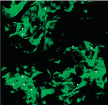
P(L/DL)LA-PHBV foams seeded with 3T3 cells at a density of 2.5⫻ 105cells/foam were examined with a
confocal microscope after staining with Acridine Or-ange on Day 14 of in vitro culture. Since the foam was too thick, only the top 350m section could be exam-ined. The observations done by confocal microscopy showed that the cells were distributed in 3D in the foam. The micrographs showed that the foam was highly porous and the distribution of the cells within the structure was quite homogeneous (Fig. 11). The shapes of the cells indicated the compatibility and resultant spreading of the cells on the polyester carri-ers.
CONCLUSION
End stage organ or tissue failure is a common prob-lem in today’s world because of our prolonged
life-Figure 11. Confocal microscopy of 3T3 seeded P(L/ DL)LA-PHBV foams on Day 14 (⫻20, arrow heads indicate cells). [Color figure can be viewed in the online issue, which is available at www.interscience.wiley.com.]
Figure 10. SEM micrographs of the top (initial seeding site) of 3T3 seeded P(L/DL)LA-PHBV foams on (a) Day 7 and (b) Day 14 (⫻350, arrow heads indicate cells).
span and severe diseases and injuries. In most cases, transplantation from a donor or a nondegradable ma-terial or device is used to restore the function of the failed organ. Because of the inherent drawbacks of these treatments (risk of infection, donor tissue scar-city, nonresponsiveness against changing environ-ment, etc.), a new expansion of the biomaterials field, tissue engineering, has started becoming the preferred approach.
In this study, biodegradable and biocompatible polyester carriers of P(L/DL)LA-PHBV were tested for the making of cell-material constructs with poten-tial for use in cornea treatment. D407 and 3T3 cells were seeded on the carriers to represent the cells of the epithelium and the stroma of the cornea, respectively, and cultured for 14 days. Characterization of these cells and the carriers was done using various micro-scopic techniques. The cell number on the carriers was quantified and the functionality of the cells was as-sessed. Although the polymers used were not ideal substrates for cell attachment, coating of their surface with fibronectin improved significantly the level of cell adhesion, brought it close to that of control TCPS. The MTS results showed that cells seeded on the poly-meric carriers attached less but proliferated signifi-cantly more than those on the TCPS. The micropat-terned surface of the polymeric films allowed alignment of the cells in the earlier days of the culture and formation of D407 cell multilayers in the later days. The microscopic observations of carriers showed that the polymeric foams had the required highly porous structure and pore interconnectivity for their utilization as cell carriers. From the micrographs of the 3T3 cells seeded on the foams, it was seen that the cells were distributed quite uniformly in the foam around the initial seeding site. However, the cell number de-creased significantly towards the core of the foam, even on Day 14 probably because of the long distance to be traversed by the cells and also that of nutrients. To obtain a more homogeneous 3D carrier and popu-lation of the complete construct with cells, the thick-ness of the foam needs to be decreased or the foam properties (pore size and porosity) to be improved. Immunohistochemical analysis proved that the cells seeded on the carriers were functional. It can, there-fore, be concluded that the P(L/DL)LA-PHBV carriers could be seriously considered as cell carriers in future studies on cornea tissue engineering.
References
1. Reim M, Kottek A, Schrage N. The cornea surface and wound healing. Prog Retin Eye Res 1997;16:183–225.
2. Ehlers N, Hjortdal J. Corneal thickness: Measurement and implications. Exp Eye Res 2004;78:543–548.
3. Maurice DM. The structure and transparency of the cornea. J Physiol 1957;136:263–286.
4. Hogan MJ, Alvarado JA, Weddel JE. Histology of the Human Eye. Philadelphia: WB Saunders; 1971.
5. Ameen DB, Bishop MF, McMullen T. A lattice model for com-puting the transmissivity of the cornea and sclera. Biophys J 1998;75:2520 –2531.
6. Mu¨ller LJ, Pels E, Schurmans LRHM, Vrensen GFJM. A new three dimensional model of the organization of proteoglycans and collagen fibrils in human corneal stroma. Exp Eye Res 2004;78:493–501.
7. Yamada J, Ksander BR, Streilein JW. Cytotoxic T cells play no essential role in acute rejection of orthotopic corneal allografts in mice. Invest Ophthalmol Vis Sci 2001;42:386 –392.
8. The Collaborative Corneal Transplantation Studies Research Group. The collaborative corneal transplantation studies (CCTS): Effectiveness of histocompatibility matching in high-risk corneal transplantation. Arch Ophthalmol 1992; 110:1392–1403.
9. Hicks CR, Fitton JH, Chirila TV, Crawford GJ, Constable IJ. Keratoprostheses: Advancing toward a true artificial cornea. Surv Ophthalmol 1997;42:175–189.
10. Chirila TV, Hicks CR, Dalton PD, Vijayasekaran S, Lou X, Hong Y, Clayton AB, Ziegelaar BW, Fitton JH, Platten S, Craw-ford GJ, Constable IJ. Artificial cornea. Prog Polym Sci 1998; 23:447– 473.
11. Chirila TV. An overview of the development of artificial cor-neas with porous skirts and the use of PHEMA for such an application. Biomaterials 2001;22:3311–3317.
12. Hicks CR, Crawford GJ, Tan DT, Snibson GR, Sutton GL, Downie N, Gondhowiardjo TD, Lam DS, Werner L, Apple D, Constable IJ.␣Cor cases: Comparative outcomes. Cornea 2003; 22:583–590.
13. Minami Y, Sugihara H, Oono S. Reconstruction of cornea in three-dimensional collagen gel matrix culture. Invest Ophthal-mol Vis Sci 1993;34:2316 –2324.
14. Germain L, Carrier P, Auger FA, Salesse C, Guerin SL. Can we produce a human corneal equivalent by tissue engineering? Prog Retin Eye Res 2000;19:497–527.
15. Griffith M, Osborne R, Munger R, Xiong X, Doillon CJ, Laycock NL, Hakim M, Song Y, Watsky MA. Functional human corneal equivalents constructed from cell lines. Sci-ence 1999;286:2169 –2172.
16. Tsai RJF, Ho YS, Chen JK. The effects of fibroblasts on the growth and differentiation of human bulbar conjunctival epi-thelial cells in an in vitro conjunctival equivalent. Invest Oph-thalmol Vis Sci 1994;35:2865–2875.
17. Chena GQ, Wua Q. The application of polyhydroxyalkano-ates as tissue engineering materials. Biomaterials 2005;26: 6565– 6575.
18. Ferreira BMP, Zavaglia CAC, Duek EAR. Films of poly(l-lactic acid)/poly(hydroxybutyrate-co-hydroxyvalerate) blends: In vitro degradation. Mater Res 2001;4:34 – 42.
19. Torun Ko¨se G, Kenar H, Hasırcı N, Hasırcı V. Macro-porous poly(3-hydroxybutyrate-co-3-hydroxyvalerate) ma-trices for bone tissue engineering. Biomaterials 2003;24: 1949 –1958.
20. Torun Ko¨se G, Korkusuz F, Korkusuz P, Purali N, O¨ zkul A, Hasırcı V. Bone generation on PHBV matrices: An in vitro study. Biomaterials 2003;24:4999 –5007.
21. Kim BS, Mooney DJ. Development of biocompatible synthetic extracellular matrices for tissue engineering. TIBTECH 1998; 16:224 –230.
22. Steele JG, Johnson G, Griesser HJ, Underwood PA. Mechanism of initial attachment of corneal epithelial cells to polymeric surfaces. Biomaterials 1997;18:1541–1551.
23. Lu L, Nyalakonda K, Kam L, Bizios R, Go¨pferich A, Mikos AG. Retinal pigment epithelial cell adhesion on novel micropat-terned surfaces fabricated from synthetic biodegradable poly-mers. Biomaterials 2001;22:291–297.
24. Dalton BA, Evans MDM, McFarland GA, Steele JG. Modula-tion of corneal epithelial stratificaModula-tion by polymer surface to-pography. J Biomed Mater Res 1999;45:384 –394.
25. Pins GD, Toner M, Morgan JD. Microfabrication of an analog of the basal lamina: Biocompatible membranes with complex topographies. FASEB J 2000;14:593– 602.
26. Nishida T, Nakagawa S, Watanabe K, Yamada KM, Otori T, Berman MB. A peptide from fibronectin cell-binding domain inhibits attachment of epithelial cells. Invest Ophthalmol Vis Sci 1988;29:1820 –1825.
27. Brunette DM. Spreading and orientation of epithelial cells on grooved substrata. Exp Cell Res 1986;167:203–217.
28. Evans MDM, McFarland GA, Taylor S, Walboomers XF. The response of healing corneal epithelium to grooved polymer surfaces. Biomaterials 2005;26:1703–1711.
29. Ban Y, Dota A, Cooper LJ, Fullwood NJ, Nakamura T, Tsuzuki M, Mochida C, Kinoshita S. Tight junction-related protein ex-pression and distribution in human corneal epithelium. Exp Eye Res 2003;76:663– 669.
30. Besseau L, Coulomb B, Lebreton-Decoster C, Giraud-Guille MM. Production of ordered collagen matrices for three-dimen-sional cell culture. Biomaterials 2002;23:27–36.
31. Rivard CH, Chaput CJ, DesRosiers EA, Yahia LH, Selmani A. Fibroblast seeding and culture in biodegradable porous sub-strates. J Appl Biomater 1995;6:65– 68.