© 2018 Eurasian Journal of Pulmonology Published by Wolters Kluwer - Medknow 167
Rarely occurrance of two diffuse
idiopathic neuroendocrine cell
hyperplasia cases
Nalan Ogan, Eylem Evrim Akpinar, Ayse Baha1, Serdar Han2, Haldun Umudum3 Abstract:
Diffuse idiopathic pulmonary neuroendocrine cell hyperplasia (DIPNECH) is a rare disease which needs a long time for diagnosis and usually defined by case reports and small series. We reported two cases, one with 67 and the other with 66‑years‑old, both of them were female. They presented with long‑lasting complaints of cough and shortness of breath. Computed tomography of patients showed multiple nodules on the basis of lungs, and additionally, mosaic perfusion was seen in the first case. Tissue samples were taken with video‑assisted thoracic surgery, and the patients were diagnosed with DIPNECH by immunohistochemical examination. We wanted to emphasize the importance of DIPNECH which is a rare clinical entity causing chronic respiratory symptoms which should be considered in differential diagnosis of multiple pulmonary nodules.
Keywords:
Diffuse idiopathic pulmonary neuroendocrine cell hyperplasia, immunohistochemical examination, thoracic computed tomography
Introduction
D
i f f u s e i d i o p a t h i c p u l m o n a r y neuroendocrine cell hyperplasia (DIPNECH) is a rare primary disorder that is detected incidentally without preexisting chronic lung disease and is considered to be neuroendocrine cell proliferation, often accompanied by constrictive obliterative bronchiolitis. It is a rare condition characterized by generalized proliferation of neuroendocrine cells confined to bronchial and bronchiolar epithelium.[1] The disease causes chronicrespiratory symptoms which usually misdiagnosed as asthma or chronic obstructive pulmonary disease. In this report, we would like to document two cases of DIPNECH.
Case Reports
Case 1
A 67‑years‑old, nonsmoker, female admitted
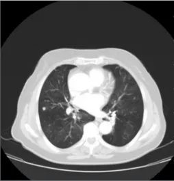
with complaints of cough for 30 years, shortness of breath, and back pain. She had a history of several hospital admissions. Her general patient history was negative for atopic diseases or allergies. The saturation level of oxygen in hemoglobin was 98% and breath sounds are natural. A chest X‑ray showed increased cardiothoracic ratio and bilateral bibasilar reticular appearance. The pulmonary function tests (PFTs) showed forced vital capacity (FVC) 1.08 L (43%), forced expiratory volume in 1 s (FEV1) of 0.72 L (36%), and FEV1/FVC: 67%. Thoracic computed tomography (CT) showed mosaic attenuation with fibroatelectatic changes in the middle lobe and lingula, multiple millimetric nodules, the largest one was 7 mm in the lower lobes [Figure 1]. Tumor and collagen tissue markers were negative. Gynecological malignancy was not considered in the genitourinary system. Thyroid US showed no evidence of malignancy in the fine‑needle aspiration biopsy taken from the heterogeneous Address for
correspondence:
Dr. Nalan Ogan, Mevlana Bulvari (Konya Yolu) No: 86-88, 06520 Ankara - Turkey. E-mail: nalanogan @gmail.com Received: 20-09-2017 Revised: 29-11-2017 Accepted: 02-01-2018 Departments of Pulmonology and
2Thoracic Surgery and 3Pathology, Faculty
of Medicine, Ufuk University, Ankara, Turkey, 1Department of
Pulmonology, Kyrenia Akcicek National Hospital, Kyrenia, Cyprus
Case Report
Access this article online
Quick Response Code:
Website:
www.eurasianjpulmonol.com
DOI:
10.4103/ejop.ejop_66_18
How to cite this article: Ogan N, Akpinar EE, Baha A,
Han S, Umudum H. Rarely occurrance of two diffuse idiopathic neuroendocrine cell hyperplasia cases. Eurasian J Pulmonol 2018;20:167-70.
This is an open access journal, and articles are distributed under the terms of the Creative Commons Attribution‑NonCommercial‑ShareAlike 4.0 License, which allows others to remix, tweak, and build upon the work non-commercially, as long as appropriate credit is given and the new creations are licensed under the identical terms.
For reprints contact: [email protected]
Ogan, et al.: Two rare cases of preinvasive malignancy
168 Eurasian Journal of Pulmonology - Volume 20, Issue 3, September-December 2018
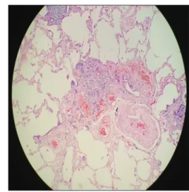
solid nodule in the lower posterior of the left lobe. Endoscopic antrum biopsy showed chronic helicobacter gastritis. Many foci of neuroendocrine cell hyperplasia, the largest was 6 mm in diameter, were detected on the wedge biopsy specimen taken by video‑assisted thoracic surgery (VATS) [Figure 2]. Immunohistochemical analysis showed that all lesions were positively stained with thyroid transcription factor‑1, CD‑56, synaptophysin (SYNP), and neuron‑specific enolase [Figure 3]. Methylprednisolone (1 mg/kg) was started and dose was tapered. The patient had apparent symptomatic relief although 6‑min walking test (MWT), spirometry, and carbon monoxide diffusion capacity (DLCO) did not show significant improvement. However, the treatment was interrupted by the patient at 3rd month because of side effects such
as ecchymosis on her face or arms; she was following up with long‑acting beta‑2‑mimetics and inhaled steroids (LABA/ICS). Informed consent has been taken from the patient.
Case 2
A 66‑year‑old female was admitted with complaints of progressive dyspnea and dry cough for 20 years. She was diagnosed as asthma before 5 years. She had neither environmental and occupational exposure nor smoking history. Reticular infiltration was observed in bilateral basalts on chest X‑ray. The PFT showed mixed pattern; FEV1: 57% (1.41 L), FVC: 50% (1.03 L), and FEV1/FVC: 73%. The DLCO was 44%. In the 6‑MWT, the distance was 300 m and desaturation was observed with exercise (Initial: 91%, 6 min: 85%). Serological markers for connective tissue disorders were all negative. CT showed bilateral diffuse millimetric nodules and subpleural reticular infiltration in bilateral bases [Figure 4]. VATS was performed. Pathologic examination of specimens showed intra‑alveolar localized neuroendocrine cell proliferation and minimal interstitial fibrosis under the bronchial epithelium and in the parenchyma [Figure 5]. On immunohistochemical study, keratin 7 was diffuse positive with chromogranin and SYNP, and keratin 20 was negative. The result was reported as DIPNECH. The patient had been started methylprednisolone 1 mg/kg then the dose was tapered every 2 weeks. The patient developed clinical and functional improvement. Due to the development of steroid‑dependent myopathy, treatment was stopped on 4th month. It has been taken
patient informed consent.
Discussion
Both of the reported cases had chronic irritating pulmonary symptoms, even one of them was diagnosed with asthma. The final diagnosis was made by VATS, and they responded to systemic steroid therapy despite systemic side effects.
DIPNECH is a rare condition that is characterized by small case series, 30 cases were involved in the largest one.[2] It is classified as a preneoplastic lesion
Figure 1: Multiple small nodules the largest 7 mm in the lower zones with mosaic
attenuation in computed tomography
Figure 2: Pulmonary neuroendocrine cell hyperplasia (H and E, ×10)
Figure 3: Carcinoid tumor cells with strong synaptophysin positivity
(synaptophysin ×10)
Ogan, et al.: Two rare cases of preinvasive malignancy
Eurasian Journal of Pulmonology - Volume 20, Issue 3, September-December 2018 169
because of the possibility of carcinoid development by the World Health Organization, defined as bronchial and bronchial epithelium‑restricted, distributed single cells, small nodules (neuroendocrine bodies), or pulmonary neuroendocrine cells. DIPNECH patients are often histopathologically diagnosed with pulmonary biopsies or resections and are shown to have increased chromogranin, SYNP, and CD56 in immunohistochemistry (C‑13).[3] In our patients,
keratin 7, chromogranin, and SYNP positivity were present. Expression of somatostatin receptor type 2 and mammalian rapamycin target signaling activation patterns has also been reported. Expression of Ki67 and p16 is seen more consistent and earlier than DIPNECH’s reactive neuroendocrine cell proliferation.[4]
DIPNECH predominantly occurs in the elderly (median age 66 years) and in women (93%) and is not associated with smoking, the demographic characteristics of our cases were consistent with the literature. PFTs may be obstructive, restrictive, or mixed.[2] Our first case was
compatible with the obstructive pattern of spirometry, and the other’s spirometry was mixed pattern.
The definitive diagnosis of DIPNECH is impossible in conventional chest radiography. CT abnormalities are not specific and include airway‑related diseases such as mosaic attenuation, bronchial wall thickening, bronchiectasis, and mucoid impaction. However, mosaic perfusion is predominant with air trapping, which is characterized by constrictive bronchiolitis and characterized by a low‑attenuation lung placed with a normally ventilated higher attenuation lung. Multiple nodules are the most common findings of DIPNECH and are usually solid or ground glass in appearance, depending on the tumor size.[5] While
the multiple nodules were predominant on CT of the reported patients, mosaic perfusion also was found in our first case. In addition, our second case had reticular infiltrations on the basis of the lungs.
The data of treatment, long‑term follow‑up, and outcomes are limited in DIPNECH patients. Therapeutic options include oral and inhaled steroids, chemotherapy, surgical lung resection and lung transplantation, and clinical observation alone in mild and stable cases. Corticosteroids have no definitive evidence of efficacy except reducing inflammatory response caused by neuropeptide‑secreting neuroendocrine cells. Conversely, cytotoxic agents are largely ineffective and not recommended. Octreotide, a somatostatin analog that has been shown to reduce the hormonal over release of neuroendocrine cells, has been used successfully, especially in the presence of somatostatin receptors.[4] However, in both of
our cases, the level of somatostatin was within normal limits. Hence, systemic steroid (1 mg/kg methylprednisolone p.o.) therapy was started the first case relieved symptomatically, and the second case showed both functional and symptomatic improvement. Unfortunately, systemic side effects of methylprednisolone did not allow continue treatment in both cases. First case’s treatment has been continuing with ICS/LABA combination to control her cough. The second case is absolutely asymptomatic and is following up without treatment. As a result, DIPNECH patients are often misdiagnosed for a long time as obstructive pulmonary disease, and definitive diagnosis needs with lung biopsies or resections. When etiology of multiple nodules is investigated, this rare clinical entity should be considered in differential diagnosis, especially in middle‑aged women with chronic cough and dyspnea that cannot be explained by other causes.
Figure 4: Bilateral diffuse millimetric nodules and subpleural reticular infiltration in
bilateral bases in high‑resolution computed tomography
Figure 5: Neuroendocrine cell proliferation and minimal interstitial fibrosis
(H and E, ×10)
Ogan, et al.: Two rare cases of preinvasive malignancy
170 Eurasian Journal of Pulmonology - Volume 20, Issue 3, September-December 2018
Declaration of patient consent
The authors certify that they have obtained all appropriate patient consent forms. In the form, the patient has given her consent for her images and other clinical information to be reported in the journal. The patient understand that names and initials will not be published and due efforts will be made to conceal identity, but anonymity cannot be guaranteed.
Financial support and sponsorship
Nil.
Conflicts of interest
There are no conflicts of interest.
References
1. Benson RE, Rosado‑de‑Christenson ML, Martínez‑Jiménez S, Kunin JR, Pettavel PP. Spectrum of pulmonary neuroendocrine proliferations and neoplasms. Radiographics 2013;33:1631‑49. 2. Carr LL, Chung JH, Duarte Achcar R, Lesic Z, Rho JY, Yagihashi K,
et al. The clinical course of diffuse idiopathic pulmonary
neuroendocrine cell hyperplasia. Chest 2015;147:415‑22. 3. Travis WD, Brambilla E, Nicholson AG, Yatabe Y, Austin JH,
Beasley MB, et al. The 2015 World Health Organization classification of lung tumors: Impact of genetic, clinical and radiologic advances since the 2004 classification. J Thorac Oncol 2015;10:1243‑60. 4. Rossi G, Cavazza A, Spagnolo P, Sverzellati N, Longo L,
Jukna A, et al. Diffuse idiopathic pulmonary neuroendocrine cell hyperplasia syndrome. Eur Respir J 2016;47:1829‑41.
5. Chassagnon G, Favelle O, Marchand‑Adam S, De Muret A, Revel MP. DIPNECH: When to suggest this diagnosis on CT. Clin Radiol 2015;70:317‑25.