I N N O V A T I V E T E C H N I Q U E S AESTHETIC
New Otoplasty Approach: A Laterally Based Postauricular
Dermal Flap as an Addition to Mustarde and Furnas to Prevent
Suture Extrusion and Recurrence
Salih Onur Basat•Ufuk Askerog˘lu•
Tolga Aksan•Brendan Alleyne•Memet Yazar•
C¸ ag˘das¸ Orman•I˙lker U¨ sc¸etin •Mithat Akan
Received: 28 May 2013 / Accepted: 14 December 2013 / Published online: 8 January 2014
! Springer Science+Business Media New York and International Society of Aesthetic Plastic Surgery 2014
Abstract Prominent ear is the most common deformity of the external ear. The major causes can be an underde-veloped antithetical fold, concha hypertrophy, and promi-nence of the ear lobule. Since Ely’s first aesthetic correction of the prominent ear in 1881, more than 200 different techniques have been described, but the choice of procedure still remains the surgeon’s preference. This report presents the laterally based posterior auricular der-mal flap technique as an adjunct to the conventional car-tilage-sparing otoplasty. An elliptical skin incision was planned according to the classic prominent ear correction technique. Instead of the excision, skin was deepithelial-ized. From the inferior border of the incision, the dermal flap was incised and elevated in a medial-to-lateral direc-tion. The posterior auricular dermal flap was used to sup-port and cover the suture material. This method was used in the treatment of 17 consecutive patients. After a follow-up period of 6–32 months (mean 16 months), the patients were evaluated in terms of the recurrence and suture line
problems. No suture line problems or recurrences were observed at the end of the follow-up period. Use of the posterior auricular dermal flap both prevents suture extru-sion and decreases recurrences.
Level of Evidence V This journal requires that authors assign a level of evidence to each article. For a full description of these Evidence-Based Medicine ratings, please refer to the Table of Contents or the online Instructions to Authorswww.springer.com/00266.
Keywords Otoplasty! Furnas ! Mustarde ! Dermal flap
Introduction
Prominent ear is the most common congenital ear defor-mity, affecting approximately 5 % of the general popula-tion [1,2]. Deficiency or prominence of the ear may cause both aesthetic and psychological sequelae in both pediatric and adult patients. Although the exact cause of the prom-inence is unclear, it is assumed that the primary factors are postural differences in newborns, muscle hypertonia, structural alterations of collagen, and possibly genetic predisposition [1].
The prominent ear contains numerous components including conchae hypertrophy, failure of antithetical folding, a wider angle than normal between the concha and the mastoid bone, lobule prominence, and a combination of these varieties [3]. Various treatments and techniques are being discussed to address these deformities. Such methods include excision, bending of the cartilage, and use of suture, scoring, and repositioning of the auricular cartilage. Surgeons who treat prominent ears should have a working knowledge of the normal ear’s anatomy and the key features of the prominent type. Additionally, it is
S. O. Basat! U. Askerog˘lu ! T. Aksan (&) ! C¸. Orman ! I˙. U¨ sc¸etin
Department of Plastic Reconstructive and Aesthetic Surgery, Okmeydanı Training and Research Hospital, Daru¨laceze Caddesi, Okmeydanı, 34384 Istanbul, Turkey
e-mail: [email protected] B. Alleyne
Case Western Reserve University, Cleveland, OH, USA M. Yazar
Department of Plastic Reconstructive and Aesthetic Surgery, S¸is¸li Etfal Training and Research Hospital, Istanbul, Turkey M. Akan
Department of Plastic Reconstructive and Aesthetic Surgery, Faculty of Medicine, Istanbul Medipol University, Istanbul, Turkey
beneficial to analyze the deformity and to create a surgical method based on the available techniques.
The main cause of deformity (e.g., cartilage elasticity) combined with the surgeon’s preference largely determines the method for surgical correction [4]. In our literature review, we found several authors reporting that the cor-rection of prominent ear with splint usage is the best method, specifically during the neonatal period. This could be due to the increased auricular cartilage elasticity in this age group [5,6].
Surgical methods generally used to correct the promi-nent ear can be classified into two groups: cartilage cutting and cartilage sparing [7, 8]. Cartilage-cutting techniques may result in several complications such as weakening of the cartilage framework, disruption of the cartilage due to scar contracture, hematoma due to the required wide dis-section, and asymmetry [9].
Due to the wide variety of complications with the carti-lage-cutting technique, cartilage-sparing methods are used more frequently. However, suture extrusion, granuloma formation on the suture lines, recurrence of the deformity due to cartilage memory, and suture fatigue are the major com-plications of the cartilage-sparing method [8,9].
In this report, we present a supportive combination technique using a cartilage-sparing suture method. We aimed to decrease the suture extrusion and recurrence rates by covering the sutures with a vascularized dermal flap and supporting the suture material.
Material and Methods
Between 2009 and 2012, 17 patients (11 females and 6 males) have undergone this combination procedure. The cases have included 16 bilateral ears and 1 unilateral affected ear. All the patients had insufficient folding of the antihelix, and 9 patients also had conchae hypertrophy with insufficient folding of the antihelix. Only Mustarde and Furnas suturing techniques were used to correct the prominence of the ears. No cartilage-cutting techniques were used.
Patients with conchae hypertrophy requiring cartilage resection were not included in this study. The mean patient age was 14.5 years (range 5–26 years). The follow-up period ranged from 6 to 32 months. During the preopera-tive evaluation, the mean distance from the most protrud-ing point of the helix to the mastoid bone was measured as 29.11 mm, and the mean concha-to-mastoid angle, mea-sured with a goniometer, was 45".
The continuous variables in the study were analyzed with the Kolmogorov–Smirnov test. The variables with Gaussian distribution are shown as average ± standard deviation, whereas the variables with non-Gaussian distri-bution are shown as median ± 25th to 75th percentile
values. In the dependent groups, for comparison of con-tinuous measurements, the paired-sample t test was used if Gaussian distribution existed. The Wilcoxon signed ranks test was used if normal distribution did not exist. All results were evaluated using the 95 % confidence interval and a p value lower than 0.05 as the significance level.
Operative Technique
With the patient under general anesthesia, a small ellipse was marked on the posterior auricular skin and then infil-trated with lidocaine and epinephrine at a concentration of 1:200,000. The marked elliptical skin area then was deepithelialized. From the inferior margin of the elliptical area, the dermal flap was incised and elevated in a medial-to-lateral fashion in the supraperichondrial plane to prepare the laterally based dermal flap.
Dissection was continued toward the scaphal area dis-tally and the mastoid bone proximally. Depending on the characteristics of the deformity, the concha-scaphal, the concha-mastoid, or a combination of both was used. For reshaping of the auricular cartilage, 3-0 and 4-0 polypro-pylene monofilament suture was used.
After reshaping of the auricular cartilage, the elevated posterior auricular dermal flap was sutured to the mastoid bone in a tensed position. We used three 4-0 polygleca-prone sutures to fix the dermal flap to the mastoid bone (Fig.1). The posterior auricular skin incision was repaired with 4-0 polyglecaprone sutures in a continuous manner. A bulky bandage with petroleum gauze was used to cover the ears, and a head bandage was applied over the dressing. The dressing was changed 24 h after the operation. The head bandage was initially maintained in place for 1 week during the day only and then for an additional 2 weeks at night. All the patients received ciprofloxacin 500 mg twice daily for 5 days.
Results
The patients were evaluated 1, 3, 6, and 12 months after the operation. The follow-up period ranged from 6 to 32 months (mean 16 months). All the patients were eval-uated for suture extrusion, formation of granulomas, and relapse of the deformity (Figs.2,3,4). We did not observe any early postoperative complications such as hematoma, infection, or skin necrosis. Neither suture extrusion nor granuloma formation was observed during the follow-up period. In one patient, an acute antithetical fold was observed, but the patient did not want to undergo any further operative repair.
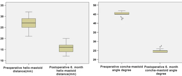
During the postoperative evaluation 6 months after surgery, the mean distance between the most protruding
point of the helix and the mastoid bone was measured as 17.22 mm, and the mean concha-mastoid angle was mea-sured as 25" (Table1). During the operation, overcorrec-tion was done to decrease the risk for protrusion of the upper third of the auricular. A range of 1–3 mm was observed in the helix-mastoid distance at 1 and 3 months. In this study, the preoperative helix-mastoid distance, the postoperative 6-month helix-mastoid distance, the preoperative concha-mastoid angle degree, and the post-operative 6-month concha-mastoid angle degree of the patients were analyzed (Table2). A significant difference was observed between the preoperative helix-mastoid dis-tance and the postoperative 6-month helix-mastoid disdis-tance (p\ 0.001), as well as between the preoperative concha-mastoid angle degree and the postoperative 6-month con-cha-mastoid angle degree (p\ 0.007) of the patients (Fig.5). No differences were observed between the 3, 6, and 12-month measurements of the helix-mastoid distance.
No recurrence of deformity was reported at the end of the follow-up period. The patients were asked whether they experienced any pain, discomfort, or sensitivity of the affected ear when laying the head toward operated ear side. No patient voiced any such concerns.
Discussion
Since the first aesthetic correction of prominent ears in 1881, more than 200 different techniques have been described by various authors [10, 11]. In general, the operative methods can be classified into two groups: car-tilage cutting and carcar-tilage sparing.
The cartilage-cutting technique potentially subjects the ear to irreversible distortion created by wound contraction and cartilage remodeling [12–18]. The postoperative stig-mata of cutting cartilage may include anterior skin
Fig. 1 a A small ellipse of posterior auricular skin was marked. b The marked elliptical skin area was deepithelialized. c The dermal flap was incised and elevated from a medial-to-lateral direction in the supraperichondrial plane. d Due to the characteristics of deformity, concha-scaphal, concha-mastoid, or a combination of both suture techniques was used to correct the prominent ear. e The posterior auricular dermal flap was sutured to the mastoid bone in a stretched position
necrosis, cartilage irregularities, sharp edges, and over-correction. Correction of these problems can be more dif-ficult and could require further revisions [2,12].
The cartilage-sparing method depends primarily on placement of permanent sutures to reshape the cartilage. The most common are Mustarde and Furnas otoplasties [19, 20]. Besides the specified advantages, the cartilage-sparing technique may result in possible complications. Recurrence rates of up to 24 % have been reported by authors using this method [8, 21]. Suture erosion, granu-loma formation over the suture lines, and pain from buried sutures are additional problems with this technique [8].
Horlock et al. [8] combined a postauricular fascia flap with Mustarde and Furnas concha-mastoid sutures. This fascia flap
Fig. 2 A 5-year-old boy referred with unilateral prominent ear (left). A laterally based postauricular dermal flap was used as an addition to the Mustarde and Furnas sutures. No recurrence or suture extrusion was reported during the 1-year follow-up period (right)
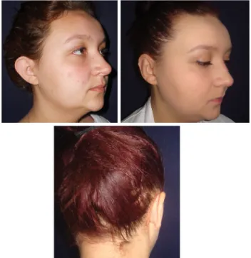
Fig. 3 A 20-year-old woman referred with bilateral prominent ears (left). A laterally based postauricular dermal flap was used as an addition to the Mustarde and Furnas sutures. No recurrence or extrusion of the suture was reported during the 1-year follow-up period (right)
Fig. 4 A 20-year-old woman referred with bilateral prominent ears (left). A laterally based postauricular dermal flap was used as an addition to the Mustarde suture. No recurrence or extrusion of the suture was reported during the 1-year follow-up period (right)
covers the sutures to prevent suture extrusion, and advance-ment of the flap acts as a supportive structure for ultimate prevention of prominence recurrence. Horlock et al. [8] reported a 0 % rate of suture extrusion and a recurrence rate of 8 % with postauricular fascia flap refinement of the cartilage-sparing technique [8]. Sinha and Richard [22] reported a similar result. These authors described the postauricular flap as a medially based flap attached to the posterior surface of the auricular cartilage.
We alternatively suggest elevation of a laterally (auric-ular base) based postauric(auric-ular dermal flap to avoid suture extrusion completely because we do not use sutures to anchor the flap to the cartilage. With our technique, we do not damage the junction between the flap and the auricular cartilage, so bending of the auricular cartilage to create the smooth antithetical fold and setback of the concha cartilage to overcome the concha hypertrophy are performed more effectively. Meanwhile, our dermal flap approach acts as an additional support to prevent asymmetry between ears whereby we can easily adjust the concha-mastoid and concha-scaphoid angles.
During the conventional suture otoplasty technique, excessive elliptical postauricular skin excision causes ten-sion along the suture lines, and this tenten-sion also causes elevation of the postauricular skin from the underlying cartilage and bone tissue. In this way, a dead space between the cartilage and postauricular skin can occur. With the usage of the laterally based postauricular dermal flap, any kind of postauricular dead space can be filled easily. This can decrease the risk of hematoma formation. Suturing of the dermal flap to the mastoid bone also can cause scar formation along the flap-mastoid junction.
With the cartilage-sparing otoplasty, suture erosion and loosening are the major causes of the recurrence. Using this technique, tension from the memory of the cartilage wears on the sutures and causes their fatigue over time, ultimately leading to erosion. By incorporating a laterally based
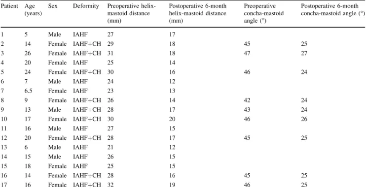
Table 1 Patient summary Patient Age
(years)
Sex Deformity Preoperative helix-mastoid distance (mm) Postoperative 6-month helix-mastoid distance (mm) Preoperative concha-mastoid angle (") Postoperative 6-month concha-mastoid angle (") 1 5 Male IAHF 27 17 2 14 Female IAHF?CH 29 18 45 25 3 26 Female IAHF?CH 31 18 47 27 4 20 Female IAHF 25 14 5 24 Female IAHF?CH 30 16 46 24 6 7 Male IAHF 24 12 7 6.5 Female IAHF 23 13 8 9 Female IAHF?CH 26 14 42 24 9 13 Male IAHF?CH 28 17 43 24 10 17 Female IAHF?CH 30 20 46 26 11 16 Male IAHF 27 15 12 20 Female IAHF?CH 28 17 45 25 13 6 Male IAHF 21 12 14 15 Male IAHF 26 15 15 18 Female IAHF 25 15 16 14 Female IAHF?CH 28 16 45 25 17 16 Female IAHF?CH 32 19 46 25
IAHF insufficient antihelical fold, CH conchal hypertrophy
Table 2 Statistical analysis Min– max Mean/ median SD/25th– 75th % tile p value Age (years) 5–26 14/5 6–24 – Preoperative helix-mastoid distance (mm) 26–32 29/11 1–83 \0.001a Postoperative 6-month helix-mastoid distance (mm) 14–20 17/22 1–79 Preoperative concha-mastoid angle (") 42–47 45 44–46 0.007b Postoperative 6-month concha-mastoid angle (") 24–27 25 24–25.5 Min minimum, max maximum, SD standard deviation
a
Paired samples t test
b
postauricular flap into the conventional suture line, tension over the sutures can be more evenly distributed and shared between the suture and the dermal tissue. Suture-only techniques house this tension only within the suture line. In
contrast to sutures, scar tissue between the dermal flap and the mastoid bone absorbs this tension over a wider area. By these mechanisms, our method can decrease recurrence rates after the operation.
Fig. 5 Statistical analysis of the pre- and postoperative 6-month measurements Fig. 6 a The dermal flap was
incised and elevated from a medial-to-lateral direction in the supraperichondrial plane. b, c Due to the characteristics of the deformity, concha-scaphal, concha-mastoid, or a combination of both suture techniques was used to correct prominent ears. c If a posterior suture is used, the distance between the helix and mastoid decreases. d If the antithetical fold is sutured anteriorly, it can be formed smoothly, and the helix-mastoid distance can be decreased (image drawn by Tolga Aksan)
After concha-scaphal and concha-mastoid sutures, if any prominence or asymmetry is identified between ears, a laterally based postauricular dermal flap could be sutured posteriorly or anteriorly to the mastoid bone to decrease or increase the concha-mastoid angle. If it is sutured poste-riorly, the concha-mastoid angle decreases. When it is sutured anteriorly, an antithetical fold can be formed smoothly while the helix-mastoid length is decreased (Fig.6). In this way, the desired symmetry can be obtained easily, and the anthelia fold also can be made more visible by adequate arrangement of the dermal flap position.
The aforementioned method can be a useful technique for overcoming one of the most significant and recurrent complications: asymmetry. If the flap is sutured posteriorly to decrease the concha-mastoid angle, minimal dead space can occur between the flap and cartilage. However, this dead space did not cause any complication in our patients. We allowed for healing to take place with the deposition of fibrotic tissue and the formation of scar tissue. Postauric-ular skin is very thin tissue, especially over the cartilage, so the dermal flap harvested from this skin tissue did not cause any fullness or bulky tissue in the retro-auricular region. During the follow-up period, we observed preservation and protection of the smooth posterior auricular sulcus.
The most important limitations of this study were the small number of the patients and the lack of a comparison between the results obtained in similar cases managed with different surgical methods. In fact, these limitations are common issues with all the correction methods for prom-inent ears.
In plastic surgery, more than 200 different correction techniques have been defined for treatment of the promi-nent ear. The multiplicity of the techniques suggests that no consensus exists in this area and that every technique can be regarded as a step toward the final goal. Consequently, it can be concluded that further comparative studies with a greater number of patients and controlled variables are needed.
Conclusion
The technique described in this report provided satisfactory clinical results in our study. The laterally based postau-ricular dermal flap is a relatively simple, controllable, and reversible method in the treatment of prominent ears. The addition of this flap to cartilage-sparing otoplasty
techniques can prevent both suture extrusion and deformity recurrence rates.
References
1. Janis JE, Rohrich RJ, Gutowski KA (2005) Otoplasty (review). Plast Reconstr Surg 115:60e–72e
2. Adamson PA, Litner JA (2006) Otoplasty technique (review). Facial Plast Surg Clin North Am 14:79–87
3. Ellis DAF, Keohane JD (1992) A simplified approach to oto-plasty. J Otolaryngol 21:66–69
4. Yazici I, Findikc¸iog˘lu F, Ozmen S, Noyan N, Yavuzer R (2009) Posterior auricular muscle flap as an adjunct to otoplasty. Aes-thetic Plast Surg 33:527–532
5. Tan ST, Abramson DL, MacDonald DM, Mulliken JB (1997) Molding therapy for infants with deformational auricular anom-alies. Ann Plast Surg 38:263–268
6. Tan ST, Shibu M, Gault DT (1994) A splint for correction of congenital ear deformities. Br J Plast Surg 47:575–578 7. Calder JC, Nassan A (1994) Morbidity of otoplasty: a review of
562 consecutive cases. Br J Plast Surg 47:170
8. Horlock N, Misra A, Gault DT (2001) The postauricular fascial flap as an adjunct to Mustardee and Furnas type otoplasty. Plast Reconstr Surg 108:1487–1490; discussion 1491
9. Hassanpour SE, Moosavizadeh SM (2010) Posterior scorring of the scapha as a refinement in aesthetic otoplasty. J Plast Reconstr Aesthet Surg 63:78–86
10. Hoehn JG, Ashruf S (2005) Otoplasty: sequencing the operation for improved result (review). Plast Reconstr Surg 115:5e–16e 11. Ely E (1881) An operation for prominence of the auricles. Arch
Otolaryngol 10:97
12. Kelly P, Hollier L, Stal S (2003) Otoplasty: evaluation, technique, and review. J Craniofac Surg 14:643–653
13. Madzharov MM (1989) A new method of auriculoplasty for protruding ears. Br J Plast Surg 42:285
14. Pitangy I, Muller P, Nelson P et al (1987) Treatment of prominent ears: a 25-years survey of the island flap technique. Aesthetic Plast Surg 11:87
15. Kaye BL (1967) A simplified method for correcting the promi-nent ear. Plast Reconstr Surg 40:44
16. Chait L, Nicholson R (1999) One size fits all: a surgical technique for correction of all types of prominent ears. Plast Reconstr Surg 104:190 17. Luckett WH (1969) A new operation for prominent ears based on
anatomy of the deformity. Plast Reconstr Surg 43:83
18. Hinderer UT, Del Rio JL, Fregenal FJ (1987) Otoplasty for prominent ears. Aesthetic Plast Surg 11:63
19. Mustardee JC (1978) Correction of prominent ears using buried mattress sutures. Clin Plast Surg 5:459
20. Furnas DW (1968) Correction of the prominent ears by concha-mastoid sutures. Plast Reconstr Surg 42:189–193
21. Tan KH (1986) Long-term survey of prominent ear surgery: a comparison of two methods. Br J Plast Surg 39:270–273 22. Sinha M, Richard B (2012) Postauricular fascial flap and suture
otoplasty: a prospective outcome study of 227 patients. J Plast Reconstr Aesthet Surg 65:367–371