Introduction
Allergic rhinitis is one of the most common chronic respiratory system diseases. This IgE mediated inflammatory disease is characterized by congestion, nasal drainage, sneezing and/or nasal itching.1The prevalence of allergic rhinitis is
17-29% in European countries and it is a global health problem whose incidence has increased during the past three decades.2,3 Incidence of this
disease in Turkey is approximately 20%.4
The diagnosis of allergic rhinitis is confirmed by serum immunoassay methods or a skin prick test, which determines specific IgEs against different antigens.1 The avoidance of these antigens is the
primary treatment method against allergic rhinitis. For the medical therapy of this disease, intranasal corticosteroids (IC) are used intermittently or continuously to overcome symptoms.5,6 IC treatment
is the most effective treatment method among patients who are unresponsive to antihistamines.7
In the literature, the use of ICs has been reported to have an approximately 20% treatment failure against allergic rhinitis.8 The cause of this
unresponsiveness is controversial and no study has to date been conducted to investigate the causes of this resistance to corticosteroids. We hypothesize that delayed hypersensitivity reactions to corticosteroids may be one of the causes of this resistance, reactions that have been reported as case reports in patients with allergic rhinitis.9,10
Hypersensitivity to inhaler corticosteroids has been reported to have an incidence of 6.6% in the largest trial conducted by Benett et al., which included 30 patients with allergic rhinitis or asthma.11
In this study, we aimed to establish the incidence and confounding factors of intranasal corticosteroid hypersensitivity among patients with allergic rhinitis in a large patient series.
H. Arslan1, Ö. Gündüz2, M. Başaran1 and S. Kocatürk1
1Ufuk University Medical School, Department of Otorhinolaryngology ; 2Ufuk University Medical School, Department
of Dermatology
Key-words. Allergic rhinitis; corticosteroids; hypersensitivity; patch test; IgE; eosinophil; prick test
Abstract. Corticosteroid hypersensitivity in allergic rhinitis. Background: intranasal corticosteroid (IC) is the most
effective treatment method in allergic rhinitis patients who are unresponsive to antihistamines. The literature reports an approximate 20% treatment failure for instances where IC is used for the treatment allergic rhinitis. Hypersensitivity reaction to corticosteroids may be one of the causes of this treatment failure.
Objective: to discover the incidence and confounding factors of corticosteroid hypersensitivity in patients with allergic
rhinitis.
Methods: after 31 patients were excluded, 150 consecutive patients who were prospectively evaluated in our outpatient
clinics with the diagnosis of allergic rhinitis and 50 age- and sex- matched healthy volunteers were included in this study. To diagnose allergic rhinitis, the symptoms of patients and a skin prick test were used. A skin patch test was used to determine corticosteroid hypersensitivity. Total IgE values and total eosinophil count were obtained for all patients. Total symptom scores were calculated for the severity of symptoms and to determine the response to therapy using intranasal corticosteroids.
Results: the incidence of corticosteroid hypersensitivity determined via the skin patch test was 14.0% (21 out of 150
patients). A difference was observed for patch test positivity results between the study and control groups (14% vs. 0%, respectively). Serum IgE levels and total eosinophil count were higher among patients who had corticosteroid hypersen-sitivity (p:0.005 and p:0.004, respectively). Patients unresponsive to intranasal corticosteroids had a higher incidence of corticosteroid hypersensitivity (71.4% vs. 4.4%, p<0.001).
Conclusion: our study is the largest to date investigating CH in patients with allergic rhinitis and patients with allergic
rhinitis have been found to have a high incidence (14%) of corticosteroid hypersensitivity, which may affect the response of patients to intranasal corticosteroid treatment.
also noted. Treatment strategy was performed according to ARIA guidelines, with first line treatment being education and allergen avoidance, the second line pharmacotherapy (first line of pharmacotherapy was antihistamines; if there was no symptom improvement within two weeks of pharmacotherapy, second line pharmacotherapy treatment was IC). Unresponsiveness to therapy criteria was accepted as indicating no improvement among symptom groups such as moderate/severe to mild or mild to no symptoms, as per ARIA guidelines.12 TSS was also calculated prior to
treatment and again after three months of IC usage. Eligible subjects were 18 years or older and allergic rhinitis was confirmed via a standard skin prick test with the following allergens: Dermatophagoides pteronyssinus, Dermatophagoides farinae, grass mixture, trees mixture, weed mixture and fungi mixture. Patients were tested one week after discontinuation of antihistamines and topical corticosteroid therapy, and at least four weeks after cessation of immunosuppressive agents.
Skin prick tests (SPT) were performed using a standard panel of the five allergen extracts used in our allergy unit, alongside histamine as positive and saline as negative controls.13 The test sites were
cleaned with alcohol then marked at least 3 cm apart to avoid overlapping reactions. The allergen was dropped and gently pricked by a sterile skin test lancet (Stallergenes, France). The test was read at 20 minutes and the largest diameter of a wheal was measured. A wheal size ≥3 mm above the negative control was defined as positive and patients with a wheal size ≥3 mm were enrolled in the study. Total IgE values were studied and eosinophilia (total eosinophil count > 400/cc) was looked for among these patients.
After 31 patients were excluded (15 patients treated with allergen avoidance, 10 patients with nasal disorders or structural abnormalities, five patients with current or a history of chronic sinusitis and one pregnant patient), 150 patients were included to the study. In the second stage of treatment, patients with intermittent symptoms used oral antihistamines as needed and did so regularly if their symptoms were persistent for two weeks. In our study, 26 of 150 patients were responsive to oral antihistamines; thus, 124 patients who were unresponsive to antihistamines had treatment involving intranasal corticosteroids twice a day. Among patients, 58 (46.8%) took budesonide, Materials and methods
The present study was approved in June 2013 by the Institutional Review Board of Ufuk University Medical School, decision number 060620131; all patients signed informed consent forms prior to entering the study.
This study included 181 consecutive patients who were prospectively evaluated in our outpatient clinics with a diagnosis of allergic rhinitis, as well as 50 age- and sex-matched healthy volunteers. The diagnosis of allergic rhinitis was made according to the “Allergic Rhinitis and its Impact on Asthma” (ARIA) guidelines.12
Subjects were excluded from the study if they were pregnant or nursing, had another clinically significant disease (particularly nasal disorders or structural abnormalities that may interfere with nasal airflow), had current or a history of chronic sinusitis (with/without polyps) or purulent postnasal drip, or if they experienced sensitivity to the study drug or its excipients. The presence of granulomatous disease (i.e., Wegener’s, sarcoidosis) or a tumour (either benign or malignant) was another exclusion criterion. Additionally, patients with allergic rhinitis whose symptoms were improved by allergen avoidance and education to the degree that they did not need to use any pharmacotherapy agents such as antihistamines or corticosteroids were also excluded.
The symptoms of nasal obstruction, nasal drainage, sneezing and/or nasal itching were asked about and the presence of at least two nasal symptoms for more than one hour per day was necessary for the diagnosis of rhinitis. Total symptom score (TSS, sum of nasal discharge/ rhinorrhoea, nasal congestion, sneezing, nasal itching and ocular symptoms) on a 4-point scale (0 = none; 3 = severe), which has been widely used in clinical studies of allergic rhinitis, was used to quantify the severity of patients’ symptoms. The symptoms were defined as moderate to severe in the presence is at least one of the followings was present: insomnia, deterioration of daily activities either at rest or during exercise, or deterioration of activities either at school or work. If none was present, symptoms were graded as mild. The disease was labelled intermittent if the symptoms were present <4 days in a week or lasted <4 weeks, otherwise it was accepted as persistent allergic rhinitis. The presence of asthma was
hours and read by the same dermatologist on the second, third and seventh days, in accordance with International Contact Dermatitis Research Group (ICDRG) guidelines. Additionally, patients were asked to return if any reaction occurred up until the tenth day of the patch test. Any positive reaction (+: weak (nonvesicular) reaction;++: strong (oedematous or vesicular) reaction; +++: extreme (bullous or ulcerative) reaction) was considered a positive reaction.
Statistical analysis
Analysis of the results was performed using the Predictive Analytics Software (PASW, Chicago, Illinois, USA), version 18.0 software for Windows. Data were tested for normal distribution using the Kolmogorov-Smirnov test. To investigate the differences between groups, a Mann-Whitney U test was used for two groups and a Kruskal-Wallis H test for more than two groups. A Spearman 27 (21.8%) took mometasone, 20 (16.1%) took
fluticasone, 16 (12.9%) took triamcinolone and 13 (10.5%) took tixocortol as the first intranasal corticosteroid.
Patch testing
A skin patch test was used to determine corticosteroid hypersensitivity in all participants. This test was performed on patients following antihistaminic therapy if the latter responded well and after corticosteroid treatment. If patients were unresponsive to IC, their treatment strategy was changed to another group agent accordingly. Patients were patch tested on the upper part of the back with IQ Ultra Chambers (Chemotechnique Diagnostics, Sweden). The names of manufactured allergens consisting of steroids are shown in Table 1. The supplier of allergens was Chemo-technique Diagnostics (Sweden) and Brial Allergen (Germany). The patch test was removed after 48
Table 1
Names and concentrations of allergens
Names of the allergens Classification24 Concentration and vehicle
**Hydrocortisone-17-butyrate A 1% alcohol
**Prednisolone A 1% petrolatum
**Tixocortol-21-pivalate A 0.1% petrolatum
*Budesonide B 0.01% petrolatum
*Triamcinolone acetonide B 1% petrolatum
**Betamethasone-17-valerate C 1% petrolatum
**Clobetasol-17-propionate D 1% petrolatum
* Intranasal corticosteroids ** Topical corticosteroids
Table 2
Patch test results and related factors
Factors Patients (n=150) Patch Test(+)(n=21) Patch Test(-)(n=129) p value Use of intranasal corticosteroids
previously for allergic rhinitis YesNo 61 (40.7%)89 (59.3%) 18 (29.5%)3 (3.4%) 43 (70.5%)86 (96.6%) < 0.001 Use of corticosteroids by any route YesNo 69 (46.0%)81 (54.0%) 20 (29.0%) 1 (1.2%) 49 (71.0%)80 (98.8%) <0.001
Asthma Yes 29 (19.3%) 6 (21%) 23(79%) 0.25
No 121 (80.7%) 8(7%) 113(93%)
Symptom frequency IntermittentPersistent 101 (67.3%)49 (32.7%) 10(9.9%)11(22.5%) 91(90.1%)38(77.5%) 0.01
Symptom severity Mild 54 (36%) 6(11.1%) 48(88.9%) 0.56
Symptoms of patients
Among patients, 54 (36%) with allergic rhinitis exhibited mild symptoms, whereas 96 (64%) patients presented with moderate to severe symptoms. These symptoms were intermittent in 101 (67%) patients and persistent in 49 (33%) patients with allergic rhinitis (Table 2). The means of baseline TSS were 0.72 ± 0.8 in the control group and 8.9 ± 2.2 in the study group (p < 0.001). Skin patch test results
The incidence of corticosteroid hypersensitivity determined by the skin patch test was 14.0% (21 patients out of 150). There was a statistically significant difference between the patch test positivity results between the study and control groups (14% vs. 0%). Additionally, none of the patients who were responsive to anti-histaminic therapy had steroid hypersensitivity. Patch test results are shown in Table 3.
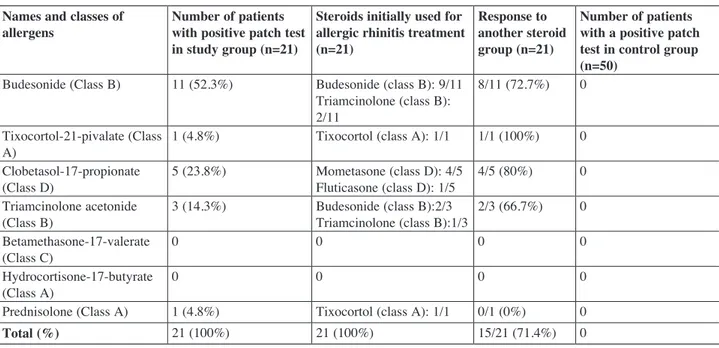
In non-responders, according to the patch test results, corticosteroid treatment was changed to another group of inhaler steroids. Out of these 21 patients, 15 (71.4%) exhibited a positive response to corticosteroid change (Table 3).
Confounding factors affecting patch test positivity Among patients, 89 (59%) were firstly diagnosed as having allergic rhinitis and these patients had correlation coefficient was used for correlation
analysis. A chi-square test was performed for categorical variables.
Results Subjects
After 31 patients were excluded (15 patients treated with allergen avoidance, 10 patients with nasal disorders or structural abnormalities, five patients with a current or history of chronic sinusitis and one pregnant patient), 150 patients [91 (61.3%) women; mean age 37.5 ± 12.0 (min-max: 18-64)] were included in this study. Fifty healthy volunteers [33 (66%) women; mean age 34.5 ± 10.8 (min-max: 20-61)] who had a negative history involving any type of corticosteroid use formed the control group. There was no significant difference between the patient and control groups in terms of gender and age (p: 0.62 and p: 0.17, respectively).
Skin prick test results
In the study group, the number and percentage of patients allergic to the tested allergens were as follows: Dermatophagoides pteronyssinus: 16 (10.7%); Dermatophagoides farina: 23 (15.3%); grass mixture: 48 (32.0%); trees mixture: 44 (29.3%); weed mixture: 4 (2.7%); fungi mixture: 15 (10%).
Table 3
Patch test results Names and classes of
allergens Number of patients with positive patch test in study group (n=21)
Steroids initially used for allergic rhinitis treatment (n=21)
Response to another steroid group (n=21)
Number of patients with a positive patch test in control group (n=50)
Budesonide (Class B) 11 (52.3%) Budesonide (class B): 9/11 Triamcinolone (class B): 2/11
8/11 (72.7%) 0 Tixocortol-21-pivalate (Class
A) 1 (4.8%) Tixocortol (class A): 1/1 1/1 (100%) 0
Clobetasol-17-propionate
(Class D) 5 (23.8%) Mometasone (class D): 4/5Fluticasone (class D): 1/5 4/5 (80%) 0 Triamcinolone acetonide
(Class B) 3 (14.3%) Budesonide (class B):2/3Triamcinolone (class B):1/3 2/3 (66.7%) 0 Betamethasone-17-valerate
(Class C) 0 0 0 0
Hydrocortisone-17-butyrate
(Class A) 0 0 0 0
Prednisolone (Class A) 1 (4.8%) Tixocortol (class A): 1/1 0/1 (0%) 0
IC therapy (2.8 ± 0.9 vs. 0.7 ± 0.5, p<0.001). Furthermore, 21 (16.9%) patients were found to be unresponsive to IC treatment and 15 (71.4%) patients unresponsive to IC were hypersensitive to corticosteroids. In contrast, 103 (83.1%) patients were responsive to IC treatment and 5.8% of these patients had corticosteroid hypersensitivity (p <0.001) (Figure 1).
Total eosinophil counts and IgE levels
Serum IgE levels in the study and control groups were, respectively, 99.9 ± 109.9 and 43 ± 13.6 (p<0.001). Total eosinophil count in the study and control groups were 146.2 ± 107.8 and 78.3 ± 16.4 (p < 0.001), respectively. The serum IgE and total eosinophil levels in patch test positive and negative patients are shown in Table 5.
Discussion
We found that delayed corticosteroid sensitization, as measured by patch testing, increased in not used IC, while 81 (54.0%) had not previously
used corticosteroids by any route (inhaled or topical). On the other hand, 61 (41%) patients had used IC, 12 (8%) had used an inhaler and 10 (6.7%) had previously used topical corticosteroids (Table 2). When concurrent use of corticosteroids by different routes were considered, a total of 69 (46.0%) patients were found to have a history of corticosteroid use (Table 2).
Confounding factors affecting patch test positivity are shown in Table 2. Accordingly, patients who had previously used IC and who had persistent allergic rhinitis were found to have a higher incidence of corticosteroid hypersensitivity, compared to their counterparts (p<0.001, p: 0.01, respectively).
Patient flow
Among the patients, 124 (17.3%) who had been unresponsive to first- and second-line pharmacotherapy had to use IC. TSS significantly decreased after three months of treatment with
Figure 1
case of unresponsiveness to medical treatment or the deterioration of symptoms. Other potential symptoms of CH may also be observed, e.g., facial erythema20, peroral dermatitis and angioedema on
the lips10,19,21 may be observed in case of CH due to
intranasal usage.
Some confounding factors may play a role in the development of CH. In our study, we found that among patients with chronic allergic rhinitis who were thought to have received IC treatment several times before, CH was more common than among patients who exhibited intermittent symptoms. Additionally, using IC treatment or corticosteroid therapy by any route any time prior to admission to our clinics was another risk factor for CH, which supported CH is more common in patients who have received IC treatment several times before. Therefore, the clinician should be careful when prescribing medication to this sub-population (chronic allergic rhinitis or past corticosteroid use) and consider CH in case of resistance to standard medical therapy. In this case, changing the corticosteroid group may be considered, but cross-reactions among different corticosteroids should also be kept in mind.18
Some factors may facilitate the development of CH. These may be related to the medication used by the patient (i.e., the ability of corticosteroid to bind arginine) or in some cases may be patient-related, e.g., recurrent high-dose corticosteroid exposure due to diseases like asthma or the presence of an atopic background in the patient.22Allergic
rhinitis is a disease with recurrent symptoms in which topical corticosteroids are commonly used for extended periods. Thus, recurrent and long-term use of these agents may explain the high incidence of CH in our patient group. However, the presence of accompanying asthma did not affect the incidence of CH in our study.
Drug-related factors may also play role in the development of CH. Budesonide, a group B corticosteroid, is one of the most common agents that clinicians encounter with inhaled CH development.15 In a recent study, Benegal et al. also
showed a 87.9% hypersensitivity to budesonide in patients with corticosteroid hypersensitivity.23
Cross-reactions between budesonide and corticosteroids in its own group (group B) and in D group have also been observed. Similarly, in a recent study including patients with asthma, half of the allergic reactions were due to budesonide.11
patients with allergic rhinitis. Patients who were unresponsive to intranasal corticosteroid treatment also had an increased incidence of corticosteroid hypersensitivity. Our study was the largest study to date performed among allergic rhinitis patients in the context of corticosteroid hypersensitivity.
Corticosteroid hypersensitivity (CH) is a rare condition that has been reported to have an incidence of 2-9%.14,15 Incidence of CH differs
according to several conditions, i.e., 1.4% in patients with asthma and 9.0% in patients with inflammatory bowel disease.16,17 This occurs as a
result of a type 4 hypersensitivity reaction and the most common occurrence of this reaction is in the form of allergic contact dermatitis. A skin patch test is the most reliable technique for the diagnosis of CH.18
In the literature, several case reports denote CH in patients using inhaled or intranasal corticosteroids.10,19 In a study performed by Bennett
et al.,11, two of 30 patients using an inhaler or
intranasal steroids were reported to have CH. The present study has to date been the largest study focusing on corticosteroid hypersensitivity in patients with allergic rhinitis or asthma. In our study, we found a 14.0% incidence of CH among 150 patients with allergic rhinitis.
In patients with allergic rhinitis, approximately 20% are reported to be unresponsive to standard medical treatment.8 We found a 16.9% incidence of
unresponsiveness to IC treatment and 71.4% of these patients were hypersensitive to corticosteroids. Therefore, we can speculate that one of the most important factors in corticosteroid resistance may be undiagnosed CH during corticosteroid treatment. The symptoms of CH such as nasal congestion, oropharyngeal irritation, pruritus or burning may confound with the symptoms of an underlying disease such as allergic rhinitis. The clinician should suspect such a condition in
Table 5
Serum IgE and total eosinophil levels among patch test positive and negative patients in the study group
Serum IgE (IU/
mL) Total eosinophil (/mm3)
Patch test positive
n: 21(14%) 168 ± 133.3 190.8 ± 111.1 Patch test negative
n: 129(86%) 88.8 ± 102.1 138.9 ± 105.9
hypersensitivity, which may affect their response to intranasal corticosteroid treatment.
References
1. Seidman MD, Gurgel RK, Lin SY, Schwartz SR, Baroody FM, Bonner JR, Dawson DE, Dykewicz MS, Hackell JM, Han JK, Ishman SL, Krouse HJ, Malekzadeh S, Mims JW, Omole FS, Reddy WD, Wallace DV, Walsh SA, Warren BE, Wilson MN, Nnacheta LC; Guideline Otolaryngology Development Group. AAO-HNSF. Clinical Practice Guideline: Allergic Rhinitis. Otolaryngol Head Neck Surg. 2015;152(2):197-206.
2. Bauchau V, Durham SR. Prevalence and rate of diagnosis of allergic rhinitis in Europe. Eur Respir J. 2004;24(5):758-764.
3. Maziak W, Behrens T, Brasky TM, Duhme H, Rzehak P, Weiland SK, Keil U. Are asthma and allergies in children and adolescents increasing? Results from ISAAC phase I and phase III surveys in Munster, Germany.
Allergy. 2003;58(7):572-579.
4. Cingi C, Topuz B, Songu M, Kara CO, Ural A, Yaz A, Yildirim M, Miman MC, Bal C. Prevalence of allergic rhinitis among the adult population in Turkey. Acta
Otolaryngol. 2010;130(5):600-606.
5. Hoyte FC, Meltzer EO, Ostrom NK, Nelson HS, Bensch GW, Spangler DL, Storms WW, Weinstein SF, Katial RK. Recommendations for the pharmacologic management of allergic rhinitis. Allergy Asthma Proc. 2014;35(Suppl 1):20-27.
6. Kennedy JL, Robinson D, Christophel J, Borish L, Payne S. Decision-makinganalysis for allergen immunotherapy versus nasal steroids in the treatment of nasal steroid-responsive allergic rhinitis. Am J Rhinol Allergy. 2014;28(1):59-64.
7. Wilson AM, O’Byrne PM, Parameswaran K. Leukotriene receptor antagonists for allergic rhinitis: a systematic review and meta-analysis. Am J Med. 2004;116(5):338-344.
8. Bousquet PJ, Bachert C, Canonica GW, Casale TB, Mullol J, Klossek JM, Zuberbier T, Bousquet J. Uncontrolled allergic rhinitis during treatment and its impact on quality of life: a cluster randomized trial. J Allergy Clin Immunol. 2010;126(3):666-668.
9. Davila-Fernández G, Vazquez-Cortés S, Chamorro-Gómez M, Elices-Apellániz A. Systemic allergic reaction due to intranasal budesonide. Allergol Immunopathol. 2012;40(6):392-393.
10. Pitsios C, Stefanaki EC, Helbling A. Type IV delayed-type hypersensitivity of the respiratory tract due to budesonide use: report of two cases and a literature review.
Prim Care Respir J. 2010;19(2):185-188.
11. Bennett ML, Fountain JM, McCarty MA, Sherertz EF. Contact allergy to corticosteroids in patients using inhaled or intranasal corticosteroids for allergic rhinitis or asthma.
Am J Contact Dermat. 2001;12(4):193-196.
12. Bousquet J, Khaltaev N, Cruz AA, Denburg J, Fokkens WJ, Togias A, Zuberbier T, Baena-Cagnani CE, Canonica GW, van Weel C, Agache I, Aït-Khaled N,Bachert In our study, CH was most commonly observed
alongside budesonide (52.3%), one of the most commonly used corticosteroids against allergic rhinitis. The ratio of budesonide-sensitivity-CH in group A corticosteroids was rare in our study; this was thought to be due to the uncommon use of group A corticosteroids in patients with allergic rhinitis.
In our study, we also found that increased total eosinophil count and total IgE levels related to type 1 hypersensitivity were associated with the presence of CH, which is in fact a type 4 hypersensitivity reaction. The serum IgE and eosinophil counts higher in patients with corticosteroid hypersensitivity may have been due to them having allergic rhinitis and therefore, exhibited in amore atopic fashion than among other patients with allergic rhinitis. The atopic background of patients with allergic rhinitis and prior usage of corticosteroids may also render them vulnerable to CH.
Steroid allergy may also be the result of previous treatments; patients with more severe symptoms were more likely to have received previous steroid treatment, to have used steroids over longer periods and thus, to have developed a hypersensitivity to steroids. This may also explain why patients with steroid resistance had higher IgE and eosinophil counts, as these tests are related to more severesymptoms. In this study, we changed the treatment strategy for non-responsive patients, who responded relatively well after changing the steroid according to skin patch test results.
There were some limitations to our study. First, the cross-sectional design of the study was unable to demonstrate the pathophysiological pathways in the development of CH in patients with allergic rhinitis. Additionally, the predictive role of the skin patch test for corticosteroid hypersensitivity in the clinical response of patients to intranasal corticosteroid therapy should also be studied, because the power of our study was low in terms of determining whether CH was a major determinant for unresponsiveness to corticosteroids. Another limitation was the absence of data regarding facial erythema and perioral dermatitis, which may be an indication for the presence of CH.
In conclusion, our study was the largest to date investigating CH in patients with allergic rhinitis. Patients with allergic rhinitis were found to have a high incidence (14%) of corticosteroid
17. Isaksson M, Bruze M, Hörnblad Y, Svenonius E, Wihl JA. Contact allergy to corticosteroids in asthma/rhinitis patients. Contact Dermatitis. 1999;40(6):327-328.
18. Vatti RR, Ali F, Teuber S, Chang C, Gershwin ME.Hypersensitivity reactions to corticosteroids. Clin Rev
Allergy Immunol. 2014;47(1):26-37.
19. Poulos GA, Brodell RT. Perioral dermatitis associated with an inhaled corticosteroid. Arch Dermatol. 2007;143(11):14-60.
20. Baeck M, Pilette C, Drieghe J, Goossens A. Allergic contact dermatitis to inhalatin corticosteroids. Eur J
Dermatol. 2010;20(1):102-108.
21. Pirker C, Misic A, Frosch PJ. Angioedema and dysphagia caused by contact allergy to inhaled budesonide. Contact
Dermatitis. 2003;49(2):77-79.
22. Ventura MT, Calogiuri GF, Muratore L, Di Leo E, Buquicchio R, Ferrannini A, Resta O, Romano A. Crossreactivity in cell-mediated and IgE-mediated hypersensitivity to glucocorticoids. Curr Pharm Des. 2006;12(26):3383-3391.
23. Berbegal L, DeLeon FJ, Silvestre JF. Corticosteroid Hypersensitivity Studies in a Skin Allergy Unit. Actas
Dermosifiliogr. 2015;106(10):816-822.
24. Coopman S, Degreef H, Dooms-Goossens A. Identification of cross-reaction patterns in allergic contact dermatitis from topical corticosteroids. Br J Dermatol. 1989;121(1):27-34.
Dr Hande Arslan, MD, Assist. Prof. of Otorhinolaryngology Ufuk University Medical School, Department of
Otorhinolaryngology Balgat 06520, Ankara, Turkey
Tel: +90 3122044383
E-mail: [email protected] C, Blaiss MS, Bonini S, Boulet LP, Bousquet
PJ, Camargos P, Carlsen KH, Chen Y, Custovic A, Dahl R, Demoly P, Douagui H, Durham SR, van Wijk RG, Kalayci O, Kaliner MA, Kim YY, Kowalski ML, Kuna P, Le LT, Lemiere C, Li J, Lockey RF, Mavale-Manuel S, Meltzer EO, Mohammad Y, Mullol J,Naclerio R, O’Hehir RE, Ohta K, Ouedraogo S, Palkonen S, Papadopoulos N, Passalacqua G, Pawankar R, Popov TA, Rabe KF, Rosado-Pinto J, Scadding GK, Simons FE, Toskala E, Valovirta E, van Cauwenberge P, Wang DY, Wickman M, Yawn BP, Yorgancioglu A, Yusuf OM, Zar H, Annesi-Maesano I,Bateman ED, Ben Kheder A, Boakye DA, Bouchard J, Burney P, Busse WW, Chan-Yeung M, Chavannes NH, Chuchalin A, Dolen WK, Emuzyte R, Grouse L,Humbert M, Jackson C, Johnston SL, Keith PK, Kemp JP, Klossek JM, Larenas-Linnemann D, Lipworth B, Malo JL, Marshall GD, Naspitz C, Nekam K,Niggemann B, Nizankowska-Mogilnicka E, Okamoto Y, Orru MP, Potter P, Price D, Stoloff SW, Vandenplas O, Viegi G, Williams D; World Health Organization; GA(2)LEN; AllerGen. Allergic Rhinitis and its Impact on Asthma (ARIA) 2008. Allergy. 2008;63(Suppl86):8-160.
13. Nyembue TD, Vinck AS, Corvers K, Bruninx L, Hellings PW, Jorissen M. Sensitization to common aeroallergens in patients at an outpatient ENT clinic. B-ENT. 2011;7(2):79-85.
14. Lutz ME, el-Azhary RA, Gibson LE, Fransway AF. Contact hypersensitivity to tixocortol pivalate. J Am Acad
Dermatol. 1998;38(5):691-695.
15. Vind-Kezunovic D, Johansen JD, Carlsen BC. Prevalence of and factors influencing sensitization to corticosteroids in a Danish patch test population. Contact Dermatitis. 2011;64(6):325-329.
16. Malik M, Tobin AM, Shanahan F, O’Morain C, Kirby B, Bourke J. Steroid allergy in patients with inflammatory bowel disease. Br J Dermatol. 2007;157(5):967-969.