ORIGINAL ARTICLE
Evaluation of the Relationships between Chronological
Age, Skeletal Maturation, Dental Maturation, and
Sagittal Jaw Relationships
Gülşilay Sayar Torun, Hüsamettin OktayDepartment of Orthodontics, İstanbul Medipol University School of Dentistry, Istanbul, Turkey
Objective: The present study aimed to determine whether there is a correlation between chronological age, skeletal maturation,
dental maturation, and ANB angle.
Methods: Lateral cephalometric, panoramic, and hand–wrist radiographs of 200 orthodontic patients were used (100 males and 100
females; mean age 13.00 and 13.70 years, respectively). Skeletal maturation was determined by two different methods: cervical ver-tebral maturation (CVM) and the hand–wrist radiography method of Grave–Brown. Dental maturation was defined by the Demirjian Index using the mandibular canine, premolars, and second molar on the left side. The ANB angle was measured on lateral cephalo-metric head films. The data were analyzed by Spearman’s rank correlation analysis.
Results: Correlation coefficients of the male and female subjects were 0.825 and 0.802 between chronological age and hand–wrist
evaluation; 0.744 and 0.778 between chronological age and CVM evaluation; 0.677 and 0.443 between chronological age and man-dibular canine development; 0.722 and 0.458 between chronological age and manman-dibular first premolar development; 0.730 and 0.517 between chronological age and mandibular second premolar development; 0.701 and 0.531 between chronological age and mandibular second molar development; and −0.183 and −0.045 between chronological age and ANB, respectively. All the correla-tions mentioned above were statistically significant (p<0.001), except for the last one.
Conclusions: High correlations were found between the chronological age, hand–wrist, and cervical vertebral maturation
evalua-tions. Chronological age was also correlated with dental maturation, particularly in mandibular second molars. There was no correla-tion between ANB and the other parameters.
Keywords: Skeletal maturation, cervical vertebral maturation, Demirjian index, dental maturation
INTRODUCTION
In dentofacial orthopedic treatments, treatment timing is a key factor for successful orthodontic therapy.¹
Deter-mination of the maturation level and growth stage of a growing patient with skeletal imbalances or dentofacial
disorders is highly important in orthodontic treatment planning.
1-3This determination is generally performed
by the evaluation of chronological age, skeletal maturation, dental maturation, height–weight, and prepubertal
maturation characteristics.
4It has been reported
1,5that chronological age has a minor or no effect on the determination of the maturation
phases of a child. Instead, biological (physiological) age may be more reliable because it includes parameters
such as somatic, sexual, skeletal, and dental maturity.
6-9Skeletal maturation shows the degree of development
of ossification in bone. The growth and maturation of a bone may be different; therefore, skeletal maturation is
more closely related to sexual maturity than to stature.
4Greulich and Pyle determined the sequence of hand and wrist bone ossification in 1950s and published a
radio-graphic atlas for the evaluation of skeletal maturity.
10Their evaluation method is still in use. In 1976, Grave and
Brown published a classification of skeletal maturation based on checking the maturity markers on hand–wrist
radiographs.
11Hand–wrist radiography is a widespread and classically used diagnostic tool in the evaluation of
Corresponding Author: Gülsilay Sayar Torun, İstanbul Medipol University School of Dentistry, Istanbul, Turkey E-mail: [email protected] - [email protected]
©Copyright 2015 by Turkish Orthodontic Society - Available online at www.turkjorthod.org
ABSTRACT
86
Received: 29.04.2015 Accepted: 11.08.2015
skeletal maturation, and an alternative to this method is cervical
vertebral evaluation on lateral cephalometric radiographs. Many
researchers have investigated cervical vertebral maturation
indi-cators and concluded that this evaluation is a reliable method for
skeletal maturation.
12-18Hassel and Farman
5stated that by briefly
looking at the cervical vertebrae on a lateral cephalometric
ra-diograph, the orthodontist can evaluate the skeletal maturity of
a patient at that point. Baccetti et al.
19published an improved
version of the CVM method for the assessment of mandibular
growth.
High correlations have generally been reported between
skel-etal and dental maturity.
8It has been suggested, however, that
racial variations also have an effect on this relationship. Ethnicity,
climate, nutrition, socioeconomic levels, and urbanization are
indicated as causative factors of these racial variations.
20The
re-lationships between dental and skeletal maturations have been
evaluated by many investigators, and it has been found that
the use of tooth calcification is more reliable than that of tooth
eruption.
8,9,19,21-26Some authors
6,26-28have claimed that the
calci-fication stages of mandibular second molars have the highest
correlation with the stages of skeletal maturity, while according
to others,
8,9,19mandibular canines have the highest correlation.
According to Choi et al.,
29the rate of skeletal maturation may
differ in different types of malocclusion. Johnston et al.
30investi-gated the relationships between skeletal maturation and
ceph-alofacial development and found a retardation in subjects with
Class II. A similar study conducted by Kim et al.
31,32revealed that
skeletal maturation in patients with Class II malocclusion begins
later than that in those with Class I or III malocclusion. Sasaki et
al.
33suggested that the skeletal disharmony can be associated
with different timings of the formation and eruption of
perma-nent teeth.
The ANB angle is recognized as a skeletal sagittal discrepancy
indicator and has become most commonly used for
measure-ment among orthodontists. This angle has a definite tendency
to decrease with increasing age.
34If a relationship can be
de-fined between skeletal maturation and ANB, this knowledge
will be useful to predict the extent of growth remaining in the
jaws. Previous reports
1,7,12,13,28have laid emphasis on the
relation-ships between chronological age, cervical vertebral maturation,
hand–wrist evaluation, and dental maturation, but no study has
evaluated the relationships between these parameters and the
sagittal relationships of the jaws to date.
METHODS
The present study sample was chosen from the files of the
Or-thodontics Department of the Dental School at Istanbul Medipol
University. Lateral cephalometric, panoramic, and hand–wrist
ra-diographs of 200 subjects (100 males, 100 females) were used in
this study. The mean age of the male and female subjects were
13.00±2.12 and 13.70±2.02 years, respectively. All of the
radio-graphs were taken by one operator using the Kodak 9000 C
sys-tem (Kodak Dental Syssys-tems, Carestream Health, Rochester, NY,
USA).
Inclusion criteria for this study were as follows: 1) Chronological
age ranging from 7 to 18 years; 2) No serious illness and
nutri-tional or hormonal problems; 3) Normal growth and
develop-ment; 4) Absence of previous history of trauma or congenital/
acquired disease to the face, neck, and hand–wrist; 5) Absence
of abnormal dental conditions, such as impaction,
transposi-tion, and congenitally missing teeth; 6) No previous orthodontic
treatment; 7) No permanent teeth extracted.
Skeletal maturation was evaluated by means of both the
im-proved version of the CVM3 (Table 1) and from hand–wrist
ra-diographs (Table 2), and the skeletal ages were rated without
any knowledge about the children’s chronological ages.
11,18,28Dental maturation was evaluated by the Demirjian Index (DI)
24on panoramic radiographs (Table 3). The mandibular left canine,
premolars, and second molar were used for this purpose.
Skele-tal classification was made on the basis of ANB angle; in skeleSkele-tal
Class I, the ANB angle was from 1 to 5 degrees, for skeletal Class II
more than 5 degrees, and for skeletal Class III less than 1 degree.
Statistical Analysis
Statistical analyses were performed by the Statistical Package for
Social Science (SPSS for Windows, version 14.0, SPSS Inc;
Chica-go, USA). The Student’s t and Mann-Whitney U tests were used
to find out the gender differences between the investigated
parameters. The Spearman’s rank correlation coefficients were
computed separately for the male and female subjects to find
out the relationships among chronological age, CVM, skeletal
maturation, dental maturation, and ANB angle.
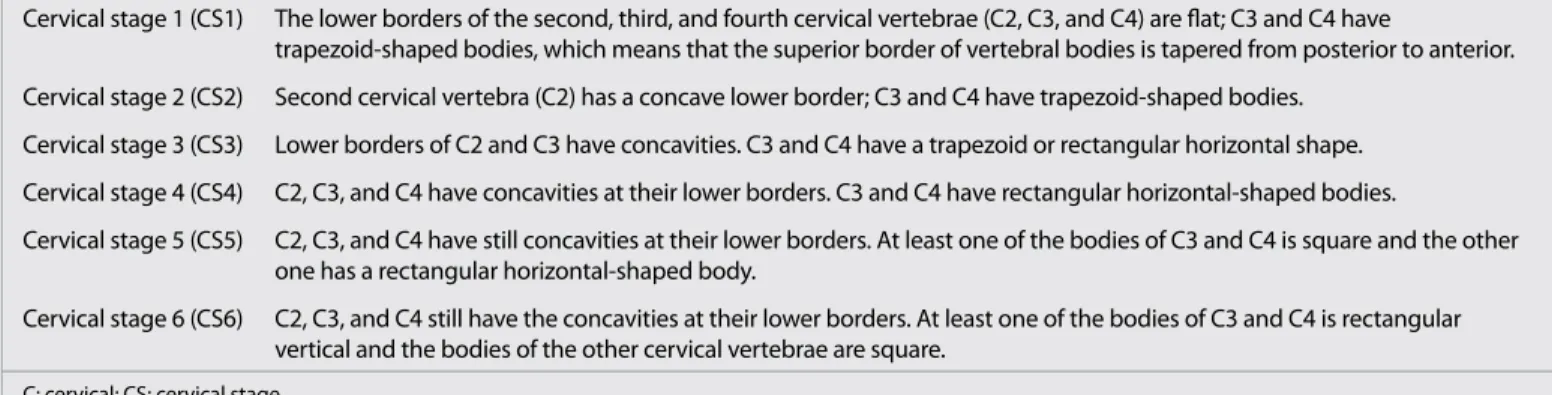
Table 1. Evaluation method of cervical vertebral maturation2
Cervical stage 1 (CS1) The lower borders of the second, third, and fourth cervical vertebrae (C2, C3, and C4) are flat; C3 and C4 have
trapezoid-shaped bodies, which means that the superior border of vertebral bodies is tapered from posterior to anterior. Cervical stage 2 (CS2) Second cervical vertebra (C2) has a concave lower border; C3 and C4 have trapezoid-shaped bodies.
Cervical stage 3 (CS3) Lower borders of C2 and C3 have concavities. C3 and C4 have a trapezoid or rectangular horizontal shape. Cervical stage 4 (CS4) C2, C3, and C4 have concavities at their lower borders. C3 and C4 have rectangular horizontal-shaped bodies.
Cervical stage 5 (CS5) C2, C3, and C4 have still concavities at their lower borders. At least one of the bodies of C3 and C4 is square and the other one has a rectangular horizontal-shaped body.
Cervical stage 6 (CS6) C2, C3, and C4 still have the concavities at their lower borders. At least one of the bodies of C3 and C4 is rectangular vertical and the bodies of the other cervical vertebrae are square.
C: cervical; CS: cervical stage
Assessing the reproducibility of the ratings was done by
reeval-uating the radiographs of 20 males and 20 females randomly
selected 6 weeks after the first evaluation, and the Spearman
Brown formula was used for this purpose.
RESULTS
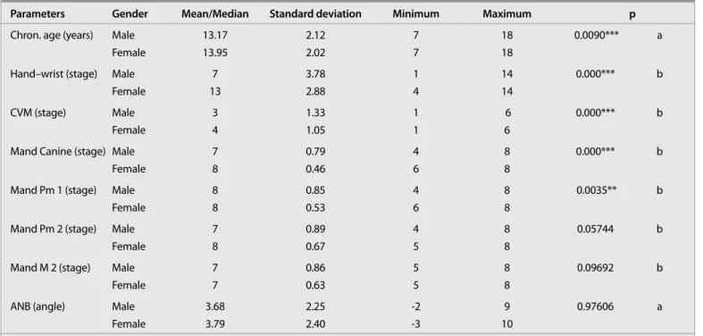
Descriptive statistics of all the variables for the male and
fe-male subjects and their comparisons are presented in Table 4.
As shown in this table, all the parameters, except for ANB and
the second molar and premolar, were statistically significant, and
they were higher in female subjects.
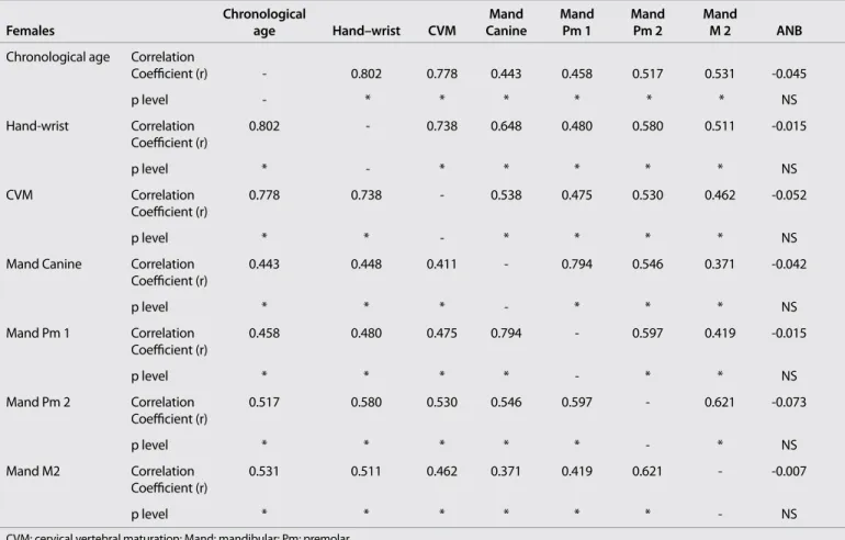
The results of correlation analysis regarding the chronological
age, skeletal maturity, and dental maturity indicators and ANB
angle are presented in Table 5 and 6 for the males and females,
respectively. As shown in these tables, the correlation
coeffi-cients for the male and female subjects were 0.825 and 0.802
be-tween the chronological age and hand–wrist evaluation; 0.744
and 0.778 between chronological age and CVM evaluation;
0.677 and 0.443 between chronological age and mandibular
ca-nine development; 0.722 and 0.458 between chronological age
and mandibular first premolar development; 0.730 and 0.517
between chronological age and mandibular second premolar
development; and 0.701 and 0.531 between chronological age
and mandibular second molar development, respectively; all of
these were statistically significant (p<0.001). There was no
cor-relation between chronological age and ANB.
Correlation coefficients between all the parameters, except for
the ANB angle, were also statistically significant (Table 5, 6).
DISCUSSION
Maturation is an important concept for orthodontists when it is
time to evaluate a growing child, especially one with dentofacial
problems. Many researchers have investigated the different
matu-ration indicators, such as chronological age, hand–wrist ossification,
cervical vertebral maturation, and dental maturation to find out if
there was a relationship between skeletal maturation and these
pa-rameters.
1,7,12,13,28In these articles, the sagittal jaw relationship
deter-mined by the ANB angle was not included in the study model.
According to the previous reports, chronological age was not
found to be sufficiently reliable in the prediction of pubertal
growth spurts because of the wide variation among patients in
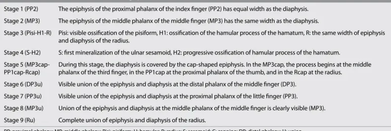
Table 2. Evaluation method of hand–wrist radiographs11,18,29Stage 1 (PP2) The epiphysis of the proximal phalanx of the index finger (PP2) has equal width as the diaphysis. Stage 2 (MP3) The epiphysis of the middle phalanx of the middle finger (MP3) has the same width as the diaphysis.
Stage 3 (Pisi-H1-R) Pisi: visible ossification of the pisiform, H1: ossification of the hamular process of the hamatum, R: the same width of epiphysis and diaphysis of the radius.
Stage 4 (S-H2) S: first mineralization of the ulnar sesamoid, H2: progressive ossification of hamular process of the hamatum.
Stage 5 (MP3cap- During this stage, the diaphysis is covered by the cap-shaped epiphysis. In the MP3cap, the process begins at the middle PP1cap-Rcap) phalanx of the third finger, in the PP1cap at the proximal phalanx of the thumb, and in the Rcap at the radius.
Stage 6 (DP3u) Visible union of the epiphysis and diaphysis at the distal phalanx of the middle finger (DP3). Stage 7 (PP3u) Visible union of the epiphysis and diaphysis at the proximal phalanx of the little finger (PP3).
Stage 8 (MP3u) Union of the epiphysis and diaphysis at the middle phalanx of the middle finger is clearly visible (MP3).
Stage 9 (Ru) Complete union of epiphysis and diaphysis of the radius.
PP: proximal phalanx; MP: middle phalanx; Pisi: pisiform; H: hamular; R: radius; S: sesamoid; C: capping; DP: distal phalanx; U: union
Table 3. Evaluation of dental maturation according to Demirjian24
Stage A Calcification of single occlusal points without fusion of different calcifications.
Stage B Fusion of mineralization points; the contour of the occlusal surface is recognizable.
Stage C Enamel formation has been completed at the occlusal surface, and dentine formation has commenced. The pulp chamber is
curved, and no pulp horns are visible
Stage D Crown formation has been completed to the level of the amelocemental junction. Root formation has commenced. The pulp
horns are beginning to differentiate, but the walls of the pulp chamber remain curved.
Stage E The root length remains shorter than the crown height. The walls of the pulp chamber are straight, and the pulp horns have
become more differentiated than those in the previous stage. In molars, the radicular bifurcation has commenced to calcify.
Stage F The walls of the pulp chamber now form an isosceles triangle, and the root length is equal to or greater than the crown
height. In molars, the bifurcation has sufficiently developed to give the roots a distinct form.
Stage G The walls of the root canal are now parallel, but the apical end is partially open. In molars, only the distal root is rated.
Stage H The root apex is completely closed (distal root in molars). The periodontal membrane surrounding the root and apex is uni
form in width throughout.
Table 4. Descriptive statistics of all variables for male and female subjects and their comparisons
Parameters Gender Mean/Median Standard deviation Minimum Maximum p
Chron. age (years) Male 13.17 2.12 7 18 0.0090*** a
Female 13.95 2.02 7 18
Hand–wrist (stage) Male 7 3.78 1 14 0.000*** b
Female 13 2.88 4 14
CVM (stage) Male 3 1.33 1 6 0.000*** b
Female 4 1.05 1 6
Mand Canine (stage) Male 7 0.79 4 8 0.000*** b
Female 8 0.46 6 8
Mand Pm 1 (stage) Male 8 0.85 4 8 0.0035** b
Female 8 0.53 6 8
Mand Pm 2 (stage) Male 7 0.89 4 8 0.05744 b
Female 8 0.67 5 8
Mand M 2 (stage) Male 7 0.86 5 8 0.09692 b
Female 7 0.63 5 8
ANB (angle) Male 3.68 2.25 -2 9 0.97606 a
Female 3.79 2.40 -3 10
Chron: chronological; CVM: cervical vertebral maturation; Mand: mandibular; Pm: premolar, M: molar Alphabetical order in Demirjian classification was converted to numerical order in statistical evaluation. a: Student’s t test, b: Mann-Whitney U test
*p<0.05, **p<0.01, ***p<0.001
Table 5. Correlation coefficients and their significance levels between chronological age and the skeletal maturity indicators, dental development,
and ANB in male subjects
Chronological Mand Mand Mand Mand
Males age Hand–wrist CVM Canine Pm 1 Pm 2 M 2 ANB
Chronological age Correlation 0.825 0.744 0.677 0.722 0.730 0.701 -0.183
Coefficient (r) p level * * * * * NS Hand–wrist Correlation 0.825 - 0.769 0.648 0.678 0.662 0.611 -0.240 Coefficient (r) p level - * * * * * NS CVM Correlation 0.744 0.769 0.538 0.522 0.557 0.501 -0.237 Coefficient (r) p level * * * * * * * NS
Mand Canine Correlation 0.677 0.648 0.538 - 0.801 0.673 0.546 -0.146
Coefficient (r) p level * * * - * * * NS Mand Pm 1 Correlation 0.722 0.678 0.522 0.801 - 0.745 0.707 -0.153 Coefficient (r) p level * * * * - * * NS Mand Pm 2 Correlation 0.730 0.662 0.557 0.673 0.745 - 0.666 -0.176 Coefficient (r) p level * * * * * - * NS Mand M2 Correlation 0.701 0.611 0.501 0.546 0.707 0.666 - -0.122 Coefficient (r) p level * * * * * * - NS
CVM: cervical vertebral maturation; Mand: mandibular; Pm: premolar *p<0.001
terms of chronological timing. The maturity level has generally
been assessed by the evaluation of hand–wrist radiographs.
4,10,15High correlations were found between chronological age and
skeletal maturation.
9,13In this study, the chronological ages of
the patients showed high correlations with both hand–wrist
os-sification and CVM in the male and female subjects. The
correla-tion coefficients of the hand–wrist evaluacorrela-tion were found higher
than that of CVM in both genders.
Some authors
11,28have suggested carrying out the evaluation of
skeletal maturity without taking any additional radiograph
be-cause of the possible danger of X-rays, and instead used cervical
vertebrae and tooth images on cephalometric and panoramic films,
respectively. CVM is an efficient method in assessing the skeletal
maturation. It has been proven by numerous studies
4,10,13that CVM
shows a high correlation with skeletal maturity. Baccetti et al.
16and
Franchi et al.
17showed that statural height was related with cervical
vertebral maturation. In this study, the CVM method modified by
Franchi et al.
2was used. The results of the present study showed
that CVM had high correlations with the hand–wrist method for
both male and female subjects. This finding was confirmed by the
previous reports, according to which the CVM method is
sufficient-ly reliable for the evaluation of skeletal maturation.
4,15,17Some authors
6,8,9,21,23-26showed a high correlation between dental
and skeletal maturity, while others
22have reported weak or
insig-nificant relationships between these parameters. In order to
inves-tigate this relationship, the Demirjian index, which was accepted
as a reliable assessment method, was used in this study. For this
purpose, radiographic images of the mandibular canine, the first
and second premolars, and the second molar teeth on the left side
were used, since the maxillary teeth had a disadvantage of
super-imposition on panoramic radiographs.
6,13,27Dental development
instead of tooth eruption was used because the previous reports
commented that eruption was an alterable situation being
affect-ed more than calcification.
24,25Developments of the first molar and
incisor teeth are completed at the early ages, and therefore they
were not included in the study model, and also third molars were
not included because of their potential for agenesis. In male
sub-jects, the best correlation was found between chronological age
and the second premolar, which could be accepted as a mild-level
relationship. In females, the best correlation couple was the
sec-ond premolar and the hand–wrist evaluation.
The results of the present study showed that the rate of
skele-tal maturation in female subjects was greater than in males. This
was an expected finding for skeletal development. Dental
de-velopments, however, did not show the same results. Although
the mean chronological age of females was greater than that of
male subjects, the developments of the second premolar and
molar teeth did not show any statistically significant difference
between the genders.
90
Table 6. Correlation coefficients and their significance levels between chronological age and the skeletal maturity indicators, dental development,
and ANB in female subjects
Chronological Mand Mand Mand Mand
Females age Hand–wrist CVM Canine Pm 1 Pm 2 M 2 ANB
Chronological age Correlation
Coefficient (r) - 0.802 0.778 0.443 0.458 0.517 0.531 -0.045 p level - * * * * * * NS Hand-wrist Correlation 0.802 - 0.738 0.648 0.480 0.580 0.511 -0.015 Coefficient (r) p level * - * * * * * NS CVM Correlation 0.778 0.738 - 0.538 0.475 0.530 0.462 -0.052 Coefficient (r) p level * * - * * * * NS
Mand Canine Correlation 0.443 0.448 0.411 - 0.794 0.546 0.371 -0.042
Coefficient (r) p level * * * - * * * NS Mand Pm 1 Correlation 0.458 0.480 0.475 0.794 - 0.597 0.419 -0.015 Coefficient (r) p level * * * * - * * NS Mand Pm 2 Correlation 0.517 0.580 0.530 0.546 0.597 - 0.621 -0.073 Coefficient (r) p level * * * * * - * NS Mand M2 Correlation 0.531 0.511 0.462 0.371 0.419 0.621 - -0.007 Coefficient (r) p level * * * * * * - NS
CVM: cervical vertebral maturation; Mand: mandibular; Pm: premolar *p<0.001.
The skeletal jaw relationships were assessed by the ANB angle.
This angle showed no correlation with any other parameters as
opposed to in previous reports.
35,36This result showed that
skel-etal maturation was similar in all sagittal skelskel-etal abnormalities.
CONCLUSION
• The highest correlations were found between
chronologi-cal age and hand–wrist evaluation in all subjects.
• A high correlation was found between the hand–wrist and
CVM methods in both genders.
• In male subjects, correlations between chronological age
and hand–wrist evaluation and between chronological
age and CVM methods were found to be higher than those
of females.
• In male subjects, the second premolars showed the
high-est correlations with chronological age, while the highhigh-est
correlations were seen between hand–wrist evaluation
and dental parameters, except for second molar in females.
• Developments of the second premolar and molar teeth did
not show any statistically significant difference between
the genders.
• There were no correlations between ANB and the other
pa-rameters.
REFERENCES
1. Baccetti T, Franchi L, McNamara JA Jr. The cervical vertebral matura-tion (CVM) method for the assessment of optimal treatment timing in dentofacial orthopedics. Seminars in Orthodontics 2005; 11: 119-29. [CrossRef]
2. Franchi L, Baccetti T, De Toffol L, Polimeni A, Cozza P. Phases of the dentition for the assessment of skeletal maturity: A diagnostic per-formance study. Am J Orthod Dentofacial Orthop 2008; 133: 395-400. [CrossRef]
3. McNamara JA Jr, Brudon WL. Orthodontics and dentofacial ortho-pedics. Ann Arbor, Mich: Needham Press; 2001. p. 78-80.
4. Hassel B, Farman AG. Skeletal maturation evaluation using cervical ver-tebrae. Am J Orthod Dentofacial Orthop 1995; 107: 58-66. [CrossRef]
5. Björk A, Helm S. Prediction of the age of maximum puberal growth in body height. Angle Orthod 1967; 37: 134-43.
6. Kumar S, Singla A, Sharma R, Virdi MS, Anupam A, Mittal B. Skeletal maturation evaluation using mandibular second molar calcification stages. Angle Orthod 2012; 82: 501-6. [CrossRef]
7. Demirjian A, Buschang PH, Tanguay R, Patterson DK. Interrelation-ships among measures of somatic, skeletal, dental, and sexual ma-turity. Am J Orthod 1985; 88: 433-8. [CrossRef]
8. Chertkow S, Fatti P. The relationship between tooth mineralization and early evidence of the ulnar sesamoid. Angle Orthod 1979; 49: 282-8. 9. Sierra AM. Assessment of dental and skeletal maturity. A new
ap-proach. Angle Orthod 1987; 57: 194-208.
10. Greulich WW, Pyle SI. Radiographic atlas of skeletal development of the hand and wrist. 2nd ed. Stanford, CA: Stanford University Press; 1959. 11. Grave KC, Brown T. Skeletal ossification and the adolescent growth
spurt. Am J Orthod 1976; 69: 611-9. [CrossRef]
12. Garcia-Fernandez P, Torre H, Flores M, Rea J. The cervical vertebrae as maturational indicators. J Clin Orthod 1998; 32: 221-5.
13. Uysal T, Ramoglu SI, Basciftci FA, Sari Z. Chronologic age and skeletal maturation of the cervical vertebrae and hand-wrist: is there a relation-ship? Am J Orthod Dentofacial Orthop 2006; 130: 622-8. [CrossRef]
14. San Román P, Palma JC, Oteo MD, Nevado E. Skeletal maturation determined by cervical vertebrae development. Eur J Orthod 2002; 24: 303-11. [CrossRef]
15. Hägg U, Taranger J. Maturation indicators and the pubertal growth spurt. Am J Orthod 1982; 82: 299-309. [CrossRef]
16. Baccetti T, Franchi L, McNamara JA Jr. The cervical vertebral matura-tion method: some need for clarificamatura-tion. Am J Orthod Dentofacial Orthop 2003; 123: 19A-20A. [CrossRef]
17. Franchi L, Baccetti T, McNamara JA Jr. Mandibular growth as related to cervical maturation and body height. Am J Orthod Dentofacial Orthop 2000; 118: 335-41. [CrossRef]
18. Björk A. Timing of interceptive orthodontic measures based on stages of maturation. Trans Eur Orthod Soc 1972; 48: 61-74. 19. Baccetti T, Franchi L, McNamara JA Jr. An improved version of the
cervical vertebral maturation (CVM) method for the assessment of mandibular growth. Angle Orthod 2002; 72: 316-23.
20. Mappes MS, Harris EF, Behrents RG. An example of regional varia-tion in the tempos of tooth mineralizavaria-tion and hand-wrist ossifica-tion. Am J Orthod Dentofacial Orthop 1992; 101: 145-51. [CrossRef]
21. Coutinho S, Buschang PH. Miranda F. Relationship between man-dibular canine calcification stages and skeletal maturity. Am J Orth-od Dentofacial Orthop 1993; 104: 262-8. [CrossRef]
22. Lewis AB, Garn SM. The relationship between tooth formation and other maturational factors. Angle Orthod 1960; 30: 70-7.
23. Engström C, Engström H, Sagne S. Lower third molar development in relation to skeletal maturity and chronological age. Angle Orthod 1983; 53: 97-106.
24. Demirjian A, Goldstein H, Tanner JM. A new system of dental age assessment. Human Biol 1973; 45: 211-27.
25. Nolla CM. The development of the permanent teeth. J Dent Child 1960; 27: 254-63.
26. Hotz R, Boulanger G, Weisshaupt H. Calcification time of permanent teeth in relation to chronological and skeletal age in children. Helv Odontol Acta 1959; 3: 4-9.
27. Krailassiri S, Anuwongnukroh N, Dechkunakorn S. Relationship be-tween dental calcification stages and skeletal maturity indicators in Thai individuals. Angle Orthod 2002; 72: 155-66.
28. Uysal T, Sari Z, Ramoglu SI, Basciftci FA. Relationships between dental and skeletal maturity in Turkish subjects. Angle Orthod 2004; 74: 657-64. 29. Choi YJ, Chung C, Kim KH. The relationship between malocclusion
and menarcheal age, and its secular trend for Korean women. Kore-an J Orthod 2012; 42: 11-6. [CrossRef]
30. Johnston FE, Hufham HP, Jr, Moreschi AF, Terry GP. Skeletal matura-tion and cepalofacial development. Angle Orthod 1965; 35: 1-11. 31. Kim KH, Baik HS, Son ES. A study on menarche and skeletal maturity
among various malocclusion groups. Korean J Orthod 1998; 28: 581-9. 32. Kim KH, Baik HS, Choy KC, Son ES. The age at onset of menarche of
women in Seoul. J Korean Dent Assoc 1998; 36: 864-71.
33. Sasaki M, Sato K, Mitani H. Tooth formation and eruption in skele-tal Class II and Class III malocclusions. Nihon Kyosei Shika Gakkai Zasshi 1990; 49: 435-42.
34. Oktay H. A comparison of ANB, WITS, AF-BF, and APDI measure-ments. Am J Orthod Dentofacial Orthop 1991; 99: 122-8. [CrossRef]
35. Reyes BC, Baccetti T, McNamara JA Jr. An estimate of craniofacial growth in Class III malocclusion. Angle Orthod 2006; 76: 577-84. 36. Armond MC, Generoso R, Falci SG, Ramos-Jorge ML, Marques LS.
Skeletal maturation of the cervical vertebrae: association with vari-ous types of malocclusion. Braz Oral Res 2012; 26: 145-50. [CrossRef]