Original Article
©Copyright 2017 by Turkish Society of Physical Medicine and Rehabilitation
The efficacy of vestibular electrical stimulation on patients with
unilateral vestibular pathologies
Ayşe Karan,1 Hasan Kerem Alptekin,2 Nalan Çapan,1 Demirhan Dıraçoğlu,1 İlknur Saral,3 Salih Aydın,4 Cihan Aksoy1
1Department of Physical Medicine and Rehabilitation, Medicine Faculty of İstanbul University, İstanbul, Turkey 2Department of Physiotherapy and Rehabilitation, Bahçeşehir University, Faculty of Health Sciences, İstanbul, Turkey 3Department of Physical Medicine and Rehabilitation, Medical Faculty of Medipol University, İstanbul, Turkey 4Department of Otolaryngology, İstanbul University, İstanbul Faculty of Medicine, İstanbul, Turkey
Received: February 2016 Accepted: April 2016
Corresponding author: Hasan Kerem Alptekin, MD. Bahçeşehir Üniversitesi Sağlık Bilimleri Fakültesi, Fizyoterapi ve Rehabilitasyon Bölümü, 34349 Beşiktaş, İstanbul, Turkey. e-mail: [email protected]
Cite this article as:
Karan A, Alptekin HK, Çapan N, Dıraçoğlu D, Saral İ, Aydın S, et al. The efficacy of vestibular electrical stimulation on patients with unilateral vestibular pathologies. Turk J Phys Med Rehab 2017;63(2):149-54.
ABSTRACT
Objectives: This study aims to investigate the efficacy of vestibular electrical stimulation (VES) in unilateral vestibular lesions including benign paroxysmal positional vertigo (BPPV).
Patients and methods: Between June 2007 and August 2007, a total of 19 patients diagnosed with BPPV were included in this study and they were randomized into two groups using the 1:1 method. Ten patients were administered medical treatment plus VES (treatment group; 1 male, 9 females; mean age 55.8 years; range 27 to 74 years), whereas nine patients were only administered medicine (control group; 2 males, 6 females; mean age 54.9 years; range 34 to 73 years). Both groups received the same medical treatment throughout the study. Vestibular electrical stimulation was performed for 30 min long twice a day, three times a week; 12 sessions in total with 80 Hz high-frequency Transcutaneous Electrical Nerve Stimulation (TENS). Before and after the treatment, patients’ severity of dizziness was assessed with Visual Analog Scale (VAS) and daily life activities with Dizziness Handicap Inventory (DHI), and their duration (sec) of single leg stance with eyes open and closed was recorded.
Results: Compared to prior to the treatment, VAS-dizziness and DHI scores, and the duration of single leg stance on one foot with eyes open and closed at the end of the treatment showed statistically significant improvement in both groups; however, although VES provided a positive contribution, we did not find a statistically significant difference between the two groups.
Conclusion: It can be concluded that VES has positive contribution to medical treatment of patients with dizziness due to unilateral vestibular lesions; however the results of this study should be further investigated with larger groups of patients.
Keywords: Unilateral vestibular pathology; vestibular electrical stimulation; vestibular rehabilitation.
Uncompensated unilateral vestibular hypofunction may derive from Ménière’s disease, viral and vascular etiologies, neuroma operation and neuritis. Unilateral vestibular lesions can be caused by inflammatory diseases, injuries of eight cranial nerve
or idiopathic processes.[1] Vestibular impairments
are usually well-treated with medical, surgical or
rehabilitative methods.[2] Electrical stimulation is
a noninvasive method used for the treatment and rehabilitation of one sided vestibular pathologies, where muscles and nerves are stimulated via surface electrodes. For this purpose, transcutaneous electrical nerve stimulation (TENS) may be used. Depolarization triggered by motor point stimulation spreads
centrally as well as peripherally. Direct stimulation of vestibular receptors is highly invasive and is not appropriate in this case thus electrical stimulation of paravertebral muscles, known as vestibular electrical stimulation (VES), is used. Electrical stimulation of the neck muscles sends proprioceptive input to the cerebrum, changes of that input result in changes of head position perception and also enhances control of
body and head position.[1]
Since there are very few studies on the effects of VES on patients with one-sided vestibular lesions, the aim of our study was to determine the efficiency of VES in patients with complaints of vertigo and dizziness due to one-sided vestibular lesions.
PATIENTS AND METHODS
Forty patients with persistent vertigo due to one-sided vestibular lesion who were visited to outpatient clinics at Istanbul University Medical Faculty, Physical Medicine and Rehabilitation Department, were screened for the study. All of the patients were referred from Istanbul University Medical Faculty Otolaryngology Department. Only 34 patients participated in the study and they were randomized into two groups using the 1:1 method. The first group was the treatment group in which VES was combined with medical treatment, the second group (control) only received medical treatment. Of the 34 patients, 17 were assigned to the control group and 17 were in the treatment group. Ten patients in the treatment group (9 females, 1 male; mean age 55.8 years; range 27 to 74 years) and 9 patients in the control group (6 females, 3 males; mean age 54.9 years; range 34 to 73 years) completed treatment and attended follow-up appointments. The reasons why the other participants did not complete the study were mainly severe vertigo and various other familial and social reasons.
Informed consent was obtained from patients before the study. The Istanbul University Medical Faculty Local Ethics Committee approved the study (reference number 2007/1298). The study was conducted in accordance with the principles of the Declaration of Helsinki.
Patients with vertigo without vestibular lesion, two-sided vestibular lesions, diabetic patients, patients with cerebrovascular accidents, with prominent anemia, cranial surgery, and cardiac pacemakers were excluded from the study. The control group was given 24 mg of betahistine dihydrochloride everyday divided into three doses for a total of seven days.
Severity of dizziness, its effects on daily life activities and its effects on balance were evaluated using a Visual Analog Scale (VAS), Dizziness Handicap Inventory (DHI), and stance time with eyes open and closed.
The Turkish validity and reliability study
of DHI was performed by Ellialtıoğlu et al.[3] The
questionnaire includes 25 questions. The possible answers are: yes, no or sometimes. The responses are scored as yes= 4, sometimes= 2, and no= 0 points. The maximum possible score is 100, which reflects a grave disability for the patient, if the total score reduces it is
considered an improvement.[4]
Vestibular electrical stimulation: A frequency of 80 Hz was administered which causes no muscle
contraction. The stimulus was a biphasic asymmetrical modulated square with a pulse width 100 µsec. That modality is commonly used as conventional TENS for analgesic effect. But in vestibular problems it has been used since 1990 as the first line treatment of acute vertigo. The pathophysiological basis of the treatment is to reduce antigravity failure and to increase proprioceptive cervical sensory substitution. Vibrations can provide specific exogenous stimulation on paravertebral muscle proprioceptors. Also this painless current may be a substitute for vibration
effect.[5,6] Electrodes sized 2 cm2 were placed parallel
to each other at a distance of 1 cm, on motor points of the trapezius muscle on the effected side and the counter side at C2-C3 paravertebral level. Each session took place for 30 min twice daily, three times a week for a total of 12 sessions. The patients laid down with their affected side placed inferiorly. Patients were instructed to fix their eyes on the impaired labyrinth. The patients were advised not to get off the bed and to
lie down on their healthy side.[1] Both groups received
the same medication during the study. Statistical analysis
Mean, standard deviation, median, minimum and maximum, frequency and ratio values were used as descriptive statistics of the data. The distribution of variables was assessed using the Kolmogorov-Smirnov test. Mann-Whitney U test was used in the analysis of quantitative data. Wilcoxon test was used to analyze repeated measures. Qualitative data was analyzed by chi-square test. If conditions weren’t acceptable for chi-square, Fisher’s exact test was used as an assessment. In the statistical analysis of all data, IBM SPSS version 22.0 software (IBM Corp., Armonk, NY, USA) was used. A p value of <0.05 was considered statistically significant.
RESULTS
Power analysis was calculated before the study. Visual Analog Scale median difference was calculated as 4; standard deviation for the before treatment group as 3, after treatment group as 2 and based on that difference a minimum 4 patients were to be enrolled from each group. Dizziness Handicap Inventory score median difference was calculated as 40 and standard deviation of the before treatment group as 30, after treatment group as 25 and based on that difference a minimum eight patients were to be enrolled for each group. As the aim of type 1 mistake was 0.05 and the power of the test was estimated as 0.80. For each group minimum 4 patients. There
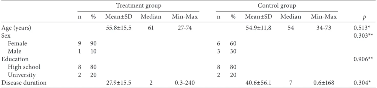
Table 1. Demographic data
Treatment group Control group
n % Mean±SD Median Min-Max n % Mean±SD Median Min-Max p
Age (years) 55.8±15.5 61 27-74 54.9±11.8 54 34-73 0.513* Sex 0.303** Female 9 90 6 60 Male 1 10 3 30 Education 0.906** High school 8 80 8 80 University 2 20 2 20 Disease duration 27.9±15.5 2 0.3-240 40.6±56.1 7 0.6±168 0.304*
SD: Standard deviation; Min: Minimum; Max: Maximum; * Mann-Whitney U test; ** Chi-square test (Fisher’s exact test).
were no significant differences between the groups in terms of age, sex, education level and disease duration (p>0.05) (Table 1, Figure 1).
No statistically significant difference was observed between groups in the VAS scores before treatment (p=0.481). Following therapy, the VAS score of the treatment group was significantly lower than the VAS score of the control group (p=0.014). Also in the treatment group, VAS score after treatment was significantly lower than before treatment (p=0.005). In the control group, VAS score after treatment was significantly lower than before treatment (p=0.027). The decrease in the difference of before and after treatment VAS scores in the treatment group was more significant than those of the control group (p=0.008) (Table 2).
The values from DHI score in the before treatment did not differ significantly in regards to both groups (p=0.103). At the end of the treatment, the treatment group DHI score values were significantly lower than control group DHI score values (p=0.009). In the treatment group, DHI score values after treatment were significantly lower than before treatment (p=0.005). In the control group, DHI score values after treatment were significantly lower than before treatment (p=0.007). The decrease in the difference of before and after treatment DHI score values in the treatment group were more significant than the control group (p=0.002) (Table 2).
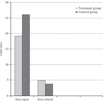
There was no statistically significant difference in time between groups regarding the eyes opened single leg stance test before treatment (p=0.581) and after treatment (p=0.805). In both the treatment (p=0.004) and the control group (p=0.000); eyes opened single leg stance test time after treatment improved significantly. In regard to both groups, the difference of eyes opened single leg stance test time before and after treatment was not significant (p=0.227) (Table 2).
Eyes closed single leg stance test did not differ significantly in both groups before (p=0.653) and after (p=0.647) treatment either. There was a statistically significant improvement in eyes closed single leg stance test regarding the treatment group (p=0.001) and control group (p=0.002) following treatment. Eyes closed single leg stance test did not differ significantly before or after treatment in both groups (p=0.294) (Table 2, Figure 2).
DISCUSSION
This study showed that vertigo symptoms due to unilateral vestibular lesions decreased in both the medical treatment and VES group. The VES treatment provided a positive contribution but the differences did not reach statistical significance. We suspect that a major limitation of our research, the low number of patients, caused that effect. Patients with vertigo feel insecure outside of their homes, so they hesitate to attend follow-up sessions in hospital. Twelve sessions lasting for four weeks were another factor that complicated the treatment adaptation. For better statistical results, we are planning a study with a larger number of patients and long-term follow-up.
One of the strengths of our research is that there are few studies about VES utilizing control groups, in which patients were followed up with medical treatment. A sham VES protocol was not used but it should be implemented as a third group in a future study.
In our study we used a protocol similar to Cesarani
et al.[1] In 1990 they treated patients with labyrinth
acute vertigo using VES. The authors presented the results as treatment studies. Patients, who did not respond to maneuvers were chosen for the study. Their patients walked or performed simple and provocative rehabilitative exercises such as the Cawthorne-Cooksey protocol during stimulation. Surface electrodes placed on the paravertebral nuchal muscles opposite the
impaired vestibular side facilitated the contralateral impaired vestibular nuclei by the crossed spinal vestibular pathway.
Mosca[2] preferred cervical electrodes with a
frequency of 100 Hz. Stimulation lasted for 30 min and this was repeated twice a week for a total number of 10 sessions. Patients with cervical vertigo, labyrinthitis or neuritis compensated well compared to the control group. The result was hypothesized to be a result of the appropriate response of the vestibuloocular reflex.
The action takes place via vestibular nuclei or by repeated decompensations mainly mediated through the cerebellum or spino-reticular connections.
Barozzi et al.[7] decided on a biphasic asymmetric
modulated square wave with a pulse width of 100 µsec and a stimulation frequency of 80 Hz. Twenty-eight patients with persistent unilateral vestibular dysfunction were allocated into two groups; in one group patients tried to practice saccadic oculomotor exercises, and VES was administered to Figure 1. Visual Analog Scale and Dizziness Handicap Inventory score differences before and after
treatment. VAS: Visual Analog Scale.
10 10 8 8 6 6 4 4 2 2 0 0
Treatment group Control group Treatment group
V isu al A nal og S cal e V isu al A nal og S cal e Control group Before treatment VAS
After treatment VAS Before treatment VASAfter treatment VAS
Table 2. Visual Analog Scale, Dizziness Handicap Inventory score and stance time comparison before and after treatment
Treatment group Control group
Mean±SD Median Min-Max Mean±SD Median Min-Max p
Visual Analog Scale score
Before treatment 6.3±3.1 8 0-10 7.2±2.8 8 3-10 0.481*
After treatment 3.1±2.3 4 0-6 6.0±1.7 7 4-8 0.014*
Differences 3.2±1.6 4 0-6 1.2±1.2 1 -1-3 0.008*
p 0.005** 0.027**
Dizziness Handicap Inventory score
Before treatment 56.0±22.6 60 24-88 55.0±26.5 52 18-96 0.103*
After treatment 19.2±17.7 14 0-50 46.4±23.2 44 16-84 0.009*
Differences 36.8±15.7 39 6-56 8.6±3.5 8 2-12 0.002
p 0.005** 0.007**
Eyes open stance in sec
Before treatment 20.4±16.0 20 4-45 27.2±12.3 30 5-50 0.581*
After treatment 40.2±35.8 25 7-120 34.4±12.1 40 10-50 0.805*
Differences 19.8±22.4 13 0-75 7.2±3.6 10 0-10 0.227*
p 0.004** 0.000**
Eyes closed stance in sec
Before treatment 6.0±5.9 3 2-20 7.4±6.4 5 2-20 0.653*
After treatment 11.5±7.7 10 3-30 11.3±8.7 8 5-30 0.647*
Differences 5.0±2.8 5 1-10 3.9±2.5 3 2-10 0.294*
p 0.001** 0.002**
Figure 2. Differences between single leg stance test eyes open and closed. 30 25 20 15 10 5 0
Eyes open Eyes closed
Ti m e ( se c) Treatment group Control group
the second group through surface electrodes on the opposite impaired side and patients were told to walk during the electrical stimulation. A comparison of the two groups showed no significant difference, revealing that both forms of therapy were effective.
Hillier and Hollohan[8] evaluated benign
paroxysmal positional vertigo (BPPV) as a vestibular dysfunction, as in our study. They emphasized that physical therapy, particularly maneuvers, show better short-term results. According to randomized controlled trials, unilateral vestibular rehabilitation
is reported as safe and with very good results.[8]
In another recent review Arnold et al.[9]
compared studies of vestibular rehabilitation for unilateral peripheral vestibular hypofunction. Seven studies were selected for inclusion. Two studies reported improvements on the dynamic gait index. The results suggested that vestibular therapy for unilateral peripheral vestibular hypofunction was effective but none of the interventions were superior. The outcomes were evaluated using the dynamic gait index or DHI. Further research should concentrate on more concrete outcomes from more objective measurements such as computerized
posturography.[9]
Galvanic vestibular stimulation (GVS) is not just used in analyzing neuroadaptive behaviors of the vestibulospinal system but also in treating several vertigo related disorders. For instance, Rizzo-Sierra
et al.[10] suggested that GVS may control motion
sickness and space adaptation syndrome. Galvanic stimulus is delivered transcutaneously at levels of 1 mA. Bilateral stimulation is effected because both the anode and cathode poles are placed on the right and left mastoid region. A small deviation towards the cathodal side is noted. A deviation forwards is noted in monopolar stimulation with cathodes alone, whereas with anodal stimulation, a backward deviation is observed. Galvanic vestibular stimulation has the potential of up-regulating the disturbed sensory-motor mismatch originated by kinetosis and space sickness by modulating the gamma-aminobutyric acid (GABA)-related ion channels neural transmission in the inner ear. It improves the signal-to-noise ratio of the afferent proprioceptive volleys, which ultimately modulates motor output and restores the disordered gait, balance, and human locomotion due to kinetosis, as well as the spatial disorientation generated by gravity transition.
Lewis et al.[11] demonstrated that electrical
stimulation of canal afferents affects the perception of head orientation, and in addition improves vestibular percepts in patients lacking normal vestibular function. An important difference was that they implanted invasive electrodes to posterior canals in rhesus monkeys. The authors suggested that a canal prosthesis could potentially improve both percepts of head orientation and postural control in patients suffering from a reduction in peripheral vestibular function.
Furthermore, Carmona et al.[12] revealed that
galvanic vestibular stimulation improves the results of vestibular rehabilitation. They examined body sway in 10 normal participants after one min of 2 mA galvanic vestibular stimulation. Stimulation affected 70% of the patients for time periods up to two hours. Compared to conventional vestibular rehabilitation, a group of 40 patients with a combination treatment of vestibular rehabilitation and galvanic vestibular stimulation, a significant improvement was noted in the second group. To eliminate a placebo effect,
“systematic” galvanic vestibular stimulation and
“nonsystematic” in a sham protocol were confronted
and no placebo effect was noted.
There are also other clues that galvanic vestibular stimulation is effective in unilateral vestibular pathologies and diseases with sporadic episodes like Ménière's disease. In one study an enhanced vestibulo-ocular reflex was observed in patients with Ménière's disease, compared to healthy individuals. These abnormalities may be a diagnostic indicator of
Ménière's disease and may also explain the nature of
Ménière's disease.[13]
To the best of our knowledge, there is not any study comparing the effects of galvanic vestibular stimulation and vestibular electrical stimulation with conventional TENS modality. Both methods are noninvasive and the principles are the same because both influence vestibular afferent system. However, GVS has both direct and indirect effects such as influencing sway pattern of the body, posture and gait respectively. On the other hand, VES was shown to modify motor neuron excitability of the
soleus muscles. Osio et al.[14] suggested H reflex was
reduced by about 80% in the left soleus and 77% in the right soleus after 10-15 min stimulation of the splenius muscle and the contralateral upper part of the trapezius. In our study we used the same protocols as stated above.
In conclusion, TENS with high frequency currents makes a significant contribution to medical therapy in patients with vertigo due to a vestibular lesion. More studies with VES, which is a painless treatment modality and without muscle contraction, should be conducted with more patients and a longer term follow-up.
Declaration of conflicting interests
The authors declared no conflicts of interest with respect to the authorship and/or publication of this article.
Funding
The authors received no financial support for the research and/or authorship of this article.
REFERENCES
1. Cesarani A, Alpini D. Vertigo and Dizziness Rehabilitation. Chapter: Treatment. In: Vestibular electrical stimulation. Berlin: Springer-Verlag; 1999. p. 112.
2. Mosca F Cervical electrostimulation in some vestibular diseases. Acta Otorhinolaryngol Ital 1994;14:525-33. [Abstract]
3. Ellialtıoğlu A, Karan A, İşsever H, Aksoy C. Validity and reliability of Turkish version of Dizziness Handicap Inventory (DHI). XVIII National Physical Medicine and Rehabilitation Congress, May 12-17, 2001, Antalya, Program and Abstract Book; 2001. p. 131.
4. Nola G, Mostardini C, Salvi C, Ercolani AP, Ralli G. Validity of Italian adaptation of the Dizziness Handicap Inventory (DHI) and evaluation of the quality of life in patients with acute dizziness. Acta Otorhinolaryngol Ital 2010;30:190.
5. Cesarani A, Alpini D, Barozzi S, Neck electrical stimulation in the treatment of labyrinth acute vertigo. Riv Ital EEG Neurof Clin 1990;13:55-61.
6. Cesarani A, Alpini D, Monti B, Raponi G, The treatment of acute vertigo. Neurological Sciences 2004;25:26-30. 7. Barozzi S, Di Berardino F, Arisi E, Cesarani A. A comparison
between oculomotor rehabilitation and vestibular electrical stimulation in unilateral peripheral vestibular deficit. Int Tinnitus J 2006;12:45-9.
8. Hillier SL, Hollohan V. Vestibular rehabilitation for unilateral peripheral vestibular dysfunction. Cochrane Database Syst Rev 2007;(4):CD005397.
9. Arnold SA, Stewart AM, Moor HM, Karl RC, Reneker JC. The Effectiveness of Vestibular Rehabilitation Interventions in Treating Unilateral Peripheral Vestibular Disorders: A Systematic Review. Physiother Res Int 2015. [Epub ahead of print]
10. Rizzo-Sierra CV, Gonzalez-Castaño A, Leon-Sarmiento FE. Galvanic vestibular stimulation: a novel modulatory countermeasure for vestibular-associated movement disorders. Arq Neuropsiquiatr 2014;72:72-7.
11. Lewis RF, Haburcakova C, Gong W, Lee D, Merfeld D. Electrical stimulation of semicircular canal afferents affects the perception of head orientation. J Neurosci 2013;33:9530-5.
12. Carmona S, Ferrero A, Pianetti G, Escolá N, Arteaga MV, Frankel L. Galvanic vestibular stimulation improves the results of vestibular rehabilitation. Ann N Y Acad Sci 2011;1233:E1-7.
13. Aw ST, Aw GE, Todd MJ, Halmagyi GM. Enhanced vestibulo-ocular reflex to electrical vestibular stimulation in Meniere's disease. J Assoc Res Otolaryngol 2013;14:49-59. 14. Osio M, Dezuanni E, Alpini A, Mangoni A, Low intensity
electrical stimulation of neck muscles modifies motor neuron excitability of the soleus muscles in man, Electroencephalography and Clinical Neurophysiology 1995;95:68-9.