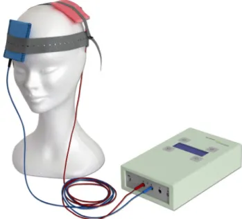
Efficacy and mechanisms of transcranial electrical stimulation in headache disorders
Tam metin
Şekil
Benzer Belgeler
Although given a high degree of flexibility, a majority of students chose working in a group at the univer- sity’s laboratory supported by course advisors as their desired
Üniversitelerin markalaflmas› konusunun içsel pazarlama ba¤lam›nda ele al›nd›¤› son bölümde öncelikle içsel pazarlama- n›n tan›m›, fonksiyonlar› ve
Bu açıdan psikiyatrik tedavi almakta olan ya da psikiyatrik tedavi başlanması planlanan hastalarda COVID-19 pan- demisinde kullanılan ilaçların psikotrop ilaçlarla
sayfalarında yer alan ‘Ankara ili Yenimahalle ilçesinde birinci basamak sağlık kuruluşuna başvuran bireylerde tütün bağımlılığı ve ilişkili risk
Katılımcıların bazı besin maddelerinin tuz o- ranları hakkındaki bilgileri ve bunları kullanma du- rumları arasındaki uyum, olgu ve çalışma gruplarına göre
Bir parafili türü olan fetiflizm, kiflinin cans›z nesneleri kullanmakla ilgili yo¤un, cinsel yönden uyar›c› fantezileri- nin, cinsel dürtülerinin ya da
Birinci basamakla ilgili bir der- lemede, hekim hasta beklentilerinin fark›nda oldu¤unda sadece hastan›n de¤il hekimin de memnuniyetinin artt›- ¤› gösterilmifl,
Yapılan analizler sonucunda girişimcilik yöneliminin, işe bağlanmanın alt boyutlarından adanma üzerindeki etkisinde algılanan örgütsel desteğin tam aracılık,
