Original Article
Can Wharton jelly derived or adipose tissue derived
mesenchymal stem cell can be a treatment
option for duchenne muscular dystrophy?
Answers as transcriptomic aspect
Eda Sun1,2*, Erdal Karaoz1,2,3*
1Histology and Embriology Department, Faculty of Medicine, İstinye University, İstanbul, Turkey; 2Center for Stem
Cell and Tissue Engineering Research & Practice, İstinye University İstanbul, Turkey; 3Center for Regenerative
Medicine and Stem Cell Research and Manufacturing, Liv Hospital, İstanbul, Turkey. *Equal contributors. Received May 11, 2020; Accepted July 8, 2020; Epub August 25, 2020; Published August 30, 2020
Abstract: Introduction: Mesenchymal stem cells (MSCs) are able to differentiate into several cell lineages including skeletal muscle. In addition to their differentiation capacities, they have the ability to transfer their content genomic information horizontally through their exosomes and fusion abilities, as we have shown in our previous clinic study on Duchenne Muscular Dystrophy (DMD) patients, dystrophin expression increased after MSC treatment. Therefore, this study aimed to compare the transcriptomic properties of Wharton’s jelly derived (WJ-) MSC and Adipose tissue (AT-) derived MSC, which are the two most preferred sources in MSC treatments applied in DMD. Methods: Both MSC cell lines obtained from ATCC (PCS-500-010; PCS-500-011) were characterized by flow cytometry then WJ-MSC and AT-MSC cell lines were sequenced via RNA-SEQ. R language was used to obtain the differentially expressed genes (DEGs) and differentially expressed miRNAs, respectively. Additionally, in order to support the results of our study, a gene expression profile data set of DMD patients (GSE1004) were acquired from Gene Expression Omnibus (GEO) database. Results: Here, we demonstrated that activated WNT signaling and downregulated TGF-β pathways under the control of decreased mir-24 which are involved in myogenic differentiation are differentially expressed in WJ-MSC. We have shown that the expression of mir-199a-5p, which is known to increase in exosomes of DMD patients, is less in WJ-MSC. Additionally, we have shown activated PI3K/Akt pathway, which is controlling mitochon-dria transfer via Tunnelling Nanotube as a new perspective in cellular therapies in myodegenerative diseases, in WJ-MSC more than in AT-MSCs. Conclusion: Summing up, WJ-MSC, which we recommend as an appropriate source candidate due to its immune-regulation properties, stands forward as a preferable source in the cellular treatment of DMD patients due to its transcriptomic aspect.
Keywords: Duchenne muscular dystrophy, gene expression, mesenchymal stem cells, cellular therapy Introduction
Duchenne Muscular Dystrophy (DMD) is an X recessive disease that affects 1/3500 males [1]. Boys with normal growth refer to the clinic at around 3 or 5 years old with symptoms such as difficulty in climbing stairs and early fatigue. The basic pathogenesis is explained by the for-mation of fibrous tissue by means of covering the connective tissue because the muscle tis-sue does not function due to the absence of dystrophin [2]. Dystrophin gene is localized at Xp21 and it is the largest gene defined in the human genome. The dystrophin protein is the
sarcolemma protein at the skeletal muscle. It is in tight contact with cell membrane proteins and muscle fibers. It is a tight bridge between the cytoplasm and cell membrane; in the ab- sence of dystrophin muscle cannot contract and fibrous [3, 4].
In a large quantity of variations, large deletions, duplications or point mutations, in the gene affect functional dystrophin protein production and cause DMD [5-8]. There is no conventional therapy that targeting all these variations. Treatment options were determined based on the type of variation. Exon-skipping is one of the
most promising therapeutic approaches that aim to restore the expression of a shorter but functional dystrophin protein [9, 10] rather than a truncated and nonfunctional protein. Allele specific oligonucleotides (ASO) are paired with the exonic splice site to removing mutated exon from pre-mRNA and produce a shorter but functional dystrophin protein in translation [11]. However, ASO therapy offers a treatment option only to the 15% of patients. Converting the nonsense mutation to missense mutation is another treatment option to produce func-tional protein [12, 13]. The therapy is based on the altering 3rd amino acid to get rid of the early stop codon to continue transcription. This treat-ment option is known as Ataluren treattreat-ment and provides a 70% reduction in plasma CK level.
There is a requirement for alternative treat-ment solutions, owing to mutation-specific treatment options are targeting just for a small group of patients. The goal for this would be to send the wild type mRNA for functional protein, rather than correcting the mutation and form-ing a truncated protein. Hence, gene transfer-ring could be a treatment option that can reach all patients, nevertheless there is no suitable vector transferring the dystrophin gene. Herein, cellular therapies are a good option for the per-sistence of treatment in the long term for of all patients via horizontal gene transfer by cellular
fusions [14, 15] or transferring exosome [16-18]. Mesenchymal Stem Cells (MSCs) promises a treatment hope for untreatable diseases with the conventional treatment protocols, such as autoimmune diseases, lung diseases, cardio-vascular diseases and neuromuscular diseas-es. Their importance in clinic stand out provid-ing the regeneration of damaged cells and sup-pression of inflammation via secreting many bioactive molecules [19].
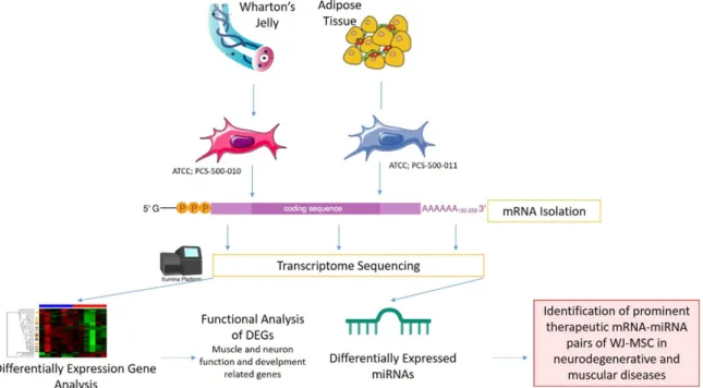
MSCs can be derived from many sources such as Wharton’s Jelly (WJ), placenta tissue, amni-otic fluid, cord blood, adipose tissue (AT), der-mis and tooth pulp [20, 21]. For the allogenic transplantations, WJ-MSC and AT-MSCs are most preferable sources for DMD. Although they are the same type of cells, gene expres-sion profiles are differentiated according to the tissue origin from which they are obtained. Different transcriptomic features will cause dif-ferentiation in mRNA, miRNA or protein cargoes that will be sent to the host cell by horizontal transfer. As a result, the effectiveness of treat-ment actually depends on the characteristics of these transfers of cells. In this study, we have compared transcriptomes of WJ- and AT- sources of MSCs to determine the most effec-tive source that can be used in DMD treatment in order to show which of them is more effective as a transcriptomic content in cellular treat-ments (Figure 1).
Materials and methods
Samples and study design
Two distinct sources derived MSC cell lines, which are WJ-MSC (PCS-500-010) and AT-MSC (PCS-500-011) were obtained from ATCC (Ma- nassas, VA). Two of these cell lines were grown in culture according to suppliers’ instructions. In order to increase the significance of the anal-ysis on cell lines, the filtering of DEGs was gen-erated via comparing the open source gene expression data which are the muscle biopsies derived from 12 DMD patients and 12 healthy controls (GSE1004) [22]. These examples were also evaluated with the GEOR2 tool [23].
Cell culture
WJ and AT derived MSC lines were cultured in T175 flasks with MSC NutriStem® XF Basal Medium and MSC NutriStem® XF Supplement Mix (Biological Industries, Cromwell, CT, USA) cell culture media supplied with 10% fetal bovine serum (FBS), 2 mM L-glutamine and streptomycin (100 mg/mL) solution at 37°C in a humidified 5% CO2 incubator and subcultured until the 3rd passage. After reaching 70% to 80% confluency, adherent cells were harves- ted with trypsinization by 0.05% trypsin-EDTA (Gibco, Germany).
Characterization of WJ-MSC and AT-MSC with cell surface marker screening
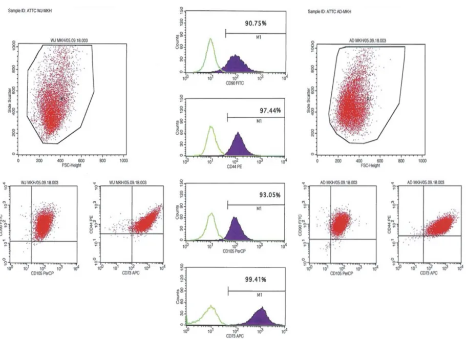
For characterization of MSCs with their cell sur-face markers, the cells were harvested at their third passages (P3), and were labeled with CD90, CD105, CD44 and CD73 (BD). The char-acterization data were acquired with Flow Cytometry analysis at FACS calibur (BD).
RNA extraction, library preparation and se-quencing
Total RNA was extracted from WJ- and AT- derived MSC lines with Purelink RNA kit (Invitrogen) according to the manufacturer’s instructions. Concentration and the Integrity Number of RNA (RIN) samples were determined with the Agilent 2100 Bioanalyzer and suitable RNA samples were selected with the RIN ≥ 7. Libraries were prepared with Illumina TruSeq RNA library kit (Illumina) and were sequenced by HiSeq2000 with 100 bp paired-end reads per sample.
Annotation and differentially expressed genes
The quality control of the raw sequence reads were analysed with FastQC v. 0.11.3 [24]. Low-quality sequences and overrepresented se- quences like adapters were trimmed by fastq-mcf module (v 1.04) [25]. Raw reads were mapped to the GRCh38 reference genome by RSEM 25 (v. 1.2.22) with TopHat aligner. For each transcript, the fragments per kilobase per million (FPKM) were detected and the reads were normalized. For the differentially expressed genes (DEGs) analysis, the thresh-old was determined as log2 FPKM and P≤0.05. DEGs were analysed in R environment EBSeq package.
Functional analysis
The result of DEG lists were submitted to Database for Annotation, Visualization, and Integrated Discovery (DAVID v.6.7) and IPA (Qiagen) software to identify their functions and the pathways that are attended.
Differentially expressed miRNAs and their tar-get prediction
Trimmed raw sequence reads were realigned by using SHiMPS aligner [26]. The counts read associated to mature miRNAs and detection of differentially expressed miRNAs were analyzed by using the R package of DESeq2 [27]. Results
Flow cytometry analysis of cell-surface markers for WJ- and AT-MSC at P3. They all expressed MSC markers including CD90, CD105, CD44 and CD73 (Figure 2).
RNA-Seq analysis
An average of 43 million 100-base long reads from each source of MSCs were mapped to the human reference genome and FPKM values were generated, which provided a measure of expression levels for each gene mapped to the transcriptome. 92 genes between WJ-MSC and AT-MSC were identified as DEGs which passed the |log2 (fold-change)|>1.5 and p Value < 0.05 filtering.
WJ-MSC has a higher regenerative capacity
We analyzed the functions of 92 DEGs. Con- sequently, the important features for stem cell
therapies that regulation of muscle cells, mus-cle development, extra cellular matrix (ECM) organization, response to growth factor and the MSC differentiation which are important for neurodegenerative diseases are significantly increased for WJ-MSCs. 35 of 92 DEGs were associated with these features and 23 of these 35 genes were also detected in DMD patients expressed contrarily. Filtering was applied with the comparison of the 35 DEGs and the DEGs of DMD patient muscle biopsy vs healthy mus-cle biopsy retrieved from GEO1004. Twenty-three of DEGs overlapped (Figure 3). The expression of 12 genes in WNT signalling and 5 genes that are play an important role in myo-genic differentiation involved in TGF-β signal-ling were detected differentially expressed. Logarithmic fold changes and p-values of the differentially expressed genes in these path-ways are presented in Table 1 in detail.
Significant miRNA expressions targeting mus-cle regulation in WJ-MSC
While mRNA data is mapping, some miRNA might include in the data. While our raw data were aligning, 110 aligned miRNAs were ma- pped, and we achieved their expression data. 24 of the 110 miRNAs identified differentially expressed. The target match analysis was
applied to these 23 miRNAs and 23 DEGs. In this analysis, experimentally observed pairs were selected whose expressions were their opposite. When this target match was investi-gated for 23 DEGs and 11 miRNAs, 2 miRNAs were targeting the muscle process functions in regeneration of WJ-MSC (Table 2).
Discussion
MSCs are a group of progenitor cells capable of differentiating into several mesenchymal lin-eages [28] and an attractive cell source for cel-lular therapies [29]. MSCs have ability to recog-nize the biological markers that secreted from the injured cell and have potential to transform into the target cells and forming the healthy tis-sue [30]. The therapeutic effects of MSCs are promoting by secreting factors and delivering genetic information to tissue-resident cells via either a paracrine effect or a direct cell-to-cell interaction. Each manufactured MSC to be used in clinical are exposed to quality control tests for the key MSC features such as cell sur-face markers, differentiation capacities, telom-erase activity [31]. Although MSCs derived from different sources share basic characteristics, they are transcriptomically different from each other. This transcriptomic difference is also important in terms of the diversity of the secre-Figure 3. Filtering workflow of whole transcriptomic data. The DEGs that both passed the filtering and placed at the intersection of MSC and DMD patients were annotated to their functional annotations and their attended pathways.
Table 1. Enriched pathway analysis and their functional categories
Functional Categories Pathway Name in PathwayRegulator Gene p-Value Log Fold change Signaling Transduction TGF-beta signaling pathway BAMBI 8.90E-19 5.2684
SMAD7 5.91E-06 1.6536
INHBA 1.52E-21 2.327
CDKN2B 2.19E-14 1.74
PITX2 1.38E-41 3.5955
WNT signaling pathway CCND1 3.80E-08 1.2589
GPC4 1.36E-42 4.3748 BAMBI 8.90E-19 5.2684 NFATC4 1.31E-10 -1.3164 SFRP4 1.49E-07 -8.1966 DKK1 3.20E-09 2.8133 SFRP2 9.50E-27 -7.8593 TCF7L1 2.43E-05 2.3311 DKK2 1.88E-08 2.1996 FZD1 5.61E-17 -2.3299 LRP5 8.96E-11 1.7443 TCF7L2 0.0030771 1.5690
Celllular Communication mTOR Signalling Pathway VEGFA 2.66E-10 1.0362
PI3K/Akt Signalling Pathway ITGA3 4.61E-14 2.9903
TNC 1.03E-19 -1.4597 NGFR 0.00023885 -7.0205 COL5A3 6.86E-14 -1.3267 LAMB1 1.63E-36 1.0533 FLT1 1.53E-42 8.3424 COMP 7.50E-61 -2.7148 VTN 5.31E-09 7.2324 CCND1 3.80E-08 1.2589 VEGFA 2.66E-10 1.0362 LAMA4 1.06E-07 -1.3721 SGK1 1.10E-05 1.5521 LAMA5 6.14E-06 3.2191 COL4A2 2.41E-266 4.4393 EPHA2 4.58E-16 2.5843 OSMR 1.47E-05 1.0598 ITGB1 1.76E-53 1.1654 VEGFB 5.22E-08 -1.0511 THBS2 1.95E-63 1.092 COL4A1 8.24E-208 5.2997 COL4A5 2.06E-08 3.5296 ITGA1 1.76E-12 3.1042
P53 signalling pathway CCND1 3.80E-08 1.2589
CD82 3.94E-05 1.1701 SERPINE1 8.52E-299 2.1331 STEAP3 9.73E-08 -1.7067 TP53I3 3.91E-07 -1.2232 CCNG2 0.00017411 1.2924 IGFBP3 1.62E-168 -2.411
tion and the genetic information that they transfer to the target cell. Therefore, the selec-tion of the appropriate source for MSCs is criti-cal in clinicriti-cal application. In this study, we iden-tified the transcriptomic patterns of MSCs derived from 2 different sources with RNA-Seq after their characterization by flow cytometry. The hierarchical clustering that based on whole transcriptome data results demonstrated its regenerative capacity, regulation of extracellu-lar matrix (ECM), immunoregulation and regula-tion of the musco-skeleton system.
In healthy skeletal muscle is able to regenerate by stem cells and precursor cells that host in its own niche. Unfortunately, DMD patient’s regen-erative capacity is depleted and they are facing with the loss muscle mass. MSCs have capable of the regulation of muscle regeneration with pathways and gene expression that came for-ward with enhancing their regenerative capaci-ties in DMD patients. One of these pathways is transforming growth factor-β (TGF-β) that is also organizing the myogenesis [32]. TGF-β is found upregulated in WJ-MSC cell line related to the AT-MSC. 5 of 80 members are differen-tially expressed. SMAD7 is upregulated and it controls the BAMBI expression and causes PITX2 upregulation. PITX2, is a transcription factor that belongs to homeobox gene family and controls both the embryonic and adult myogenesis [33]. In the adult myogenesis PITX2 expression was detected in proliferating satellite cells and they promote the differentia-tion of satellite cell-derived myoblasts [34, 35]. As a transcriptional factor, PITX2 can control different pathways simultaneously. One of the negatively regulated mechanisms under the PITX2 is mir-31. The main role is the degrada-tion of the dystrophin mRNA. Since mir-31 expression is repressed, mRNA will be tran-scripted and resulting in increase of dystrophin protein [36]. Another mechanisms is under the control of PITX2 is the suppression of miR-106b/miR-503/miR-23b/miR-15b pathway. It stimulates the increasing of CCND1, CCND2 and MYF5, that functions in triggering cell
pro-liferation and myogenic commitment [36]. PITX2 revealed as an appropriate candidate regulator in DMD treatments both the compen-sating the dystrophy expression and increasing the cell proliferation and myogenic commit-ment. So, these two mechanisms may provide the improvement of muscle regeneration. TGF-β has another face as a master regulator of fibrosis [37]. Previous studies indicated that the upregulated TGF-β signaling causes both pathological fibrosis in connective tissue [38-40] and the inflammation in DMD patients [41]. The over expression of both PITX2 and the mir-24 in WJ-MSCs may be serving more anti-inflammatory effects in comparison with the AT-MSC. So, these results can be guide us as strong clues that WJ-MSC might be an appropri-ate stem cell source for cell treatments [36, 42] (Figure 4; Table 1).
Another prominent pathway is WNT signaling and it is found upregulated in WJ-MSC cell line related to the AT-MSC. 11 of 140 members are differentially expressed these are on duty in the β-catenin dependent canonical pathway. The effect of canonical pathway on muscle differen-tiation was determined by evaluating gain of function and loss of function mutations of β-catenin on muscle precursors [43]. In the absence of β-catenin, the muscle differentia-tion is weakened; in the high amount of β-catenin the premature differentiation was observed. In other words, β-catenin dependent WNT signaling is necessary to muscle tissue repairing and skeletal muscle differentiation. BAMBI, CCND1, DKK1 and TCF7L2 were detect-ed upregulatdetect-ed in WJ-MSCs. These elements both have crucial roles in the canonical WNT pathway and are effective in muscle differentia-tion. Additionally, MYOD and MYF5 are also key regulators in the satellite cells, so their dysregu-lated expressions may cause an injury [44]. These two fundamental genes are under the control of the canonical WNT pathway; MYOD is a direct target of BAMBI [45]; MYF5 and CCND1 are co-expressed genes [46]. BAMBI encodes a transmembrane glycoprotein that roles in sig-Table 2. Fold change of miRNAs that pass-through filtering that have functions is muscle regeneration Predicted miRNAs Log Fold Change Functions in the muscle regeneration
hsa-mir-24 ↑ 1.987 Regulation of TGF-B signalling during the skeletal muscle differentiation hsa-mir-199a-5p ↓ -1.929 Exosomal miRNA cargo during the muscle fibrosis
naling transduction in myogenesis. Zhang et al. have detected the knockdown of BAMBI via siRNA causes the downregulation of MYOD which forms the myotubes in C2C12 cells [47]. So, the inhibition of BAMBI expression effects the myotube formation and myogenic differen-tiation by dysregulation the MYOD expression. Another study showed that, upregulation of MYF5 expression in satellite cells also elevat-ing the CCND1 gene and it initiates the myogen-esis by providing proliferation [46]. Besides all these, TCF7L2 regulates the cell cycle check-points via binding p53 promoter [48]. TCF7L2 has an inhibitory role on p53 expression post-transcriptionally by histone deacetylases [49]. TCF7L2 provides proliferation relatively via evading of apoptosis. Besides apoptosis, TCF7L2 is also important in the organization stabilization of connective tissue. Connective tissue is the well-organized niche for the transi-tion of progenitors to differentiated mature cells. In normal muscle homeostasis, fibro-blasts have high TCF7L2 expression and they regulating the muscle fiber development by generating suitable molecular and cellular niche for muscle progenitors [50]. However, when the TCF7L2 expression balance has been disturbed, muscle fibrosis can be observed. So, the high expression of TCF7L2 in WJ-MSC can supply these niche features and it may reverse the muscle injury (Figure 4; Table 1).
Aberrant miRNA expression may cause fibro-genesis [38] by dysregulation the key signaling pathways. Regulation of both TGF-β and WNT signaling is controlling not only by the members of the pathway but also regulating with epigen-etic regulators. mir-24 has been upregulated in WJ-MSCs and regulates the signaling transduc-tions as well [51]. Its effect on myogenesis was described via mir-24 antisense treatment on myoblasts. When mir-24 was suppressed in myoblasts, both the myotubule formation and the differentiation was decreased. When the treatment reversed to elevation of mir-24, the expression of myogenic markers returned to normal level and differentiation was compen-sated (Figure 4).
miRNAs are the one of the most important bio-cargos transferred by exosomes that affecting gene expression [52, 53]. Zanotti et al. com-pared with the healthy myoblasts and DMD fibroblasts derived exosomes and they demon-strated high level expression of miR-199a-5p which has profibrotic effect via dysregulation collagen production [54]. Again in the same study, it has also been suggested that increased mir-199a-5p may be an early prognostic marker for DMD patients due to its fibrotic properties [55]. Parallelly, mir-199a has been reduced in WJ-MSCs. In cellular therapies, the effects of Figure 4. Complete evaluation and functional roles DEGs of whole results of the study.
exosomes are known to be through horizontal transfers they send to host cells [56]. In our previous study, we have also demonstrated this transfer with increased dystrophin expression in DMD patients at the post WJ-MSC transplant term [31]. Besides the exosome transfer, Tunneling Nanotube (TNT) organelle transfer between the host and transplanted cells approach is emerging [57]. So, it was under-stood that horizontal transfer consists of not only exosomes but also included TNT-mediated mitochondria transfer. During fibrosis forma-tion, cells get stressed and p53 pathway is acti-vated. Activation of p53 triggered the activa-tion of PI3K/AKT pathway and then the mTOR pathway like the domino effect and induced the tubular formation [58]. Mitochondria transfer via TNT, is contributing to alterations in the bio-energetic dysfunctions of recipient cells [59] which is important for the DMD patient [60]. In cellular therapies, MSCs are a suitable candi-date for mitochondria transfer via TNT [61]. As our transcriptomic finding, WJ-MSC has upregu-lated p53, PI3K/Akt and mTOR signaling and it is an appropriate candidate source of MSC for cellular therapy for DMD patients (Figure 4). In our previous study, we also suggested WJ-MSCs as an alternative source for clinical use which is a readily available tissue without an ethical concerns and the weak immunosup-pressive properties [62] and we demonstrated its effectiveness as a therapeutic on DMD patients in our another study too [31]. As a con-clusion, we supported our previous finding that WJ-MSC is a suitable candidate in the cellular treatment of DMD patients with transcriptomic and pathway analyses. As the study was per-formed on cell lines, repeating these findings with patients in a large cohort would strength-en our results.
Acknowledgements
We thank Elif Sozen Kucukkara her excellent technical assistance at cell culture and flow cytometry.
Disclosure of conflict of interest None.
Address correspondence to: Dr. Erdal Karaoz, İstinye University, Maltepe Mahallesi Edirne Çırpıcı Yolu No.9 Cevizlibağ-Topkapı-İstanbul 34010 Turkey. E-mail: [email protected]
References
[1] Emery AE. Population frequencies of inherited neuromuscular diseases-a world survey. Neu-romuscul Disord 1991; 1: 19-29.
[2] Klingler W, Jurkat-Rott K, Lehmann-Horn F and Schleip R. The role of fibrosis in Duchenne muscular dystrophy. Acta Myol 2012; 31: 184-195.
[3] Ibraghimov-Beskrovnaya O, Ervasti JM, Leveille CJ, Slaughter CA, Sernett SW and Campbell KP. Primary structure of dystrophin-associated gly-coproteins linking dystrophin to the extracellu-lar matrix. Nature 1992; 355: 696-702. [4] Gao QQ and McNally EM. The dystrophin
com-plex: structure, function, and implications for therapy. Compr Physiol 2015; 5: 1223-1239. [5] Zhang T, Liu S, Wei T, Yong J, Mao Y, Lu X, Xie J,
Ke Q, Jin F and Qi M. Development of a com-prehensive real-time PCR assay for dystrophin gene analysis and prenatal diagnosis of Chi-nese families. Clin Chim Acta 2013; 424: 33-38.
[6] Guo R, Zhu G, Zhu H, Ma R, Peng Y, Liang D and Wu L. DMD mutation spectrum analysis in 613 Chinese patients with dystrophinopathy. J Hum Genet 2015; 60: 435-442.
[7] Prior TW and Bridgeman SJ. Experience and strategy for the molecular testing of Duchenne muscular dystrophy. J Mol Diagnostics 2005; 7: 317-326.
[8] Wang Y, Yang Y, Liu J, Chen XC, Liu X, Wang CZ and He XY. Whole dystrophin gene analysis by next-generation sequencing: a comprehensive genetic diagnosis of Duchenne and Becker muscular dystrophy. Mol Genet Genomics 2014; 289: 1013-1021.
[9] Pramono ZA, Takeshima Y, Alimsardjono H, Ishii A, Takeda S and Matsuo M. Induction of exon skipping of the dystrophin transcript in lymphoblastoid cells by transfecting an anti-sense oligodeoxynucleotide complementary to an exon recognition sequence. Biochem Bio-phys Res Commun 1996; 226: 445-449. [10] Long C, Li H, Tiburcy M, Rodriguez-Caycedo C,
Kyrychenko V, Zhou H, Zhang Y, Min YL, Shel-ton JM, Mammen PPA, Liaw NY, Zimmermann WH, Bassel-Duby R, Schneider JW and Olson EN. Correction of diverse muscular dystrophy mutations in human engineered heart muscle by single-site genome editing. Sci Adv 2018; 4: eaap9004.
[11] Aartsma-Rus A and Krieg AM. FDA approves eteplirsen for Duchenne muscular dystrophy: the next chapter in the eteplirsen saga. Nucleic Acid Ther 2017; 27: 1-3.
[12] NCT01826487. Phase 3 study of ataluren in patients with nonsense mutation duchenne muscular dystrophy. Https://ClinicaltrialsGov/ Show/Nct01826487 2013.
[13] Politano L, Nigro G, Nigro V, Pilus G, Papparella S, Paciello O and Comi LI. Gentamicin adminis-tration in Duchenne patients with premature stop codon. Preliminary results. Acta Myol 2003; 22: 15-21.
[14] Nygren JM, Jovinge S, Breitbach M, Säwén P, Röll W, Hescheler J, Taneera J, Fleischmann BK and Jacobsen SE. Bone marrow-derived he-matopoietic cells generate cardiomyocytes at a low frequency through cell fusion, but not transdifferentiation. Nat Med 2004; 10: 494-501.
[15] Kouris NA, Schaefer JA, Hatta M, Freeman BT, Kamp TJ, Kawaoka Y and Ogle BM. Directed fu-sion of mesenchymal stem cells with cardio-myocytes via VSV-G facilitates stem cell pro-gramming. Stem Cells Int 2012; 2012: 414038.
[16] Karp JM and Leng GS. Mesenchymal stem cell homing: the devil is in the details. Cell Stem Cell 2009; 4: 206-216.
[17] Porada CD and Almeida-Porada G. Mesenchy-mal stem cells as therapeutics and vehicles for gene and drug delivery. Adv Drug Deliv Rev 2010; 62: 1156-1166.
[18] Lai RC, Yeo RW, Tan KH and Lim SK. Exosomes for drug delivery - a novel application for the mesenchymal stem cell. Biotechnol Adv 2013; 31: 543-551.
[19] Meregalli M, Andrea F and Torrente Y. Mesen-chymal stem cells as muscle reservoir. J Stem Cell Res Ther 2011; 01.
[20] Karaoz E, Ayhan S, Okçu A, Aksoy A, Bayazit G, Osman Gürol A and Duruksu G. Bone marrow-derived mesenchymal stem cells co-cultured with pancreatic islets display β cell plasticity. J Tissue Eng Regen Med 2011; 5: 491-500. [21] da Silva Meirelles L, Chagastelles PC and
Nar-di NB. Mesenchymal stem cells reside in virtu-ally all post-natal organs and tissues. J Cell Sci 2006; 119: 2204-2213.
[22] Haslett JN, Sanoudou D, Kho AT, Bennett RR, Greenberg SA, Kohane IS, Beggs AH and Kun-kel LM. Gene expression comparison of biop-sies from Duchenne muscular dystrophy (DMD) and normal skeletal muscle. Proc Natl Acad Sci U S A 2002; 99: 15000-15005. [23] Sean D and Meltzer PS. GEOquery: a bridge
between the Gene Expression Omnibus (GEO) and bioconductor. Bioinformatics 2007; 23: 1846-1847.
[24] Andrews S. FastQC: a quality control tool for high throughput sequence data. Http://Www-BioinformaticsBabrahamAcUk/Projects/Fa- stqc/ 2010; http://www.bioinformatics.babra-ham.ac.uk/projects/.
[25] Aronesty E. Comparison of sequencing utility programs. Open Bioinforma J 2013; 7: 1-8. [26] David M, Dzamba M, Lister D, Ilie L and Brudno
M. SHRiMP2: sensitive yet practical short read
mapping. Bioinformatics 2011; 27: 1011-1012.
[27] Anders S and Huber W. Differential expression analysis for sequence count data. Genome Biol 2010; 11: R106.
[28] Caplan a I. Mesenchymal stem cells. J Orthop Res 1991; 9: 641-50.
[29] Conrad C and Huss R. Adult stem cell lines in regenerative medicine and reconstructive sur-gery. J Surg Res 2005; 124: 201-208. [30] Fu Y, Karbaat L, Wu L, Leijten J, Both SK and
Karperien M. Trophic effects of mesenchymal stem cells in tissue regeneration. Tissue Eng Part B Rev 2017; 23: 515-528.
[31] Dai A, Baspinar O, Yeşilyurt A, Sun E, Aydemir Çİ, Öztel ON, Capkan DU, Pinarli F, Agar A and Karaöz E. Efficacy of stem cell therapy in am-bulatory and nonamam-bulatory children with Duchenne muscular dystrophy - Phase I-II. De-gener Neurol Neuromuscul Dis 2018; 8: 63-77. [32] Pirskanen A, Kiefer JC and Hauschka SD. IGFs,
insulin, Shh, bFGF, and TGF-β1 interact syner-gistically to promote somite myogenesis in vi-tro. Dev Biol 2000; 224: 189-203.
[33] Hernandez-Torres F, Rodríguez-Outeiriño L, Franco D and Aranega AE. Pitx2 in embryonic and adult myogenesis. Front Cell Dev Biol 2017; 5: 46.
[34] Ono Y, Boldrin L, Knopp P, Morgan JE and Zam-mit PS. Muscle satellite cells are a functionally heterogeneous population in both somite-de-rived and branchiomeric muscles. Dev Biol 2010; 337: 29-41.
[35] Knopp P, Figeac N, Fortier M, Moyle L and Zam-mit PS. Pitx genes are redeployed in adult genesis where they can act to promote myo-genic differentiation in muscle satellite cells. Dev Biol 2013; 377: 293-304.
[36] Vallejo D, Hernández-Torres F, Lozano-Velasco E, Rodriguez-Outeiriño L, Carvajal A, Creus C, Franco D and Aránega AE. PITX2 enhances the regenerative potential of dystrophic skeletal muscle stem cells. Stem Cell Rep 2018; 10: 1398-1411.
[37] Meng XM, Nikolic-Paterson DJ and Lan HY. TGF-β: the master regulator of fibrosis. Nat Rev Nephrol 2016; 12: 325-338.
[38] Bowen T, Jenkins RH and Fraser DJ. MicroR-NAs, transforming growth factor beta-1, and tissue fibrosis. J Pathol 2013; 229: 274-285. [39] Ishitobi M, Haginoya K, Zhao Y, Ohnuma A,
Mi-nato J, Yanagisawa T, Tanabu M, Kikuchi M and Iinuma K. Elevated plasma levels of transform-ing growth factor β1 in patients with muscular dystrophy. Neuroreport 2000; 11: 4033-4035. [40] Song Y, Yao S, Liu Y, Long L, Yang H, Li Q, Liang
J, Li X, Lu Y, Zhu H and Zhang N. Expression levels of TGF-β1 and CTGF are associated with the severity of duchenne muscular dystrophy. Exp Ther Med 2017; 13: 1209-1214.
[41] Chen YW, Nagaraju K, Bakay M, McIntyre O, Rawat R, Shi R and Hoffman EP. Early onset of inflammation and later involvement of TGFβ in Duchenne muscular dystrophy. Neurology 2005; 65: 826-834.
[42] Ismaeel A, Kim JS, Kirk JS, Smith RS, Bohan-non WT and Koutakis P. Role of transforming growth factor-β in skeletal muscle fibrosis: a review. Int J Mol Sci 2019; 20: 2446.
[43] Rudolf A, Schirwis E, Giordani L, Parisi A, Lep-per C, Taketo MM and Le Grand F. β-catenin activation in muscle progenitor cells regulates tissue repair. Cell Rep 2016; 15: 1277-1290. [44] Rudnicki MA, Schnegelsberg PN, Stead RH,
Braun T, Arnold HH and Jaenisch R. MyoD or Myf-5 is required for the formation of skeletal muscle. Cell 1993; 75: 1351-1359.
[45] Fujimaki S, Kuwabara T and Takemasa T. Wnt regulates satellite cell conversion after volun-tary running. Adv Exerc Sport Physiol 2014; 20: 89.
[46] Panda AC, Abdelmohsen K, Martindale JL, Di Germanio C, Yang X, Grammatikakis I, Noh JH, Zhang Y, Lehrmann E, Dudekula DB, De S, Becker KG, White EJ, Wilson GM, De Cabo R and Gorospe M. Novel RNA-binding activity of MYF5 enhances Ccnd1/Cyclin D1 mRNA trans-lation during myogenesis. Nucleic Acids Res 2016; 44: 2393-2408.
[47] Zhang Q, Shi XE, Song C, Sun S, Yang G and Li X. BAMBI Promotes C2C12 myogenic differen-tiation by enhancing wnt/β-catenin signaling. Int J Mol Sci 2015; 16: 17734-17745.
[48] Zhou Y, Zhang E, Berggreen C, Jing X, Osmark P, Lang S, Cilio CM, Göransson O, Groop L, Ren-ström E and Hansson O. Survival of pancreatic beta cells is partly controlled by a TCF7L2-p53-p53INP1-dependent pathway. Hum Mol Genet 2012; 21: 196-207.
[49] Roose J and Clevers H. TCF transcription fac-tors: molecular switches in carcinogenesis. Biochim Biophys Acta 1999; 1424: M23-37. [50] Mathew SJ, Hansen JM, Merrell AJ, Murphy
MM, Lawson JA, Hutcheson DA, Hansen MS, Angus-Hill M and Kardon G. Connective tissue fibroblasts and Tcf4 regulate myogenesis. De-velopment 2011; 138: 371-384.
[51] Sun Q, Zhang Y, Yang G, Chen X, Zhang Y, Cao G, Wang J, Sun Y, Zhang P, Fan M, Shao N and Yang X. Transforming growth factor-β-regulated miR-24 promotes skeletal muscle differentia-tion. Nucleic Acids Res 2008; 36: 2690-2699. [52] Valadi H, Ekström K, Bossios A, Sjöstrand M,
Lee JJ and Lötvall JO. Exosome-mediated transfer of mRNAs and microRNAs is a novel mechanism of genetic exchange between cells. Nat Cell Biol 2007; 9: 654-9.
[53] Zomer A, Vendrig T, Hopmans E, van Eijnd-hoven M, Middeldorp J and Pegtel DM. Exo-somes: fit to deliver small RNA. Commun Integr Biol 2010; 3: 447-450.
[54] Zanotti S, Gibertini S, Blasevich F, Bragato C, Ruggieri A, Saredi S, Fabbri M, Bernasconi P, Maggi L, Mantegazza R and Mora M. Exo-somes and exosomal miRNAs from muscle-derived fibroblasts promote skeletal muscle fi-brosis. Matrix Biol 2018; 74: 77-100.
[55] Hrach HC and Mangone M. miRNA profiling for early detection and treatment of duchenne muscular dystrophy. Int J Mol Sci 2019; 20: 4638.
[56] Ratajczak J, Miekus K, Kucia M, Zhang J, Reca R, Dvorak P and Ratajczak MZ. Embryonic stem cell-derived microvesicles reprogram he-matopoietic progenitors: evidence for horizon-tal transfer of mRNA and protein delivery. Leu-kemia 2006; 20: 847-856.
[57] Rustom A, Saffrich R, Markovic I, Walther P and Gerdes HH. Nanotubular highways for in-tercellular organelle transport. Science 2004; 303: 1007-1010.
[58] Wang Y, Cui J, Sun X and Zhang Y. Tunneling-nanotube development in astrocytes depends on p53 activation. Cell Death Differ 2011; 18: 732-742.
[59] Torralba D, Baixauli F and Sánchez-Madrid F. Mitochondria know no boundaries: mecha-nisms and functions of intercellular mitochon-drial transfer. Front Cell Dev Biol 2016; 4: 107. [60] Hughes MC, Ramos SV, Turnbull PC, Rebalka
IA, Cao A, Monaco CMF, Varah NE, Edgett BA, Huber JS, Tadi P, Delfinis LJ, Schlattner U, Simpson JA, Hawke TJ and Perry CGR. Early my-opathy in Duchenne muscular dystrophy is as-sociated with elevated mitochondrial H2O2 emission during impaired oxidative phosphory-lation. J Cachexia Sarcopenia Muscle 2019; 10: 643-661.
[61] Wang J, Li H, Yao Y, Zhao T, Chen YY, Shen YL, Wang LL and Zhu Y. Stem cell-derived mito-chondria transplantation: a novel strategy and the challenges for the treatment of tissue in-jury. Stem Cell Res Ther 2018; 9: 106. [62] Karaoz E, Cetinalp Demircan P, Erman G,
Gun-gorurler E and Eker Sariboyaci A. Comparative analyses of immunosuppressive characteris-tics of bone-marrow, Wharton’s Jelly, and adi-pose tissue-derived human mesenchymal stem cells. Turk J Haematol 2017; 34: 213-225.