Elif Damla Arısan
Sand
Narçin Palavan-Ünsal
İstanbul Kültür University, Molecular Biology and Genetics Department 34156 Istanbul-Turkey
Abstract
Mitochondria is a gatekeeper of cell death with dual function in survival and death decision. Mitochondria is also an important center for reactive oxgen species (ROS) production within most mammalian cells. The bulk of mitochondrial ROS generation can occur at relatively high rates compared to cytosolic ROS production and is primarily determined by metabolic conditions. This event contributes to mitochondrial damage in a range of pathologies and is also important in redox signalling from the organelle to the rest of the cell. Mitochondria targetting strategies in the therapeutical options are promising to increase the ROS generation to induce the apoptosis in cancer cells. Identification of molecular basis of therapeutic potential of mitochondrial ROS generation may provide more successful therapeutic tools in cancer therapy.
Key Words: Mitochondria, reactive oxygen species,
cell death, cancer
Introduction
The investigation of reactive oxygen species (ROS)-induced DNA mutations, protein inactivation in the physiological processes, gains importance. Although ROS are not only damaging agents, they are also taking important roles in evolutionary perspective, signalling pathways and regulation of metabolic events in the cells. Therefore, continuous formation and removal of ROS in the cells is not only a threat but also means of conveying information.
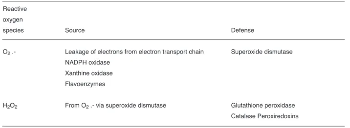
Molecular oxygen is critical for high organized body stuctures due to aerobic respiration. It is relatively not reactive but oxygen derivatives more prone to participate in chemical reactions are formed during aerobic metabolism and in the enviroment. One the main ROS is superoxide radical ion produced in vivo. Superoxide radical has both reducing and oxidizing properties (Table 1) (Bartosz, 2009). However, the other well known hydrogen peroxide radical is a weak oxidant attacking mainly thiols. Peroxynitrite is produced following reaction of superoxide with nitric oxide. Peroxynitrite is not a common one but it is a strong oxidizing, nitrating and nitrosylating agent. Hydroxyl radical is generated following hydrogen peroxide reactions with transition metals. Hydroxyl radical reacts rapidly and indiscriminately with biomolecules including nucleic acids, proteins, carbohydrates.
Mitochondria and
apoptosis: Mitoptosis
Apoptotic cell death is mainly placed in three different major places in the cells. One of them is plasma membrane which accepts the extracellular death signals and transfer these signals to the other important point, nucleus for cell death regulation. The
Reviews
Therapeutical potential of mitochondrial reactive oxygen
species in carcinogenesis
Advances in Molecular Biology (1-2): 1-8, 2009
İstanbul Kültür University, Printed in Turkey www.advmolbiol.org
SCorrespondence Author:
İstanbul Kültür University, Molecular Biology and Genetics Department 34156 Istanbul-Turkey
E-mail: [email protected]
last main place is mitochondria which are responsible for intrinsic cell death regulation in the presence of caspases and Bcl-2 family members. Following dysregulation of mitochondrial membrane potantial (MMP), cell death inducers are released into the cytosol and form a new complex structure, apoptosome to finalize the cleavage of cellular macromolecules in the cell.
Mitochondria are also referred as a garden or gatekeeper of cell death with dual function in survival and death decision. Mitochondria is a garden because pro-apoptotic and anti-apoptotic Bcl-2 family members reside on the membranes of mitochondria. The ratio between pro-apoptotic and anti-apoptotic Bcl-2 family members determine the cell fate as death or survival. Upon activation of Bcl-2 homology-3 (BH3) only proteins, pro-apoptotic multidomain members of the Bcl-2 family, fully insert into them mitochondrial outer membrane (MOMP), oligomerize and form protein-permeable channels which leads to MOMP permeabilization. It was suggested that MMP appears to be a more valuable marker for cell demise, than caspase activation which is induced by apoptotic stimuli or PARP cleavage, a last event for cell death or, DNA laddering. Genotoxic effects of DNA-damaging drugs, growth factor withdrawal or irradiation may increase MMP loss in the cells. However it is not clear whether mitochondria can act as enhancer of caspase activity, or downstream
caspases can contribute to cytochrome c release or apoptosis inducing factor (AIF) release and loss of MMP (Aouacheriaet al., 2007)
Mitoptosis is the term of self elimination of mitochondria in the cells and massive mitoptosis may result in apoptosis (Gogvadzeet al., 2009a). MMP is determined by two factors: outer membrane stability and inner membrane structure. Skommer et al., 2007 indicated that whether inner membrane partakes in outer membrane of mitochondria, a permeability transition pore (PTP) opens following by osmotic flux of water and molecules up to 1.5 kDa. Following equilibration of ions between the mitochondria matrix and cytoplasm, PTP complex consists of adenine nuclear translocator in inner membrane, cyclophilin D in the matrix and VDAC (voltage dependent anion channel) and peripheral benzodiazepin outer membrane. Continuous opening of PTP leads to MMP loss and osmotic swelling of the matrix, often sufficient to distort the structure of cristae and to rupture the outer mitochondrial membrane (Skommer et al., 2007).
In spite of the exact process accounting for MOMP, three discernible and not mutually exclusive mechanisms were proposed as below to understand how cell death process occurs (Figure 1) (Skommer et al., 2007):
Table 1. Endogenous sources of some radical oxygen species and scavenging strategies.
Reactive oxygen
species Source Defense
O2.- Leakage of electrons from electron transport chain Superoxide dismutase
NADPH oxidase Xanthine oxidase Flavoenzymes
H2O2 From O2.- via superoxide dismutase Glutathione peroxidase
1. Release of mitochondria-residing death factors that promote caspase-dependent (holocytochrome c, Smac/DIABLO (second mitochondrial apoptosis-activating protein) and Omi/HtrA2) or caspase independent (AIF (apoptosis inducing factor), endonuclease G and Omi/HtrA2) cell death. This mechanism still is not clear. There are controversial studies whether these proteins are co-released during apoptosis.
2. Impaired mitochondrial functions crucial for cell survival. Here, it is well established that Cyt c holds a fundamental role in cellular respiration, transfer of electrons from complex III to complex IV. Generally it is assumed that mitochondrial damage is the point of no return of death machinery; impaired mitochondrial apoptosis is an accumulating process and not all or nothing phenomenon. It was shown that lack of the respiratory functions during early apoptosis can be reversed when Cyt c is added.
3. Generation of ROS. Mitochondria are the place for generation of ROS which leads lipid peroxidation, calcium mobilization, mitochondrial permeability transition, ATP depletion, protein oxidation, loss of electron transport and/or DNA damages. Their over-existence than required concentration triggers cell defense and the last death
mechanism. Contrary, ROS can induce nuclear factor kappa B (NFκB) activation or inhibition of caspases to protect cells.
The mitochondrial cell death event is thought to be controlled by the relative balance of opposing Bcl-2 family members and their mutual interactions. However, despite extensive investigation, the precise biochemical mechanisms by which the three subgroups of Bcl-2 family members control mitochondrial apoptosis are still unclear (Skommeret al., 2007). BH3-only proteins function upstream of the multidomain proteins and act as sensors connecting multiple cytotoxic signals to the core apoptotic pathway. It was shown that Bax and Bak knock-out cells do not undergo MOMP and become resistant to various death stimuli. Mechanistically, Bax and Bak are thought to homooligomerize into pores in the MOM and cause Cyt c and other proteins release into cytosol (Viktorsson et al., 2005; Skommer et al., 2007). In healthy cells, Bak is held in an inactive and it is placed into MOM at monomeric state thorough its association with VDAC-2. However, Bax may lay dormant in the cytosol through interactions with several proteins, including Ku-70, 14-3-3 and humanin peptide (Guoet al., 2003; Sawada et al., 2003; Breckenridge and Xue, 2004; Tsurutaet al.,
Mitochondrial reactive oxygen species in carcinogenesis
Arısan and Ünsal 3
Figure 1. Schematic model of mitochondria reactive oxygen species in apoptosis. Various death signals can lead apoptosis and due
2004). Following apoptotic stimuli, BH3-only protein dependent translocation of Bax is induced and then placed into MOM. Bax and Bak homooligomers are formed here and change the intermembrane space proteins conformation. Bcl-2 and Bcl-xLare localized on mitochondria and antagonize the functions of Bax, Bak and other BH3-only proteins. It was shown that increased association between Bcl-2 and Bak is observed following binding of tBid to Bak. Hence, it is proposed that Bcl-2 may play a role in regulation of Bax and Bak oligomerization. It is probable that anti-apoptotic proteins regulate mitochondrial apoptosis not only via interaction with BH3-only proteins, but also through heterodimerization with Bax and Bak and prevention of their insertion or/and oligomerization within MOM. Following this action, mitochondria outer membrane permeabilized and intermembrane space proteins leak out. However, induction of intermembrane space proteins occur one step before membrane permeabilization and these proteins differs upon upstream check point signals (Breckenridge and Xue, 2004). Briefly, Bcl-2 family proteins are key regulators of the mitochondrial or intrinsic apoptotic pathway, including or preventing the release of Cyt c, AIF, Smac/DIABLO, Endonuclease G, translocates to the nucleus and causes oligonucleosomal DNA fragmentation, and Omi/HtrA2 that reside in the intermembrane space of mitochondria in healthy cells. Mitochondrial proteins cause caspase-dependent cell death include Cyt c
which triggers caspase-9 activation by binding and activating the Apaf-1, and Smac/DIABLO, which potentiates caspase activation by binding IAP and blocking their caspase-inhibitory activity. However, the existence of a redundant molecule or molecules capable of compensating for the loss of Smac function has been suggested by gene deletion studies (Adams and Cory, 2007; Skommeret al., 2007).
Mitochondria, cell death and
reactive oxygen species
Mitochondria are also an important center for reactive oxgen species (ROS) production within most mammalian cells. The bulk of mitochondrial ROS generation occurs at the electron transport chain (ETC), as a by-product of respiration. As a result, this generation can occur at relatively high rates compared to cytosolic ROS production and is primarily determined by metabolic conditions. This event contributes to mitochondrial damage in a range of pathologies and is also important in redox signalling from the organelle to the rest of the cell (Murphy, 2009). Recent knowledge about how mitochondria produce ROS is vital to understand a range of currently important biological and biomedial topics. The first report about ROS production due to respiration at mitochondria was shown in 1966 (Jensen, 1966). Following this discovery, isolated mitochondria produce H2O2 (Boveris and Chance,
1973; Chanceet al., 1979), it was confirmed that this H2O2arose from the dismutation of superoxide (O2•−) generated within mitochondria. The parallel discovery that mitochondria contain their own superoxide dismutase, MnSOD, confirmed the biological significance of mitochondrial O2•− production (Weisiger and Fridovich, 1973). Within mitochondria, O2•− is produced by the one-electron reduction of O2. Therefore it is the kinetic and thermodynamic factors underlying the interaction of potential one-electron donors with O2 that control mitochondrial ROS production (Murphy, 2009).
Intracellular generation of ROS per se is an inevitable (and sometimes physiologically important) process. To counter it, mitochondria, and cells in general, possess numerous ROS defense systems. It should have been implicit that the true source of oxidative stress is not the ROS generation per se but spatiotemporal imbalance of ROS production and detoxification, and, yet, until recently, even the capacity of mitochondrial ROS defence was unknown.
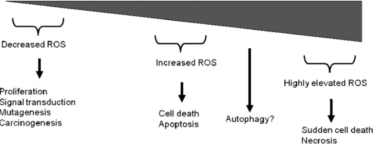
In fact ROS are not all accidental and unwanted byproducts. This cellular process orchestrate many critical events in the cells. One of the main event is the modulation of cell death decision. Mitochondria is one of the central place which regulate cell death and survival decision under the control of Bcl-2 family members. Excessive production of ROS is a typical sign of apoptosis (Figure 2). For these reason, it has been shown that the increased oxidative stress induced-cellular changes in cells following treatment of various cytotoxic drugs. Although, some chemotherapeutics such as taxanes or platinium derived agents have structurally dissimilarities, they acted on different cellular targets and induced oxidative stress by generation of ROS in cancer cells. It is proposed that increased ROS formation induce cytotoxic responses in these cells (Timuret al., 2005).
The sustained mitochondrial inhibition due to apoptotic induction would also potentiate ROS formation, leading to the depletion of antioxidant defenses (thiols such as gluthation) and initiation of lipid peroxidation. Lipid peroxidation could cause structural damage to membranes, including those
which form mitochondria, and potentiate their dysfunction. This sequential process can modulate several proteins and structures to finalize cell death decision. H2O2 generation which triggers ROS generation and lipid peroxidation was contributed to apoptosis induced by physiological concentrations of several drugs. Therefore, there are a number of studies supporting the hypothesis oxidative stress might modulate the cytotoxicity of several cytotoxic agents. However, the mechanisms by which ROS mediate the apoptotic effect of drugs and the identity of specific ROS involved in the process are unclear (Timuret al., 2005). It was shown that the regulation of anti-oxidant systems and specific contribution of ROS might be related to apoptotic machinary players such as Bcl-2 family members. Previous studies have shown that O2- might have role in Bcl-2 expressional regulation. Downregulation of superoxide dismutase decreases Bcl-2 expression and increases cisplatin-induced cell death (Ueta et al., 1999). Since superoxide dismutase acts by dismutation of O2•- to form H2O2 and since H2O2 is a major source of OH• production, i.e., via Fenton-like reactions, it is possible that other oxidative species may play a role. Similar these findings, it was proposed that H2O2 may be a common regulator of Bcl-2 expression under various pathological conditions. Since aberrant expression of Bcl-2 has been associated with several human cancers, and linked with drug-insensitive phenotype, ROS mediated Bcl-2 regulation might be important in the understanding of cancer prognosis (Wanget al., 2008).
Mitochondria is a target for
cancer chemotherapy
A wide array of human malignancies share molecular alterations that directly or indirectly linked to mitochondria. Since mitochondria occupy at a strategic position between bioenergetic/biosynthetic metabolism and cell death regulation, they are emerging as privileged targets for the development of novel chemotherapeutic agents. Some significant molecular determinants are used for efficient therapeutical tools which repair the impaired mitochondrial apoptotic events. Therefore mitochondriotoxic substances are designed on the
Mitochondrial reactive oxygen species in carcinogenesis
basis of molecular features of cancer cells (Galluzzi et al., 2009; Gogvadze et al., 2009a, b).
One of the main characteristics of cancer cells is rapid proliferation rate. This feature is required high maintaince of oxygen, but when the cell proliferation rate is high, oxygen consumption resulted as hypoxia. Hypoxia induced gene expression and regulation profile induce vascularization to maintain required oxygen. Low amount of oxygen decreases mitochondrial activity, for this reason cell activates survival signalling pathways (e.g. metabolic pathways). The insufficient activity of mitochondria to provide enough energy for cell survival under hypoxia conditions, results in elevated glycolysis ratio which leads to induced-hypoxia-inducing factor (HIF-1). As mentioned before by Godvadze et. al., (2009) HIF-1 expression regulates the regulation of vascularization, survival mechanism of the cells and invasion. HIF-1 was shown not only induce the metabolic activation of cells by utilization of glycolysis, HIF-1 supresses mitochondrial functions of the cells.
Recent studies about mitochondria targetting therapeutic tools, such as small molecules are generally related with Bcl-2 family members which reside generally on the mitochondria membranes. Anti-apoptotic Bcl-2 family members are overexpressed in the majority of cancer cases. Furthermore overexpression of anti-apoptotic Bcl-2 family members may also inhibit the MOMP potential due to inactivation of pore-forming Bax and Bak. Therefore targetting these proteins in the cancer therapy gains more importance everyday (Skommer et al., 2007).
Another key characteristic feature of cancer cells is an elevated level of ROS. ROS are generated continously in the cells and play important roles in a variety of processes. Normally, about 2 % mitochondrially consumed oxygen is converted into the superoxide anion. However this situtation is controlled by many kinds of enzymes and water or lipid soluble antioxidants. In some cases, ROS generation can not be controlled by these mechanisms and excess ROS amount can lead to
deleterious mutations which induce carcinogenesis or abnormal metabolic processes (Chanceet al., 1979; Aouacheria et al., 2007; Skommer et al., 2007; Bartosz, 2009; Galluzziet al., 2009; Gogvadze et al., 2009a, b)
A number of chemotherapeutics was shown to be induced anti-tumoral activity via ROS-mediated cell killing properties. It is proposed that elevated levels of ROS induction may induce some signalling signature molecules and regulate the impaired apoptotic machinary. Here the most interesting point is that antioxidant defence molecules such as mitochondria residing superoxide dismutases can overcome destroying effect of ROS in the cells, therapeutical agents may decrease the transcriptional level of antioxidant players. However, studies are controversial to underlie the molecular mechanism of antioxidant players in the apoptotic function (Gogvadze et al., 2009a). Therefore, it should be highlighted that the efficiency of cancer therapy might be regulated by the modulation of redox potential of cells following chemotherapeutics application. Furthermore, in order to reveal the therapeutic efficiency of any molecule, molecular basis of regulation of antioxidant players or their suppression by any co-stimuli in the cancer progression should be investigated.
In conclusion, major steps of the cell death mechanism is orchestrated in mitochondria. Mitochondria is the one of main ROS production place due to respiration responsibilities in the cells. It should be highlighted that the identification of detailed information about molecular basis of ROS may provide more successful therapeutical tools in cancer therapy.
References
Adams JM and Cory S. Bcl-2-regulated apoptosis: mechanism and therapeutic potential. Curr Opin Immunol. 19(5): 488-496, 2007.
Aouacheria A, Cibiel A, Guillemin Y, Gillet G and Lalle P. Modulating mitochondria-mediated apoptotic cell death through targeting of Bcl-2 family proteins. Recent Pat DNA Gene Seq. 1(1): 43-61, 2007.
Bartosz G. Reactive oxygen species: destroyers or messengers? Biochem Pharmacol. 77(8): 1303-1315, 2009.
Boveris A and Chance B. The mitochondrial generation of hydrogen peroxide. General properties and effect of hyperbaric oxygen. Biochem J. 134(3): 707-716, 1973.
Breckenridge DG and Xue D. Regulation of mitochondrial membrane permeabilization by BCL-2 family proteins and caspases. Curr Opin Cell Biol. 16(6): 647-652, 2004.
Chance B, Sies H and Boveris A. Hydroperoxide metabolism in mammalian organs. Physiol Rev. 59(3): 527-605, 1979.
Galluzzi L, Morselli E, Kepp O, Vitale I, Rigoni A, Vacchelli E, Michaud M, Zischka H, Castedo M and Kroemer G. Mitochondrial gateways to cancer. Mol Aspects Med, 2009.
Gogvadze V, Orrenius S and Zhivotovsky B. Mitochondria as targets for cancer chemotherapy. Semin Cancer Biol. 19(1): 57-66, 2009a.
Gogvadze V, Orrenius S and Zhivotovsky B. Mitochondria as targets for chemotherapy. Apoptosis. 14(4): 624-640, 2009b.
Guo B, Zhai D, Cabezas E, Welsh K, Nouraini S, Satterthwait AC and Reed JC. Humanin peptide suppresses apoptosis by interfering with Bax activation. Nature. 423(6938): 456-461, 2003. Jensen PK. Antimycin-insensitive oxidation of succinate
and reduced nicotinamide-adenine dinucleotide in electron-transport particles. I. pH dependency and hydrogen peroxide formation. Biochim Biophys Acta. 122(2): 157-166, 1966.
Murphy MP. How mitochondria produce reactive oxygen species. Biochem J. 417(1): 1-13, 2009.
Sawada M, Sun W, Hayes P, Leskov K, Boothman DA and Matsuyama S. Ku70 suppresses the apoptotic translocation of Bax to mitochondria. Nat Cell Biol. 5(4): 320-329, 2003.
Skommer J, Wlodkowic D and Deptala A. Larger than life: Mitochondria and the Bcl-2 family. Leuk Res. 31(3): 277-286, 2007.
Mitochondrial reactive oxygen species in carcinogenesis
Timur M, Akbas SH and Ozben T. The effect of Topotecan on oxidative stress in MCF-7 human breast cancer cell line. Acta Biochim Pol. 52(4): 897-902, 2005.
Tsuruta F, Sunayama J, Mori Y, Hattori S, Shimizu S, Tsujimoto Y, Yoshioka K, Masuyama N and Gotoh Y. JNK promotes Bax translocation to mitochondria through phosphorylation of 14-3-3 proteins. Embo J. 23(8): 1889-1899, 2004.
Ueta E, Yoneda K, Yamamoto T and Osaki T. Manganese superoxide dismutase negatively regulates the induction of apoptosis by 5-fluorouracil, peplomycin and gamma-rays in squamous cell carcinoma cells. Jpn J Cancer Res. 90(5): 555-564, 1999.
Viktorsson K, Lewensohn R and Zhivotovsky B. Apoptotic pathways and therapy resistance in human malignancies. Adv Cancer Res. 94: 143-196, 2005. Wang L, Chanvorachote P, Toledo D, Stehlik C, Mercer
RR, Castranova V and Rojanasakul Y. Peroxide is a key mediator of Bcl-2 down-regulation and apoptosis induction by cisplatin in human lung cancer cells. Mol Pharmacol. 73(1): 119-127, 2008.
Weisiger RA and Fridovich I. Superoxide dismutase. Organelle specificity. J Biol Chem. 248(10): 3582-3592, 1973.