Abstract
In this study, the specific polymerase chain reaction has been standardized and evaluated for the direct diagnosis of Brucella canis in vaginal swab samples from dogs. The specific primer sets are directed to the 16S-23S rRNA inter-space region of Brucella spp. and the deletion of 351 bp in BMEI1426-BMEI1427 in B. canis. A total of 21 references and field strains and 35 vaginal swab samples were used for the evaluation of the polymerase chain reaction. It found that polymerase chain reaction is positive for B. canis DNA indicated by only amplification of 214 bp product. It detected at least 2.7 x 101 CFU/g of bacteria diluted in vaginal swab samples indicates that the polymerase chain reaction can be used as a practical alternative for bacterial isolation. The novel polymerase chain reaction provides a simple and rapid for the detection of B. canis in clinical and field samples in one step and in short time about 24 h.
Keywords: Specific PCR, Brucella canis, Vaginal swab samples
Köpeklerde Brucella canis’in Teşhisi Amacıyla Yeni Polimeraz
Zincir Reaksiyonu Metodunun Geliştirilmesi
Özet
Bu çalışmada, köpeklerin vajinal sıvap örneklerinden Brucella canis (B. canis)’in direk teşhisi amacıyla spesifik polimeraz zincir reaksiyonu metodu standardize edildi ve değerlendirildi. Brucella spp.’in 16S-23S inter-space bölgesine ve B. canis’in BMEI1426-BMEI1427 gen bölgesinde ki 351 bp’lik delesyon alanına özgü primer çiftleri seçildi. polimeraz zincir reaksiyonu yönteminin değerlendirilmesinde toplam 21 referans ve saha suşu ile 35 vajinal sıvap örneği kullanıldı. Sadece 214 bp’lik amplifiye polimeraz zincir reaksiyonu ürününün görüntülenmesi B. canis için pozitif olarak kabul edildi. Vajinal sıvap örneklerinden en az DNA tespit limiti yeni metot için 2.7 x 101 CFU/g bakteri olarak bulundu. Bu yöntem bakteriyel izolasyon metoduna pratik bir alternatif olabilir. Yeni polimeraz zincir reaksiyonu testi B. canis’in klinik veya saha örneklerinden 24 saat sürede ve tek adımda hızlı ve basit tespitini sağlamaktadır.
Anahtar sözcükler: Spesifik PZR, Brucella canis, Vajinal sıvap örneği
A Novel Polymerase Chain Reaction to Detect
Brucella canis in Dogs
Zeki ARAS
1
Mehmet TAŞPINAR
2İbrahim AYDIN
31 2 3
Department of Microbiology, Faculty of Veterinary Medicine, Aksaray University, TR-68100, Campus, Aksaray - TURKEY Department of Medical Biology, Faculty of Medicine, Yuzuncu Yil University, TR-65080 Campus, Van - TURKEY
Department of Obstetrics and Gynaecology, Faculty of Veterinary Medicine, Selçuk University, TR-42075 Campus Konya - TURKEY
INTRODUCTION
Brucella (B.) canis is main etiologic agent of canine
brucellosis and induces various reproductive failures in dogs and in human. It was first isolated from dogs in 1966 by Leland Carmichael. Canine brucellosis is an important disease because it causes the great economic losses in commercial breeding kennels and threats public health. The other smooth Brucella species (B. abortus and B.
melitensis) have occasionally been isolated from dogs in
various regions of the world [1]. Canine brucellosis is found
most of the world but Australia and New Zealand appear
to be free [1] and the global prevalence of the disease is
unknown [2]. Normally, B. canis infections in humans have
been seen through either laboratory accidents or contact with positive dogs [1,3,4].
The main clinical findings of canine brucellosis are reproductive failure such as late abortion, birth of weak offspring, epididymitis, orchitis, and testicular atrophy. Lymphadenitis is a common finding in both sexes, affecting most lymph nodes in the body [1,5]. The only definitive
method for diagnosis of canine brucellosis is based on the isolation of B. canis from various clinical samples [3,6,7].
İletişim (Correspondence) +90 382 2882941
[email protected]
KafKas Universitesi veteriner faKUltesi Dergisi JoUrnal Home-Page: http://vetdergi.kafkas.edu.tr
online sUbmission: http://vetdergikafkas.org
Research Article
Kafkas Univ Vet Fak Derg 21 (2): 169-172, 2015
DOI: 10.9775/kvfd.2014.11977
170
A Novel Polymerase Chain ...
However, microbiological culture method has some dis-advantages such as it is time consuming, includes complex tests, requires skilled personnel and is hazardous for laboratory workers [1,6,7]. Generally, serology can be used
for diagnosis of infection but cross-reactions between B.
canis and other bacteria can occur in some serological
tests. Nonspecific agglutination reactions also cause false- positive results in some dogs [4,8,9].
In fact, the polymerase chain reaction (PCR) is a rapid, very specific, highly sensitive, and inexpensive technique for Brucella DNA detection. Hence, it is an alternative to bacteriological isolation for direct diagnosis of canine brucellosis [10,11]. Several studies were carried out in recent
years in order to standardize PCR assays for the detection of B. canis DNA in various clinical samples including canine blood, semen, vaginal swab, blood serum, lymphoid tissue [11-16]. However, the PCR assays were used Brucella
genus-specific primer pairs that were directed to the 16S-23S rRNA interspace and/or the virB2 gene regions of Brucella spp. The Bruce-ladder multiplex PCR assay has been developed as a rapid and one-step molecular test for identification, typing of Brucella species and enhancing to distinguish between B. suis and B. canis [17,18]. However,
this method is rarely used for direct detection of Brucella species DNA in clinical samples, because it was designed for bacterial isolates [19]. It is recently reported that,
Bruce-ladder multiplex assay does not yield ideal results when DNA is extracted directly from clinical samples because of uneven amplification pattern [19].
With the above consideration in mind, the aim of this study is to use the novel PCR assay to detect B. canis in pure bacterial culture and vaginal swab samples, for the first time. The primers are directed to the 16S-23S rRNA inter-space region of Brucella spp. and the deletion of 351 bp in BMEI1426-BMEI1427 in B. canis [15,20]. It proved that the
PCR assay discriminates B. canis from other Brucella species and also detects the other Brucella species as Brucella spp.
MATERIAL and METHODS
Bacterial Strains
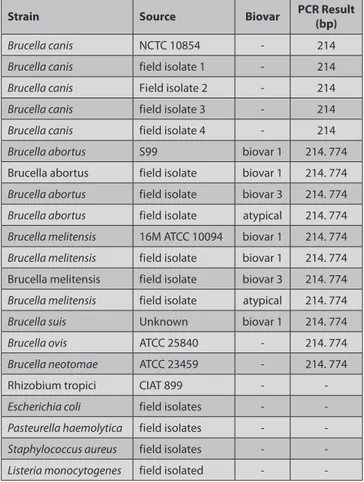
Strains examined in this study are lisited in Table 1.
Brucella isolates were classified according to standard
microbiological procedures, as described by Alton et al.[3].
Dogs
Vaginal swab samples were collected from 35 bitches from the city’s pound of Konya Province of west-central Turkey in the years 2012. Breeds included Boxer (2), Golden Retriever (1), Kangal (3), and Mongrel (29). The ages of the dogs were in the range of 1 to 10 years. Clinical signs that suggested canine brucellosis were investigated in these dogs by questionnaire forms that were obtained from the Veterinarian of the city pound. These data stated that
generalized lymphadenitis, metritis, vaginal discharge, still-birth, osteomyelitis, uveitis, lethargy, decreased appetite, weight loss, and hyperthermia were seen and some of the dogs were reported to have received antibiotics.
Vaginal swab samples were collected in duplicate from the 35 bitches by sterile swabs. One of the swab samples was placed in a tube containing 2 mL of Brucella Broth (Sigma, B3051, MO, USA) for bacterial isolation. The other sample was put into a tube containing 2 mL of TE buffer (10mM Tris-HCl pH 8.0, 1mM disodium EDTA pH 8.0) and was kept at -20°C until used for the PCR assay.
Vaginal swab samples from three dogs were contaminated with B. canis NCTC 10854 (supplied by Refik Saydam Hifzissihha Institute, Ankara, Turkey) to determine the detection limit of the PCR assay. The dogs were previously diagnosed as negative to canine brucellosis by clinical, serological and culture examinations. Tested samples were transported to the laboratory under cool condition. The study protocol was approved by Selcuk University Veterinary Faculty Ethical Committee (2007/24).
Bacteriological Examinations
Samples were processed using the method described by Alton et al.[3]. Vaginal swab sample was immediately
cultured onto Blood Agar Base (Oxoid, CM0271, Hampshire, Table 1. Strains examined in this study and PCR pattern
Tablo 1. Bu çalışmada kullanılan suşlar ve elde edilen PZR bantları
Strain Source Biovar PCR Result (bp)
Brucella canis NCTC 10854 - 214
Brucella canis field isolate 1 - 214
Brucella canis Field isolate 2 - 214
Brucella canis field isolate 3 - 214
Brucella canis field isolate 4 - 214
Brucella abortus S99 biovar 1 214. 774
Brucella abortus field isolate biovar 1 214. 774
Brucella abortus field isolate biovar 3 214. 774
Brucella abortus field isolate atypical 214. 774
Brucella melitensis 16M ATCC 10094 biovar 1 214. 774
Brucella melitensis field isolate biovar 1 214. 774 Brucella melitensis field isolate biovar 3 214. 774
Brucella melitensis field isolate atypical 214. 774
Brucella suis Unknown biovar 1 214. 774
Brucella ovis ATCC 25840 - 214. 774
Brucella neotomae ATCC 23459 - 214. 774
Rhizobium tropici CIAT 899 -
-Escherichia coli field isolates -
-Pasteurella haemolytica field isolates -
-Staphylococcus aureus field isolates -
-171 ARAS, TAŞPINAR AYDIN UK) plates containing 5% defibrinated sheep blood with
an antibiotic mixture. The cultures were incubated at 37°C under aerobic conditions for 7 days. B. canis were identified by morphological and biochemical characteristics.
DNA Extraction
First, reference and field strains were grown in Brain Heart Infusion Broth (Oxoid, CM225, Hampshire, UK). Bacteria were killed by addition of 0.5% formaldehyde. After that DNA was extracted using the protocol provided in Promega Wizard Genomic DNA purification Kit (Promega, A1120, WI, USA). DNA concentration was determined spectrophotometrically (Eppendorf, Model 6131, Germany) by absorbance readings in the range of 260 to 280 nm.
B. canis DNA from vaginal swab samples was extracted
using the protocol reported by Leal-Klevezas et al.[21]. Two
mL of Tris EDTA (TE) buffer containing swab samples was used for this purpose. The DNA samples were kept at -20°C until used as templates for amplification.
PCR Assay
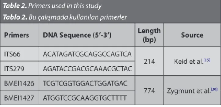
The two primer sets of the 16S-23S rRNA inter-space region of Brucella spp.[15] and the deletion of 351 bp in
BMEI1426-BMEI1427 in B. canis [20] used in this study are
recorded in Table 2.
The amplification reaction mixture was prepared in a total volume of 50 µL containing 5 µL of 10 x PCR buffer, 250 µM each of the four dNTPs (Fermentas, Vilnius, Lithuania), 1.5 mM MgCl2, 1.5 U of Taq DNA polymerase
(Fermentas, Vilnius, Lithuania), 0.5 µM of each primer (IDT, USA) and 5 µL of template DNA. The amplifications were performed in a thermal cycler (Ependorf, Mastercycler
gradient, Germany) with the following steps: 1 × 7 min at
95ºC, 30 × 45 s at 95ºC, 45 s at 63ºC, 120 s at 72ºC, and a final extension at 72ºC for 5 min. DNA extracted from
B. canis NCTC 10854 and nuclease free water was served
as positive and negative controls, respectively. The PCR product (10 µL) was further analyzed by electrophoresis on 2% agarose gel, and the gel was stained with ethidium bromide (1.5 µg/mL) and photographed. Reactions were considered positive for B. canis when they yielded unique PCR product of 214 bp but products of 214 and 774 bp were accepted positive for the other Brucella species.
Determination of Detection Limit of the PCR
Detection limit of the PCR assay was evaluated using B.
canis NCTC 10854 reference strain. The suspension of the
72 h culture of B. canis was prepared in sterile saline and 10-fold dilutions (10-1 to 10-10) for determine of
colony-forming unit (CFU) and concentration of undiluted B. canis culture calculated as 2.1 x 10⁷ CFU/mL by microbiological
culture method. To determine the sensitivity of the assay, decreasing numbers of B. canis culture (methanol killed) were added to the 1 mL of the vaginal swab samples (obtained by three number of non-infected bitches). The final concentrations of B. canis in mixture were 2.1 x 10⁷,
2.1 x 10⁶, 2.1 x 10⁵, 2.1 x 10⁴, 2.1 x 10³, 2.1 x 10² and 2.1 x 101
CFU/g. DNA extraction was performed with all dilutions of mixture, as described previously [21]. Then DNA samples
processed by PCR assay as described above.
RESULTS
The PCR assay was evaluated with 21 reference and field isolates (Table 1). All B. canis strains exhibited unique PCR band of 214 bp. However, other Brucella species such as B. abortus, B. melitensis, B.ovis, B. suis and B. neotomae showed 2 bands of 214 and 774 bp. No PCR products were amplified with DNAs from bacteria genetically related to
Brucella such as Rhizobium tropici and other non-Brucella
organism commonly associated with animals (Table 1). To determine the analytical sensitivity of the assay, decreasing numbers of B. canis were added to the vaginal swab samples. A positive PCR product for B. canis always achieved with different amounts containing at least 2.7 x 101 CFU/g of vaginal swab samples. The limit of PCR
detection of B. canis was determined to be 2.7 x 101 CFU/g
at least.
A total of 35 vaginal swab DNA samples were tested by the novel PCR assay. Namely, B. canis DNA was not detected from any vaginal swab samples in addition to that the samples were bacteriological negative.
DISCUSSION
Microbiological culture method and serological tests are widely used for diagnosis of canine brucellosis. The isolation of agent from different tissues of dogs is still considered as the gold standard for the definitive diagnosis of infection [3,7,22,23]. These methods have some
disadvantages, while the PCR assay is fast, simple, highly sensitive and specific for detection of B. canis [11,15]. A very
specific, highly sensitive and reliable diagnostic PCR assay for B. canis is very important for controlling the spread of infection in animal population and public health. In the present study, a species-specific PCR assay was designed and evaluated for detection and differentiation of B. canis in vaginal swab samples.
Table 2. Primers used in this study Tablo 2. Bu çalışmada kullanılan primerler
Primers DNA Sequence (5’-3’) Length (bp) Source
ITS66 ACATAGATCGCAGGCCAGTCA 214 Keid et al.[15] ITS279 AGATACCGACGCAAACGCTAC BMEI1426 TCGTCGGTGGACTGGATGAC 774 Zygmunt et al.[20] BMEI1427 ATGGTCCGCAAGGTGCTTTT
172
A Novel Polymerase Chain ...
The primers that used in the novel PCR assay were directed to the 16S-23S rRNA inter-space region of Brucella spp. and the deletion of 351 bp in BMEI1426-BMEI1427 in B. canis [15,20]. This PCR assay discriminated B. canis from
other Brucella species in a single reaction. The specificity of the species-specific PCR assay was explored with bacteria related to Brucella including Rhizobium tropici, Escherichia
coli, Pasteurella haemolytica, Staphylococcus aureus and Listeria monocytogenes (Table 1) and it demonstrated a
remarkable good specificity. However, B. canis is main etiological agent of canine brucellosis but the other smooth Brucella species (B. abortus and B. melitensis) have occasionally been isolated from dogs [1]. A major advantage
of the present assay is that it can directly identify B. canis at species level and also detected all other Brucella species as Brucella spp. in clinical samples.
In our study, decreasing numbers of B. canis were added to the vaginal swab samples to determine the analytical sensitivity of the assay. The novel PCR assay detected at least 2.7 x 101 CFU/g of bacteria diluted in vaginal swab
sample. This good analytical sensitivity agreed with the analytical sensitivity of Brucella genus-specific PCR assay from vaginal swab samples as previously described [14].
The degree of sensitivity of PCR assay is a key issue for its effective use in detection of brucellosis [24]. In our
study, to detect the sensitivity and specificity of PCR assay, a total of 35 vaginal swab samples were tested with the PCR and culture methods. All samples were found negative for B. canis by two methods and in a full consistence with data between the PCR assay and culture method, the gold standard method for direct diagnosis of B. canis.
Finally, we concluded that, the novel species-specific PCR assay has been developed for the direct diagnosis of B.
canis in vaginal swab samples from dogs. It was proved that
the PCR is highly specific and sensitive for detection of B.
canis. This technique open a new gate to detect alternative
for bacterial isolation. We strongly believe that the PCR assay provides a simple and rapid tool for detection of
B. canis in one step and one day. It is suitable for routine
diagnosis of this disease and can be used for confirmation of B. canis cultures.
REFERENCES
1. Wanke MM: Canine brucellosis. Anim Reprod Sci, 82-83, 195-207, 2004. 2. Corbel M: Brucellosis: An overview. Emerg Infect Dis, 3, 213-221, 1997. 3. Alton GG, Jones LM, Angus RD, Verger JM: Techniques for the
brucellosis laboratory. 149-153, INRA, Paris, 1988.
4. Köylü Ö, Aras Z, Uçan US: Seroprevalance of Brucella canis infection in a
risky human population in province Konya. Turkish J Infect, 23, 73-77, 2009.
5. Carmichael LE, Kenney RM: Canine abortion caused by Brucella canis.
J Am Vet Med Assoc, 152, 605-616, 1968.
6. Aras Z, Uçan US, Ateş M: Identification and biotyping of Brucella
strains. Eurasian J Vet Sci, 25, 51-59, 2009.
7. Aras Z, Uçan US: Diagnosis of Brucella canis by Polymerase Chain
Reaction. In, DeGiovine VM (Ed): Dogs: Biology, Behavior and Health
Disorders. 201-218, Nova Science Publishers, New York, 2011.
8. Lucero NE, Escobar GI, Ayala SM, Lopez G: Sensitivity and specificity
of an indirect enzyme-linked immunoassay for the diagnosis of Brucella
canis infection in dogs. J Med Microbiol, 51, 656-660, 2002.
9. Uçan US, Aras Z, Zorlutuna M: Detection of canine brucellosis by a
rapid agglutination test using Rhizobium tropici as antigen. Revue Med
Vet, 161, 51-56, 2010.
10. Bricker BJ: PCR as a diagnostic tool for brucellosis. Vet Microbiol,
90, 435-446, 2002. DOI: 10.1016/S0378-1135(02)00228-6
11. Aras Z, Uçan US: Detection of Brucella canis from inguinal lymph nodes
of naturally infected dogs by PCR. Theriogenology, 74, 658-662, 2010. DOI: 10.1016/j.theriogenology.2010.03.023
12. Kim S, Lee DS, Suzuki H, Watari M: Detection of Brucella canis
and Leptospira interrogans in canine semen by multiplex nested PCR. J
Vet Med Sci, 68, 615-618, 2006.
13. Keid LB, Soares RM, Vasconcellos SA, Chiebao DP, Megid J, Salgado VR, Richtzenhain LJ: A polymerase chain reaction for the detection
of Brucella canis in semen of naturally infected dogs. Theriogenology, 67, 1203-1210, 2007. DOI: 10.1016/j.theriogenology.2007.01.003
14. Keid LB, Soares RM, Vasconcellos SA, Chiebao DP, Salgado VR, Megid J, Richtzenhain LJ: A polymerase chain reaction for detection of
Brucella canis in vaginal swabs of naturally infected bitches. Theriogenology,
68, 1260-1270, 2007. DOI: 10.1016/j.theriogenology.2007.08.021
15. Keid LB, Soares RM, Vieira NR, Megid J, Salgado VR, Vasconcellos SA, da Costa M, Gregori F, Richtzenhain LJ: Diagnosis of canine brucellosis:
Comparison between serological and microbiological tests and a PCR based on primers to 16S-23S rDNA interspacer. Vet Res Commun, 31, 951-965, 2007.
16. Keid LB, Soares RM, Vasconcellos SA, Salgado VR, Megid J, Richtzenhain LJ: Comparison of a PCR assay in whole blood and serum
specimens for canine brucellosis diagnosis. Vet Rec, 167, 96-99, 2010. DOI: 10.1136/vr.c3811
17. Garcia-Yoldi D, Marin CM, de Miguel MJ, Munoz PM, Vizmanos JL, Lopez-Goni I: Multiplex PCR assay for the identification and
differentiation of all Brucella species and the vaccine strains Brucella
abortus S19 and RB51 and Brucella melitensis Rev.1. Clin Chem, 52,
779-781, 2006. DOI: 10.1373/clinchem.2005.062596
18. Lopez-Goni I, Garcia-Yoldi D, Marin MC, De Miguel MJ, Barquero-Calvo E, Guzman-Verri C, Albert D, Garin-Bastuji B: New
Bruce-ladder multiplex PCR assay for the biovar typing of Brucella suis and the discrimination of Brucella suis and Brucella canis. Vet Microbiol, 154, 152-155, 2011. DOI: 10.1016/j.vetmic.2011.06.035
19. Sánchez-Jiménez MM, Ortiz-Román LF, Castrillón-Salazar LL, Giraldo-Echeverri CA, Olivera-Angel M: Application of a polymerase
chain reaction test for the detection of Brucella canis from clinical samples of canines and humans. Rev Colom Cienc Pecua, 27, 3-11, 2014.
20. Zygmunt MS, Blasco JM, Letesson JJ, Cloeckaert A, Moriyon I:
DNA polymorphism analysis of Brucella lipopolysaccharide genes reveals marked differences in O-polysaccharide biosynthetic genes between smooth and rough Brucella species and novel species-specific markers.
BMC Microbiol, 13, 92-95, 2009. DOI: 10.1186/1471-2180-9-92
21. Leal-Klevezas DS, Martinez-Vazquez IO, Lopez-Merino A, Martinez- Soriano JP: Single-step PCR for detection of Brucella spp. from blood
and milk of infected animals. J Clin Microbiol, 33, 3087-3090, 1995.
22. Büyük F, Şahin M: Investigation of Brucella species from various
samples of aborted cattle in Kars province (Turkey) by cultural and molecular methods and epidemiological analysis of cases. Kafkas Univ
Vet Fak Derg, 17, 809-816, 2011.
23. Erdenliğ Gürbilek S, Baklan EA, Aksoy HY, Stack J: Comparative
production of Rapid Slide Agglutination Test (RSAT) antigen used in serological diagnosis of Brucella canis in different culture media in the fermenter. Kafkas Univ Vet Fak Derg, 20, 273-278, 2014. DOI: 10.9775/ kvfd.2013.10019
24. Mukherjee F, Jain J, Patel V, Nair M: Multiple genus-specific markers
in PCR assays improve the specificity and sensitivity of diagnosis of brucellosis in field animals. J Med Microbiol, 56, 1309-1316, 2007. DOI: 10.1099/jmm.0.47160-0