Long-term antibacterial properties of fluoride-releasing orthodontic bonding adhesives
DDS,aSertaç Aksakalli, DDSc
aInönü University, Faculty of Dentistry, Department of Orthodontics , Malatya.
bSelçuk University, Faculty of Selçuklu Medicine, Department of Microbiology, Konya. cSelçuk University, Faculty of Dentistry, Department of Orthodontics , Konya.
Received: 27 September 2011 Accepted: 25 October 2011
ABSTRACT
Objectives: The aim of this study was to compare antibacterial properties of 3 different fluoride-releasing
orthodontic adhesives during three months by using the direct contact test (DCT).
Materials and Methods: The materials tested in the present study were Transbond Plus (3M Unitek, Monrovia,
USA), Light Bond (Reliance Ortho Prod. Inc. Itasca, IL, USA), and Kurasper F (Kuraray Medical Inc., Okayama, Japan). Eight specimens of each material type were prepared for estimation. For the DCT, wells of 96-microtitre plates were coated with each of three bonding adhesives. Eight uncoated wells, using identical inocula size, served as a positive control (bacteria with medium). A Streptococcus mutans suspension was placed on the surface of each specimen that was held for 1 hour at 37C. Bacterial growth was monitored for 16 hours with a temperature-controlled microplate spectrophotometer. The kinetics of the growth in each well was recorded continuously at 650 nm in every 30 minutes. Three additional measurements were performed on these tested materials after aging for 1 week, 1 month and 3 months. One-way ANOVA and Tamhane’s T2 multiple comparison tests were applied to the data. The level of significance was set as p<0.05.
Results: The DCT showed that there were significant differences found in freshly mixed samples between four
groups (p<0.001, F=20.901). Freshly mixed samples of Transbond Plus showed antibacterial activity (p<0.001). Kurasper F and Light Bond did not differ from positive control. In the measurements of aged specimens, all materials showed an increase in the logarithmic growth rate of Streptococcus mutans.
Conclusion: Only Transbond Plus showed antibacterial properties in freshly mixed specimens. However, none
of the tested orthodontics composites showed antibacterial properties after aging.
Key Words: Antibacterial agents, direct contact test, fluorides, orthodontic adhesives, streptococcus mutans.
---INTRODUCTION
Orthodontic bands and brackets are highly susceptible to biofilm formation that threatens the integrity of the teeth and the soft tissues by means of decalcification and periodontal disease. Professional tooth cleaning, local application of fluorides and use of antimicrobial mouth rinses are basic strategies to prevent the clinical side-effects of the fixed orthodontic treatment.1,2 Enamel decalcification can
---Firat OZTURK
İnönü Universitesi, Dişhekimliği Fakültesi Ortodonti AD
44280, Malatya, Türkiye Tel: +90 422 3410106 Fax: +90 422 3410108 e-mail: [email protected]
also be reduced using sealants.3
Streptococcus mutans is considered to be
the primary organisms responsible for enamel demineralization. There is a significant increase in the salivary and plaque levels of these acidogenic, aciduric bacteria in patients undergoing fixed appliance treatment.4-8 Studies have shown that decalcification is a significant risk in orthodontic patients with rates reported from 15% to 75%.9
Because none of the preventive strategies have the potential to inhibit bacterial adhesion on the bracket surfaces, incipient carious lesions have been demonstrated in vivo around the brackets after only 4 weeks.10-12 The results of decalcification vary from no evident
change to white spots on the enamel of teeth, or even cavitation. They appear as unsightly lesions on previously sound teeth at the end of orthodontic treatment.
Fluoride is the most anticariogenic agent known to date.12 A variety of mechanisms are involved in the anticariogenic effects of fluoride, including the inhibition of demineralization, the enhancement of remineralization, the prevention of pellicle and plaque formation, and the inhibition of microbial growth and metabolism.13
Researches have recommended that topical fluorides are likely to decrease decalcification during active orthodontic phase.14,15 One method of fluoride application is to incorporate it into the adhesive.14 Fluoride-releasing bonding materials not only decrease decalcification, but also stimulate the development of a calcium fluoride layer on enamel surfaces adjacent to the brackets. This layer has a potential reserve to release fluoride ions slowly during the demineralization and remineralization processes. It also serves as a barrier against acid challenge.16,17
Resin composites may contain fluoride in a variety of forms, such as inorganic salts, leachable glasses or organic fluoride. Thereby, not only the amount of fluoride, but also the type and particle size of the fluoridated filler, the type of resin, silane treatment and porosity might be important factors contributing to fluoride release.18,19 The direct contact test (DCT) quantitatively measures the effect of direct and close contact between the test microorganism and the tested materials, regardless of the solubility and diffusibility of their components.20
Some studies21-24 have presented the antibacterial properties of different resin materials. There have been no reports that investigated the long-term antibacterial
orthodontic adhesives by using the DCT during 3-month aging period.
MATERIALS AND METHODS The fluoride-releasing adhesives used in this study are shown in Table 1.
Streptococcus mutans (Refik Saydam
National Public Health, 676, Ankara,Turkey) was grown aerobically to late logarithmic or early stationary phase from frozen stock cultures in brain hearth infusion (BHI) broth containing 0.5% bacitracin for 48 hours at 37ºC before applying it to the specimens according to the experimental design.
Direct Contact Test (DCT)
The DCT20 is based on the turbidometric determination of bacterial growth in 96-well microtitre plates (96-well, flat-bottom Nunclon; Nunc, Copenhagen, Denmark). The kinetics of the outgrowth in each well was recorded continuously at 650 nm in every 30 minutes, using a temperature-controlled spectrophotometer (µquant, Bio-Tek Instruments Inc., Winooski VT, USA).
In all wells, the sidewall was coated with evenly measured amount of tested materials [Transbond Plus (3M Unitek, Monrovia, USA), Light Bond (Reliance Ortho Prod. Inc. Itasca, IL, USA) and Kurasper F (Kuraray Medical Inc., Okayama, Japan)] while the plate was held vertically. Light-cured primers were brushed on and applied to the sidewalls of the wells. A thin film thickness was provided with care. To ensure a steady surface area, a thin coat adhesive was applied with a micro-brush. The test samples were light polymerized due to the manufacturers’ recommendations. Special care was taken to avoid the material flow to the bottom of the well, which would interfere with the light path through the
position. After incubation for 1 hour in a humid atmosphere at 37°C, evaporation of the liquid was evident and direct contact between bacteria and the tested material surfaces ensured. Then, 220 µL of BHI broth was added to each of the wells and the plate was placed in the spectrophotometer. The bacterial
outgrowth was estimated after direct contact with the tested material on the basis of the changes in the readings of optical density at 650 nm, which were recorded automatically by the spectrophotometer every 30 minutes for 16 hours.
Table 1. Orthodontic composites used in this study.
Brand Components Chemical Composition Manufacturer Lot
number Light Bond
Light bond Bisphenol A Diglycidylmethacrylate. Urethane Dimethacrylate,
Triethyleneglycol Dimethacrylate, Hydrofluoride Methacrylate
Reliance Ortho Prod. Inc., Itasca, IL, USA
0911076
Paste Bisphenol A Diglycidylmethacrylate, Silica-crystalline, Silica, Fused Amorphous Silica, Sodium Fluoride
09066775
Kurasper F
F bond
Methacryloyl Fluoride-MMA copolymer,
2-Hydroxyethyl-Methacrylate, Silanated Colloidal Silica, Sodium Fluoride, Dimethacrylates
Kuraray Medical Inc., Okayama, Japan
00041C
Paste Dimethacrylates, Silanated Barium Glass filler, Silica filler
00052B Transbond Plus Transbond plus Self-Etching Primer
Water, Methacryloyl Phosphate Monomer, Phosphine Oxide, Fluoride Complex 3M Unitek, Monrovia, USA 361980 Transbond Plus Paste
Silane-treated Quartz, Glass reacted with Hydrolyzed Silane, Polyethylene Glycol Dimethacrylate, Citric Acid Dimethacrylate Oligomer, Silane-Treated Silica, Bisphenol A Diglycidyl Ether Dimethacrylate
Eight uncoated wells, using identical inocula size, served as a positive control (bacteria with medium) so that bacterial outgrowth could be monitored in the absence of the tested material. To show no microbial growth was derived from BHI broth, the negative control group consisted of a set of wells containing 220 µL of BHI broth.
Serial measurements were performed after the tested materials were aged for 1 week, 1 month and 3 months. Aging was arranged with 250 µL of phosphate-buffered saline (PBS), which was replaced every 48 hours.
Statistical Analyses
Bacterial growth curves were evaluated for each well. Using the equation y=ax+b, a regression line on the ascending linear portion of the curve was estimated. This equation provided the value of the slope corresponding to the growth rate. Descriptive statistics, including the mean, standard deviation, minimum and maximum values were calculated for each of the three groups. Shapiro-Wilks normality and Levene’s variance homogeneity tests were applied to the data. The data did not show normal distribution and there were not homogeneity of variances among the groups. One-way ANOVA and Tamhane’s T2 multiple comparison tests were applied to the data.
The level of significance was set as p<0.05. All statistical analyses were performed using the Statistical Package for Social Sciences, version 13.0 for Windows (SPSS for Windows 13.0; SPSS, Chicago, Illinois, USA).
RESULTS
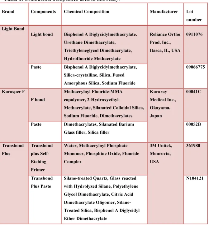
Figure 1-4 present the growth of
Streptococcus mutans in a 96-well
microtitre plate. Each point on the growth curve is the average of the optic density measured in eight wells at any given time. Each curve includes 32 measurements taken within 16 hours. There is no significant microbial growth observed in the negative control group in all measurements (p=0.007, fresh sample;
p<0.001, 1 week; p=0.001, 1 month; p<0.001, 3 months).
There are significant differences in freshly mixed samples between four groups (p<0.001, F=20.901). Freshly mixed samples of Transbond Plus showed antibacterial activity (p<0.001). Kurasper F (p=0.878) and Light Bond (p=0.747) did not differ from positive control (Figure 1).
In the serial experiments with 1 week, 1 month and 3 month- aged specimens, all materials showed an increase in the logarithmic growth rate of Streptococcus
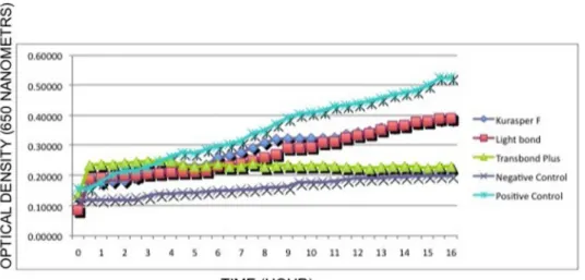
Figure 2. Bacterial growth after direct contact with 1 week aged material. Each point on the growth curve represents the average optical density measured at 650 nm in eight wells.
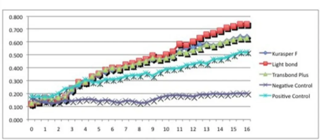
Figure 3. Bacterial growth after direct contact with 1 month aged material. Each point on the growth curve represents the average optical density measured at 650 nm in eight wells.
Figure 4. Bacterial growth after direct contact with 3 month aged material. Each point on the growth curve represents the average optical density measured at 650 nm in eight wells.
DISCUSSION
The oral situations of orthodontic patients experiences some changes, such as pH reduction, increased accumulation of food particles available for Streptococcus
mutans collection.5 Such changes may
contribute to the development of the decalcification lesions frequently observed at the end of orthodontic treatments.5,25 The high prevalence of post-orthodontic treatment decalcifications is caused by the increment of Streptococcus mutans adjacent to the orthodontic appliances.4,26
Streptococcus mutans has been widely
used for testing the antimicrobial activity of restorative materials in dentistry.20,27,28 Growth of the Streptococcus mutans was investigated in the present study using the DCT. Both the agar diffusion test (ADT) and the DCT has been used to analyze the antibacterial properties of dental materials.6,23,27 Lewinstein et al.21 found that the DCT was more effective than ADT in order to detect the antibacterial properties. With a temperature-controlled spectrophotometer and the appropriate software, the DCT allows the researchers to evaluate the number of viable bacteria at the end of the direct contact incubation period using calibration growth curves.
Dental plaque is a microbial biofilm formed by organisms tightly bound to a solid substrate and to each other by means of an exopolymer matrix. Bacteria exhibit different properties when contained within a biofilm.7,8 Such biofilms are characterized by several cell layers, with bacteria stratification arranged by metabolism and aerotolerance. When the orthodontic bands and brackets are applied, new attachment sites for microorganisms would be created, and the appliances may lead to opportunities for non-oral microorganisms to be maintained longer or even to colonize in the mouth.29All
While the amount of fluoride, type and particle size of the fluoridated filler, the type of resin, silane treatment, and porosity might be important factors contributing to fluoride release18,19 and these may differ from brand to brand, in the present study three different fluoride-releasing composites. The analyses of our measurements revealed that Transbond Plus had antibacterial activity in only fresh material. The antibacterial characteristics of orthodontic adhesives were compared in only one study in the scientific literature. Matalon et al.22 examined different types of orthodontic materials (cement-composite and those that release fluoride and not) and evaluated the antibacterial properties of conventional glass ionomer cement (CX-Plus), resin forced glass ionomer cement (GC Fuji Ortho LC), composite resin (Transbond XT) and fluoride-releasing composite resin (Transbond Plus) using the DCT. The researchers stated that while GC Fuji Ortho LC and Transbond Plus showed the antibacterial properties in fresh material, Transbond Plus did not show the antibacterial properties after aging. Our findings are similar to the result of Matalon et al.22
To obtain the antibacterial effects, on tooth structures, several methods such as bonding systems containing antimicrobial agents,30,31 fluoridated toothpastes, mouth rinses and gels have been proposed. However, toothpastes, mouth rinses and compliance and provide only intermittent protection against demineralization.32 Systematic usage of fluoride varnish seems to reduce lesion formation in orthodontic patients.33 Besides, fluoride releasing cements, elastomeric modules and chains could be administered during fixed
34
we aimed to compare the effectiveness of
adhesives have been shown to reduce incipient carious lesions in patients wearing fixed appliances.14,35 Fluoride has some important effects, including the inhibition of demineralization, the enhancement of remineralization, and the inhibition of microbial growth and metabolism.13 Remineralization by releasing fluoride is important, but the antibacterial property of fluoride is a direct strategy for eliminating the cause of dental caries.36 The antibacterial properties of Durelon, Ketac-cem, and Harvard cements were evaluated by using the DCT.21 The authors found that Durelon and Harvard cement had significant antibacterial effects. The antibacterial activity of several glass ionomer cements, dentin bonding systems and luting agents were investigated using the ADT. Marked antibacterial activity was revealed with the glass ionomer cement, whereas amalgams, composites, luting agents, and bonding systems did not affect the bacterial growth.37 Feuerstein et al.24 indicated that four different tested self-etching adhesive systems (AdheSe, Adper Prompt L-Pop, Clearfil Protect Bond and Xeno III) had a bactericidal effect on Streptococcus
mutans within 16 hours by using the DCT.
fresh material was evaluated for 16 hours. (one week, one month and three months later) were performed. No bacterial growth inhibition was determined during aging periods. The levels of fluoride release at a constant rate are required for effectiveness. The fluoride release was high on the first day, fell rapidly over the next day, then gradually decreased to a nearly constant level by the end of the third day.17Because orthodontic patients have routine examining appointments, elastomeric ligature ties impregnated with fluoride would be a solution to provide a long-term low-dose fluoride release.38This method of fluoride delivery would eliminate any need for patient compliance and would replace fluoride at each orthodontic visit.
Fluoride-releasing materials may act as a fluoride reservoir and may increase the fluoride level in saliva and plaque. The results of the studies, which compared the rates of fluoride release over time from the orthodontic materials, showed that the mean fluoride releasing rate declined with time.39,40
The fluoride-releasing rate of resin materials can increase after the topical fluoride is applied. Resin materials can recharge the fluoride and release it again into the environment. In general, materials with a higher initial rate of fluoride release have a higher recharge capability.19,41 However, fluoride release from aged and re-fluoridated specimens did not reach the initial rate of fluoride release.41
CONCLUSIONS
1. Freshly mixed samples of Transbond Plus showed the antibacterial activity.
2. None of the materials showed the antibacterial activity after aging protocol.
ACKNOWLEDGEMENT
The authors thank 3M Unitek, Reliance, and Kuraray for providing the adhesives tested in this study.
REFERENCES
1. Demling A, Elter C, Heidenblut T, Bach Fr-W, Hahn A, Schwestka-Polly R, Stiesch M, Heuer W. Reduction of biofilm on orthodontic brackets with the use of a polytetrafluoroethylene coating. Eur J Orthod 2010;32:414-418.[CrossRef]
2. Tufekci E, Casagrande ZA, Lindauer SJ, Fowler CE, Williams KT. Effectiveness of an essential oil mouthrinse in improving oral health in orthodontic patients. Angle Orthod 2008;78:294-298.[CrossRef]
3. Buren JL, Staley RN, Wefel J, Qian F. Inhibition of enamel demineralization by an enamel sealant, Pro Seal: an
in-vitro study. Am J Orthod Dentofacial
Orthop 2008;133:88-94.[CrossRef]
In the present study, bacterial growth in For the aged material three measurements
4. Kocak MM, Ozcan S, Kocak S, Topuz O, Erten H. Comparison of the efficacy of three different mouthrinse solutions in decreasing the level of
Streptococcus mutans in saliva. Eur J
Dent 2009;3:57-61.
5. Rosenbloom RG, Tinanoff N. Salivary
Streptococcus mutans levels in
patients before, during, and after orthodontic treatment. Am J Orthod Dentofacial Orthop 1991;100:35-37.
[CrossRef]
6. Duran I, Sengun A, Hadimli HH, Ulker M. Evaluation of antibacterial effectiveness of desensitizers against oral bacteria. Eur J Dent 2008;2:43-47.
7. Poeta P, Igrejas G, Gonçalves A, Martins E, Araújo C, Carvalho C, Rodrigues J, Vinué L, López M, Torres C. Influence of oral hygiene in patients with fixed appliances in the oral carriage of antimicrobial-resistant
Escherichia coli and Enterococcus
isolates. Oral Surg Oral Med Oral Pathol Oral Radiol Endod 2009;108:557-564.[CrossRef]
8. Sbordone L, Bortolaia C. Oral microbial biofilms and plaque related diseases: microbial communities and their role in the shift from oral health to disease. Clin Oral Invest 2003;7:181-188.[CrossRef]
9. Uysal T, Amasyali M, Koyuturk AE, Sagdic D. Efficiency of amorphous calcium phosphate-containing orthodontic composite and resin modified glass ionomer on demineralization evaluated by a new laser fluorescence device. Eur J Dent 2009;3:127-134.
10. Ahn SJ, Lim BS, Lee SJ. Prevalence of cariogenic streptococci on incisor brackets detected by polymerase chain reaction. Am J Orthod Dentofacial
months after bracket placement. Am J Orthod Dentofacial Orthop 2006;130:275.e17-22.[CrossRef]
12. O’Reilly MM, Featherstone JDB. Demineralization and remineralization around orthodontic appliances: an in vivo study. Am J Orthod Dentofacial Orthop 1987;92:33-40.[CrossRef]
13. Wiegand A, Buchalla W, Attin T. Review on fluoride-releasing restorative materials-fluoride release and uptake characteristics, antibacterial activity and influence on caries formation. Dent Mater 2007;23:343-362. [CrossRef]
14. Rogers S, Chadwick B, Treasure E. Fluoride-containing orthodontic adhesives and decalcification in patients with fixed appliances: A systematic review. Am J Orthod Dentofacial Orthop 2010;138:390.e1-390.e8.[CrossRef]
15. Chadwick BL, Roy J, Knox J, Treasure ET. The effect of topical fluorides on decalcification in patients with fixed orthodontic appliances: a systematic review. Am J Orthod Dentofacial Orthop 2005;128:601-606.[CrossRef]
16. Mota SM, Enoki C, Ito IY, Elias AM, Matsumoto MAN. Streptococcus mutans counts in plaque adjacent to
orthodontic brackets bonded with resin-modified glass ionomer cement or resin-based composite. Braz Oral Res 2008;22:55-60.[CrossRef]
17. Rix D, Foley TF, Banting D, Mamandras A. A comparison of fluoride release by resin-modified GIC and polyacidmodified composite resin. Am J Orthod Dentofacial Orthop 2001;120:398-405.
18. Dijkman GE, de Vries J, Lodding A, Arends J. Long-term fluoride release of visible light-activated composites in
of fluoride-releasing materials. Biomaterials 2003;24:2451-2461.
[CrossRef]
20. Weiss EI, Shlhav M, Fuss Z. Assessment of antibacterial activity of endodontic sealers by a direct contact test. Endod Dent Traumatol 1996;12:179-184.[CrossRef]
21. Lewinstein I, Matalon S, Slutzkey S, Weiss EI. Antibacterial properties of aged dental cements evaluated by direct-contact and agar diffusion tests. J Prosthet Dent 2005;93:364-371.
[CrossRef]
22. Matalon S, Slutzky H, Weiss EI. Antibacterial properties of 4 orthodontic cements. Am J Orthod Dentofacial Orthop 2005;127:56-63.
[CrossRef]
23. Beyth N, Yudovin-Fearber I, Domb AJ, Weiss EI. Long-term antibacterial surface properties of composite resin incorporating polyethyleneimine nanoparticles. Quintessence Int 2010;41(10):827-835.
24. Feuerstein O, Matalon S, Slutzky H, Weiss EI. Antibacterial properties of self-etching dental adhesive systems. J Am Dent Assoc 2007;138:349-354. 25. Stratemann MW, Shannon IL. Control
of decalcification in orthodontic patients by daily self-administered application of a water-free 0.4 per cent stannous fluoride gel. Am J Orthod. 1974;66:273-279.[CrossRef]
26. Zimmer BW, Rottwinkel Y. Assessing patient-specific decalcification risk in fixed orthodontic treatment and its impact on prophylactic procedures. Am J Orthod Dentofacial Orthop 2004;126:318-324.[CrossRef]
27. Herrera M, Carrion P, Baca P, Liebana J, Castillo A. In vitro antibacterial activity of glass-ionomer cements. Microbios 2001;104:141-148.
28. Tobias RS. Antibacterial properties of dental restorative materials: a review. Int Endod J 1988;21:155-160.
[CrossRef]
29. Ristic M, Vlahovic M, Svabic M, Sasic M, Zelic O. Clinical and microbiological effects of fixed orthodontic appliances on periodontal tissues in adolescents. Orthod Craniofac Res 2007;10:187-195.
[CrossRef]
30. Imazato S, Torii M, Tsuchitani Y, McCabe JF, Russell RR. Incorporation of bacterial inhibitor into resin composite. J Dent Res 1994;73:1437-1443.
31. Fan C, Chu L, Rawls HR, Norling BK, Cardenas HL, Whang K. Development of an antimicrobial resin-a pilot study. Dent Mater 2011;27(4):322-328.
[CrossRef]
32. Spencer CG, Campbell PM, Buschang PH, Cai J, Honeyman AL. Antimicrobial effects of zinc oxide in an orthodontic bonding agent. Angle Orthod 2009;79(2):317-322.
[CrossRef]
33. Øgaard B, Larsson E, Henriksson T, Birkhed D, Bishara SE. Effects of combined application of antimicrobial and fluoride varnishes in orthodontic patients. Am J Orthod Dentofacial Orthop 2001;120:28-35.
34. Hu W, Featherstone JD. Prevention of enamel demineralization:an in-vitro study using light-cured filled sealant. Am J Orthod Dentofacial Orthop 2005;128:592-600. [CrossRef]
35. Benson PE, Parkin N, Millett DT, Dyer FE, Vine S, Shah A. Fluorides, orthodontics and demineralization. A systematic review. J Orthod 2005;32:102-114.[CrossRef]
36. Eminkahyagil N, Korkmaz Y, Gokalp S, Baseren M. Shear bond strength of orthodontic brackets with newly developed antibacterial self-etch adhesive. Angle Orthod 2005;75:843-848.
37. Prati C, Fava F, Di Gioia D, Selighini M, Pashley DH. Antibacterial effectiveness of dentin bonding systems. Dent Mater 1993;9:338-343.
38. Baturina O, Tufekci E, Guney-Altay O, Khan SM, Wnek GE, Lindauer SJ. Development of a sustained fluoride delivery system. Angle Orthod 2010;80(6):1129-1135.[CrossRef]
39. Cacciafesta V, Sfondrini MF, Tagliani P, Klersy C. In-vitro fluoride release rates from 9 orthodontic bonding adhesives. Am J Orthod Dentofacial Orthop 2007;132:656-662.[CrossRef]
40. McNeill CJ, Wiltshire WA, Dawes C, Lavelle CL. Fluoride release from new light-cured orthodontic bonding agents. Am J Orthod Dentofacial Orthop 2001;120:392-397.[CrossRef]
41. Attar N, Turgut MD. Fluoride release and uptake capacities of fluoride-releasing restorative materials. Oper Dent 2003;28:395-402.