EXPERIMENTAL STUDY
Effect of fl uid resuscitation on acute lung injury in a rat
model of sepsis
Erdogan A
1, Erdogan MA
2, Kara AY
2, Bora S
3, Yigitturk G
4, Erbas O
5 Izmir Cigli Regional Training Hospital, Department of Emergency Medicine, Izmir, Turkey.ABSTRACT
AIM: Sepsis is a systemic infection reaction and intravascular volume therapy plays a crucial role in it’s treatment. Acute respiratory distress syndrome (ARDS) occurs in the lungs, the most affected organ. This study aimed to investigate the different effects of fl uid therapy on ARDS caused by sepsis.
METHOD: To form a sepsis model, cecal ligation and puncture (CLP) procedure were performed on 44 adult rats. Divided into six groups; normal, CLP group, those treated with 40 ml/kg 0.9 % NaCl, 3 % NaCl (hypertonic saline), Ringer Lactate and Hydroxyethyl starch. After 24 hours treatments, histopathological examination of the lungs were done, and the plasma levels of CRP, TNF-α and IL-6 and paO2 were measured.
RESULTS: The scores of all histological parameters of the group treated with hypertonic saline were signifi cantly lower than of the other groups (p < 0.001). Likewise, according to the arterial blood gas results, paO2 was signifi cantly higher (p < 0.01) in the hypertonic saline group compared to the other groups, and paCO2 was signifi cantly lower (p < 0.01). CRP, TNF-α and IL-6 levels of infl ammatory markers were also signifi cantly lower in hypertonic saline groups compared to other groups (p < 0.001).
CONCLUSIONS: Our study shows that treatment with hypertonic saline reduces the progression of ARDS in sepsis (Tab. 3, Fig. 4, Ref. 49). Text in PDF www.elis.sk
KEY WORDS: ARDS, infl ammation, lung injury, sepsis, volume replacement fl uids.
1Izmir Cigli Regional Training Hospital, Department of Emergency
Medi-cine, Izmir, Turkey, 2Izmir Katip Celebi University, Faculty of Medicine,
Department of Physiology, Izmir, Turkey, 3Izmir Ataturk Training and
Research Hospital, Department of Emergency Medicine, Izmir, Turkey,
4Mugla Sitki Kocman University, Faculty of Medicine, Department of
Histology and Embryology, Mugla, Turkey,and 5 Istanbul Bilim
Univer-sity, Faculty of Medicine, Department of Physiology, Istanbul, Turkey Address for correspondence: M.A. Erdogan, Assoc Prof, Izmir Katip Celebi University, Faculty of Medicine, Department of Physiology, Izmir, Turkey. Phone: +90.543.3818677
Introduction
Sepsis, which is an erratic response of the host to infection, is a life threatening disease with a globally high mortality rate (1). Severe sepsis is a suspected or documented presence of infection with signs of systemic response which is accompanied by organ dysfunction (2). Mortality rates from serious sepsis exceed 25 % to 45 % in the case of septic shock, even with optimal treatment (2). Therefore, in order to perform the clinical management of sepsis in the best way, the resuscitation method should be selected as the most appropriate procedure.
Systemic infl ammatory response syndrome, which usually be-gins with severe infection or acute injury, is a common problem in sepsis (3, 4). This syndrome has also caused multiple organ
dysfunction, including the lungs, liver, heart and kidneys (3, 5). The lungs are the most commonly affected organ in multiple or-gan dysfunction syndrome due to sepsis, and this syndrome takes the form of acute respiratory distress syndrome (ARDS) with lung injury (6, 7).
Although there are signifi cant progresses in our experience about the pathophysiology of sepsis, treatments are still limited to aggressive fl uid resuscitation, antibiotics, vasopressor admini-stration and supportive care (8). Therefore, it is highlighted that there is a need to develop new therapeutic strategies for this syn-drome. Intravenous (IV) fl uid resuscitation is a critical compo-nent of the ideal treatment plan for serious sepsis patients (2). Consequently, the effect of various types of IV fl uids has also been a popular subject of interest in sepsis studies and research (2). For all that it is still controversial about the use of optimum fl uid replacement especially with regard to the kidney function. For example, though in European emergency clinics, gelatin and hydroxyethyl starch are generally used combination with crystal-loids for fl uid resuscitation, in North America crystalcrystal-loids can be used frequently (9‒12).
In sepsis induced ARDS and/or Acute Lung Injury (ALI), the lungs are the most severely infl amed and damaged areas (13, 14). The main cause of damage in ARDS is the destroying of the alveo-lar epithelium and pulmonary capilalveo-lary endothelium by activated neutrophils, leukocytes, and macrophages, which form a large
number of infl ammatory mediators, interleukins (ILs), including tumor necrosis factor (TNF)-α (15). Thus, infl ammatory processes must be evaluated regarding this information. Fluid resuscitation plays an essential role in septic tissue hypoperfusion therapy of hypovolemia (16). For this reason, this study was planned to re-search the effects of volume replacement by IV fl uids on sepsis induced ARDS model in rats.
Materials and methods
Animals
In our study, 44 Sprague Dawley albino mature 200‒220 g male rats were used. We received local Animal Ethics Commit-tee’s approval. And then all experimental procedures were per-formed according to the stated rules in the Guide for the Care and Use of Laboratory Animals adopted by the National Institutes of Health and ARRIVE guidelines. Animals were fed and kept in temperature ‒ controlled steel cages (21 ± 2 °C) light-dark period for 12 hours.
Experimental procedures
In order to create a sepsis model, cecal ligation and puncture (CLP) procedure were performed on 44 rats as previously de-scribed (16, 17). 8 rats were excluded from the study because they died. We randomly divided the rats into six groups. We designed the working groups as follows: Group 1: normal (non-operative and orally fed control, n = 6); Group 2: CLP (untreated group, n = 6); Group 3: CLP and 40 ml/kg 0.9 % NaCl i.p (n = 6), Group 4: CLP and 40 ml/kg 3 % NaCl i.p. (n = 6), Group 5: CLP and 40 ml/kg Ringer Lactate i.p (n = 6), Group 6: CLP and 40 ml/kg Hydroxyethyl starch (6 % HES 130/0.4) i.p. We anesthetized the rats by intraperitoneally administering a combination of ketamine hydrochloride (80 mg/kg) and xylazine hydrochloride (7 mg/kg) according to the CLP procedure. Aseptically, the cecum was reached by applying 3 cm laparotomy from the midline of the rat. It was tightly tied at the base of the cecum with a 3.0 silk suture and pierced with a 22-gauge needle. A small amount of stool was then squeezed out of the puncture site. The cecum was placed back into the peritoneal cavity and the laparotomy incision was closed with 4-0 polyglactin sutures. Treatment was started in the fi rst hour of the surgical pro-cedure. All volume replacement therapies were given equally every 12 hours a day in two sections. All treatments were admi-nistered for 24 hours. When we ended the study, the rats were euthanized, biochemi-cal analysis was performed from the blood sample taken by cardiac puncture, and his-topathological examination was performed by pneumonectomy.
Measurement of Arterial blood gas
Carotid artery blood (0.2 mL) of rats was taken and paO2 and paCO2 levels were evaluated.
Measurement of plasma CRP, TNF-α and IL-6 levels
Biochemical analysis was performed from blood samples collected by cardiac puncture after the sacrifi ce procedure. Blood samples were centrifuged at 3000 rpm for 10 minutes at room temperature and then stored at ‒20 °C. Commercially available enzyme-linked immunosorbent assay (ELI-SA) kits were used to evaluate CRP, TNF-a, and IL-6 levels. We identifi ed all samples in duplicate according to the manufacturer’s instructions.
Histopathological examination of lung
All rats were anesthetized by ketamine (40 mg/kg, Alfamine, Alfasan International B.V., The Netherlands) and xylazine (4 mg/kg, Alfazyne, Alfasan International B.V., The
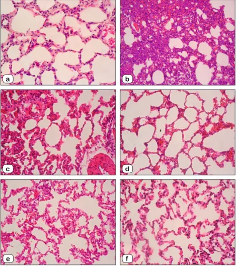
Fig. 1. Lung Tissue. Hematoxylin and Eosin (H&E) stain, X 20 magnifi cation. a: Control group, normal alveolar and interstitial structure, b: CLP group, c: CLP and 0.9 % NaCl group, d: CLP and 3 % NaCl group, e: CLP and Ringer Lactate group, f: CLP and HES group.
a b
c d
Netherlands) i.p. Lungs were removed for histological examination and fi xed with formalin. During the examination, it was stained with hematoxylin/eosin. All stages were taken by the Olympus C-5050 digital camera mounted on the Olympus BX51 microscope. The basic histopathological lung injury score was calculated as used in previous publications (18). In a nutshell, histopathological lung damage was assessed by scoring alveolar congestion (AC), hemorrhage (H), infi ltration or aggregation of leukocytes in air spaces/vessel walls (AL), perivascular/interstitial edema (PE), and thickness of the alveolar wall/hyaline membrane formation (TA) (18). For all cases, the signifi cance was graded on a scale of 1 (0‒25 %), 2 (25‒50 %), 3 (50‒75 %), and 4 (75‒100 %).
Statistical analysis
The data obtained from the subjects were compared with one-way analysis of variance (ANOVA). Post-hoc Tukey HSD test was used for comparison of the groups. Differences in control and treatment values were determined using Student’s t-test. The limit of statistical signifi cance was assigned to p < 0.05.
Results
Hitopathological examination of lung
Lung tissue histopathology changes are shown in Figure 1. His-topathological indicators of ARDS (hemorrhage, alveolar
conges-tion, infi ltration/ aggregation of leukocytes in air spaces-vessel walls, perivascular-in-terstitial edema, and thickness of the alveo-lar wall-hyaline membrane formation) were observed in the CLP group. In all groups, alveolar congestion was lowest in the group treated with 3 % NaCl (p < 0.001), whereas it was highest in the 0.9 % NaCl treated group (p < 0.05). Hemorrhage and thickness of the alveolar wall-hyaline membrane for-mation scores were decreased in CLP + 3 % NaCl and CLP + HES groups compared to CLP + 0.9 % NaCl and CLP + Ringer Lactate groups (p < 0.001). Infi ltration-aggregation of leukocytes in air spaces-vessel walls score in the CLP + 3 % NaCl and CLP + Ringer Lactate groups were found to be lower than those in the CLP + 0.9 % NaCl and CLP + HES groups (p < 0.001). Perivascular/interstitial edema was also found to be decreased in CLP + 3 % NaCl, CLP + Ringer Lactate and CLP + HES groups compared to the CLP group (p < 0.001). Overall, the score of all histological parameters about lung injury decreased substantially in the CLP + 3 % NaCl group relative to other groups (Tab. 1).
Arterial blood gas analysis
When we examine blood gasses, in all groups PaO2 were meaningfully lower than in the control group, however, PaCO2 was meaningfully higher (p < 0.001). Likewise, paO2 in the CLP + 3 % NaCl group was signifi cantly higher than in the other groups (p < 0.01), moreover, paCO2 was signifi cantly lower in this group (p < 0.01) (Tab. 2).
Plasma CRP, IL-6, TNF-α levels
Serum CRP, IL-6 and TNF-α levels of all groups are shown in Table 3. As shown in Figure 2, serum CRP levels were signifi -cantly higher in the CLP group compared to the control group (p < 0.0001). Among all groups, rats treated with 3 % NaCl had signifi -cantly lower CRP levels than other treatment groups (p < 0.001).
Plasma levels of TNF-α and IL-6 in all groups are shown in Figure 3 and 4. The plasma levels of TNFα and IL6 were signifi -cantly higher in the CLP-sepsis group compared with control group (p < 0.0001). In the same pattern, there were signifi cant inhibitions in TNF-α and IL-6 levels in the CLP + 3 % NaCl group, compared to the other groups. TNF-α levels were signifi cantly lower in the CLP + 3 % NaCl and CLP + HES groups, while IL-6 levels were signifi cantly lower in the CLP + 3 % NaCl and CLP + 0.9 % NaCl groups compared to the other treatment groups (p < 0.001).
AC H AL PE TA
Control group 0.3±0.2 0.5±0.2 0.6± 0.3 0.5±0.2 0.3±0.2
CLP group 4.3±0.4* 3.3±0.3* 4.3±0.4* 3.8±0.4* 4.3±0.4*
CLP and 0.9% NaCl group 3.5±0.2# 2.0±0.3# 3.3±0.3# 2.3±0.2# 3.1±0.4#
CLP and 3% NaCl group 1.6±0.1## 1.7±0.4## 1.3±0.2## 1.5±0.2## 1.6±0.4##
CLP and Ringer Lactate group 2.6±0.3# 2.2±0.3# 1.5±0.2## 1.7±0.2## 2.2±0.3#
CLP and HES group 2.0±0.3## 1.8±0.2## 3.0±0.2# 1.8±0.3## 2.0±0.4## * p < 0.0001, compared with Control group, # p < 0.05, compared with CLP group, ## p < 0.001, compared with CLP
group. Alveolar congestion (AC), hemorrhage (H), infi ltration or aggregation of leukocytes in air spaces/vessel walls (AL), perivascular/interstitial edema (PE), and thickness of the alveolar wall/hyaline membrane formation (TA) Tab. 1. Changes in histopathological lung injury scores.
paO2 (mmHg) paCO2 (mmHg)
Control group 121.±5 1.7 36.6±1.3
CLP group 72.3±5.6* 55.3±4.2*
CLP and 0.9% NaCl group 94.8±7.4# 44.5±4.7#
CLP and 3% NaCl group 110.±6 4.7## 39.5±3.3##
CLP and Ringer Lactate group 95.6±5.4# 45.1±1.3#
CLP and HES group, 99.3±7.3## 43.5±1.8# * p < 0.001, compared with Control group, ## p < 0.01, compared with CLP group, #
p < 0.05, compared with CLP group Tab. 2. Arterial blood gas analysis.
Control group CLP group CLP and 0.9% NaCl group, CLP and 3% NaCl group CLP and Ringer Lactate group CLP and HES group CRP (mg/dl) 0.36±0.02 0.79±0.03* 0.57 ±0.02## 0.43 ±0.02## 0.8±0.04 0.63±0.03# TNF-α (pg/ml) 24.54±11.28 212.38±34.65* 122.38±19.76## 87.60±15.80## 154.67±14.58 107.99±37.52# IL-6 (pg/ml) 40.53±7.56 524.5±28.4* 228.4±37.8 150.7±25.3## 456.3±48.5 234.0±38.4#
* p < 0.0001, compared with Control group, # p < 0.05, compared with CLP group, ## p < 0.001, compared with CLP group
Discussion
Sepsis is a complicated clinical syndrome with a loud mor-bidity and mortality rate, with the development of progressive damage to multiple organ systems far from the site of infection (19). First and most severely affected organ is the lung. Thefore, the most common fatal complication of sepsis is acute re-spiratory distress syndrome (ARDS) (20, 21). As sepsis-induced ARDS is the fi rst step in developing multi-organ dysfunction syndrome in critically ill patients, its prevention is an important therapeutic target.
Mediators that cause infl ammatory events in sepsis increase the microvascular permeability and cause leakage in capillaries, causing an increase in interstitial fl uid, protein loss and tissue oedema. That is, sepsis is related to intravascular volume defi -ciency due to venous pooling, vasodilatation and endothelial bar-rier disruption (22, 23). Due to impaired albumin synthesis and increased loss from the transcapillary membrane, intravascular colloid osmotic pressure (COP) decreases, and the preservation of intravascular volume is impaired. Therefore, sepsis causes organ dysfunction due to insuffi cient tissue perfusion and oxygenation, resulting in arterial hypotension with reduced cardiac preload and output (24). In this clinical event, fl uid resuscitation is the most important thing to restore and maintain the intravascular volume required for microcirculation and maintenance of tissue perfusion (25). As known, severe or extended hypotension is associated with bad clinical outcomes in patients with sepsis, while fast recovery of organ perfusion seems to be correlated with lower mortality rates (26). Nevertheless, there is no consensus on which type of fl uid should be used as fi rst-line therapy in resuscitation of septic shock patients.
Many studies have shown that fl uid therapy is important in ARDS due to sepsis. However, positive fl uid balance has also been shown to harm ARDS patients. Therefore, the key to treatment is to fi nd the balance between pulmonary edema while achieving improvement in tissue perfusion (35). So, we planned this study to investigate which fl uid is more effective in lung injury.
Acute respiratory distress syndrome includes microvascular lung damage, including accumulation of leukocyte in the tissue, pulmonary edema and infl ammation (23). When we compared pulmonary edema, which suggests a protective effect on the lung, among the groups we used fl uid therapy, we found that hypertonic saline was the best preventive measure. The edema interferes with lung function, changes the surfactant and deteriorates the barrier
Fig. 2. The effect of volume replacement therapies on serum CRP levels. The results are expressed as mean ± S.E.M. * p < 0.0001, com-pared with Control group, # p < 0.05, comcom-pared with CLP group, ## p < 0.001, compared with CLP group.
Fig. 3. The effect of volume replacement therapies on serum TNF-α levels. The results are expressed as mean ± S.E.M. * p < 0.0001, com-pared with Control group, # p < 0.05, comcom-pared with CLP group, ## p < 0.001, compared with CLP group.
Fig. 4. The effect of volume replacement therapies on serum IL-6 levels. The results are expressed as mean ± S.E.M. * p < 0.0001, com-pared with Control group, # p < 0.05, comcom-pared with CLP group, ## p < 0.001, compared with CLP group.
0.9 600 0.8 # 500 '2 0.7 '2
~
0.6~
400 C. 0.5 ## C.~
~ 300 ## # _§_ 0.4s
Ol ## [l_ 0.3 (!) 200 o:: 0.2 ...'.ı (.) 100 0.1 O.Oo
300 '2 250~
200 C.~
150 ## #s
o 100u.
z 1- 50o
lesions of the alveolocapillary. Any effort to decrease edema could therefore have benefi cial effects on respiratory function. Similar results have been found in other studies (27).
The infl ammatory mechanism introduced during ARDS is characterized by the release of infl ammatory mediators and the infl ow of leukocytes in the pulmonary tissue (28). In our study, we also observed an increase in leukocyte infi ltration in the untreated CLP group. We found that leukocyte infi ltration in the lungs of groups treated with 0.9 % NaCl and HES could not be adequately prevented, but treatment with hypertonic saline signifi cantly re-duced the content of leukocytes. Supporting this, Oliveira et al suggests that the most interesting benefi cial effect of hypertonic saline is the reduction of SIRS and attenuated MODS, especially in sepsis (29).
In consideration of perfusion, there was a study in the rat sepsis model caused by peritonitis (CASP) which determined that administration of the hydroxyethyl starch (HES) solutions provided no additional benefi t on improvement of microcircula-tion in comparison to crystalloid solumicrocircula-tions (30). Similarly, with regard to organ failure, it has been shown that albumin had no advantage over Ringer’s Lactate (RL) on effi cacy in reducing ventilator induced lung injury in an endotoxic rat model of sepsis (31). Nonetheless, there was also another study in rat cecal liga-tion and puncture (CLP) model of sepsis which showed that un-balanced crystalloid solution (0.9 % NaCl) was more effective on the development of acute kidney injury (AKI) and survival time when compared to a balanced crystalloid (Plasma-Lyte) (32). In another study in an endotoxic rat sepsis model it has been report-ed that synthetic colloid solution Hextend rreport-educreport-ed mortality and lessened metabolic acidosis when compared to resuscitation with the saline or RL (33).
Unlike complicated fi ndings from small-animal models, ex-periments in large animal models usually support the administra-tion of colloid soluadministra-tions in sepsis. Qui, et al. studied the effects of crystalloids (0.9 % NaCl and 7.5 % NaCl) or colloid (HES) so-lutions, on levels of vascular endothelial growth factor (VEGF), capillary permeability, and development of pulmonary edema in a sepsis dog model (2). Shown in studies, HES was more ef-fective than crystalloid solutions in reducing tissue expression of VEGF, capillary permeability, and lung edema levels (34). However, there was another study performed in a sheep model of peritonitis induced sepsis which showed that even though the albumin and HES could improve the cardiac output, delivery of oxygen and reduce the blood lactate levels, there was no change in survival when compared with the administration of the crys-talloid, RL (35).
In addition to the importance of clinical results associated with the amount and form of fl uid resuscitation in septic care, the fundamental mechanisms to mediate these responses must also be examined. Recently, it has been shown that Nitric Oxide (NO) had a main role in sepsis pathophysiology. As it is known, the endo-thelium displays a vasodilatory response to fl uid loading in order to tolerate increased blood fl ow and endothelial shear stress. In a hamster model of lipopolysaccharide (LPS)-induced sepsis, it has been demonstrated that enhancing or ameliorating NO
bioavail-ability throughout sepsis may improve arteriole dilation, functional capillary density, and survival albeit reducing leukocyte endothelial interactions (2, 36). Vasopressin 1 receptor (V1R) is another player that has an important role in volume resuscitation therapy. It has been determined that in an endotoxic rat model, volume resusci-tation can cause vasopressin release to enable the necessary V1R during the periods of low vasopressin secretion for an increased blood pressure in response to the fl uids (37). The same pathway was also active and served a function after volume resuscitation in the CLP rat model of severe sepsis (38).
With a regard to infl ammatory mechanisms, it has been shown that circulating TNF-α levels were substantially decreased in vo-lume resuscitated animals compared to controls in the endotoxic rat sepsis model (2). The reduced TNF-α correlated with de-creased total peripheral resistance (TPR), high cardiac beats, and higher survival rate (39). C-reactive protein (CRP) is an acute-phase protein, which increase in blood rapidly in response to in-fection, trauma, ischemia, burns, and other infl ammatory condi-tions (40). Because of these results, plasma CRP levels have been evaluated for a long time as an important biomarker for evaluating systemic infl ammation (41). CRP levels have been shown to have prognostic/diagnostic signifi cance in a great range of diseases, with the inclusion of sepsis, pneumonia, coronary artery disease, stroke and diabetes (42‒44). Some studies indicate that CRP may be an indication of progressive organ damage (45‒47). ARDS is an infl ammatory process and many studies have centered on determining the function of infl ammatory plasma mediators in this disease and how they correlate with phenotypes and clinical outcomes (18, 48, 49). While literature studies associating CRP levels with prognoses in other diseases have been performed, there is little knowledge of the CRP function in ARDS and ALI patients. In our study, we also found a signifi cant decrease in CRP, TNF-α and IL-6 levels in the CLP + 3 % NaCl group when com-pared with the other groups. Therefore, this may be the fi rst data about CRP and proinfl ammatory cytokine levels in sepsis-induced ARDS/ALI and since infl ammatory processes have important role in sepsis, the effects of volume resuscitation on CRP levels and its underlying mechanisms need to be investigated in detail.
In our study, we showed that hypertonic saline was the most effective fl uid for volume resuscitation. It reduced the lung inju-ry parameters histologically, improved functional parameters in analysis of arterial blood gas and also had some anti-infl ammatory effects with regard to CRP, TNF-α and IL-6 levels.
Conclusion
We demonstrate that hypertonic saline 3 % NaCl-treatment signifi cantly mitigates lung injury, with quantifi able suppression of injury parameters on lung histochemistry, increase in paO2 and reduction in paCO2 in the arterial blood gas analysis, and also de-crease in CRP, TNF-α and IL-6 levels. We also found that colloid solution HES can be more effective when compared with other crystalloids, 0.9 % NaCl and Ringer Lactate. Our fi ndings repli-cate earlier published fi ndings which demonstrated that hyper-tonic saline could ameliorate the ARDS/ALI in another rat model
of sepsis and add to the body of evidence that hypertonic saline may serve as a fi rst line therapy. Further studies are needed before these fl uids can be suggested as a fi rst line treatment and also for clarifi cation of the underlying mechanisms of these different re-suscitation fl uids in sepsis-related ARDS/ALI.
References
1. Stevenson EK, Rubenstein AR, Radin GT, Wiener RS, Walkey AJ. Two decades of mortality trends among patients with severe sepsis: a com-parative meta-analysis. Crit Care Med 2014; 42: 625‒631.
2. Cornelius DC, McCalmon M, Tharp J, Puskarich M, Jones AE. Fluid Resuscitation in Animal Models of Sepsis: A Comprehensive Review of the Current State of Knowledge. Arch Emerg Med Crit Care 2016; 1 (1): 1002. 3. Singer M, Deutschman CS, Seymour CW et al. The third interna-tional consensus defi nitions for sepsis and septic shock (sepsis-3). JAMA 2016; 315: 801‒810.
4. Blanco J, Muriel-Bombin A, Sagredo V et al. Incidence, organ dys-function and mortality in severe sepsis: A Spanish multicentre study. Crit Care 2008; 12: R158.
5. Nesseler N, Launey Y, Aninat C, Morel F, Malledant Y, Seguin P. Clinical review: The liver in sepsis. Crit Care 2012; 16: 235.
6. Baue AE, Durham R, Faist E. Systemic infl ammatory response syn-drome (SIRS), multiple organ dysfunction synsyn-drome (MODS), multiple organ failure (MOF): Are we winning the battle? Shock 1998; 10: 79–89. 7. Ware LB, Matthay MA. The acute respiratory distress syndrome. N Engl J Med 2000; 342: 1334–1349
8. Dellinger RP, Levy MM., Rhodes A. Surviving Sepsis Campaign: In-ternational guidelines for management of severe sepsis and septic shock, 2012. Intensive Care Medicine 2013; 39: 165–228.
9. Patel A, Laffan MA, Waheed U, Brett SJ. Randomised trials of hu-man albumin for adults with sepsis: systematic review and meta-anal-ysis with trial sequential analmeta-anal-ysis of all-cause mortality. BMJ 2014; 349: 4561.
10. Farrugia A, Bansal M, Balboni S, Kimber MC, Martin GS, Cas-sar J. Choice of Fluids in Severe Septic Patients ‒ A Cost-effectiveness Analysis Informed by Recent Clinical Trials. Rev Recent Clin Trials 2014; 9: 21‒30.
11. Perner A, Haase N, Wetterslev J et al. Comparing the effect of hy-droxyethyl starch 130/0.4 with balanced crystalloid solution on mortal-ity and kidney failure in patients with severe sepsis (6S‒Scandinavian Starch for Severe Sepsis/Septic Shock trial): study protocol, design and rationale for a double-blinded, randomised clinical trial. Trials 2011; 12 (1): 1‒9.
12. Ertmer C, Rehberg S, Van Aken H, Westphal M. Relevance of nonalbumin colloids in intensive care medicine. Best Pract Res Clin An-aesthesiol 2009; 23: 193‒212.
13. Compton CN, Franko AP, Murray MT, Diebel LN, Dulchavsky SA. Signaling of apoptotic lung injury by lipid hydroperoxides. J Trauma 1998; 44: 783–788.
14. Xiao X, Yang M, Sun D, Sun S. Curcumin protects against sepsis-induced acute lung injury in rats. Journal of Surgical Research 2012; 176 (1): e31‒9.
15. Rivers EP, Jaehne AK, Eichhorn-Wharry L, Brown S, Amponsah D. Fluid therapy in septic shock. Curr Opin Crit Care 2010; 16: 297‒308.
16. Erbas O, Taskiran D. Sepsis-induced changes in behavioral stereotypy in rats; involvement of tumor necrosis factor-alpha, oxidative stress, and dopamine turnover. J Surg Res, 2014; 186 (1): p. 262‒8.
17. Bostanci H, Dikmen K, Comu FM, Arslan M, Kucuk A. Investiga-tion of the effects of thymoquinone on erythrocyte deformability in sepsis treatment which created by cecal perforation in rat. Bratisl Med J 2018; 119 (3): 152‒155.
18. Iraz M, Iraz M, Eşrefoglu M, Aydin MŞ. Protective effect of\beta-glucan on acute lung injury induced by lipopolysaccharide in rats. Turk J Med Sci 2015; 45 (2): 261‒267.
19. Neviere RR, Ceepinskas G, Madorin WS et al. LPS pretreatment ameliorates peritonitis-induced myocardial infl ammation and dysfunction: role of myocytes. Am J Physiol 1999; 277: H885.
20. Kollef MH, Schuster DP. The acute respiratory distress syndrome. N Engl J Med 1995; 332: 27–37.
21. Imm A, Carlson RW. Fluid resuscitation in circulatory shock. Crit Care Clin 1993; 9: 313–333.
22. Force AD, Ranieri VM, Rubenfeld GD, Thompson BT, Ferguson ND, Caldwell E. Acute respiratory distress syndrome. Jama 2012; 307 (23): 2526‒33.
23. Marx G. Fluid therapy in sepsis with capillary leakage. Eur J Anaes-thesiol 2003; 20 (6): 429‒442.
24. Boldt J. Volume therapy in the intensive care patient – we are still confused, but. Intensive Care Med 2000; 26: 1181–1192.
25. Jones AE, Brown MD, Trzeciak S et al. Emergency Medicine Shock Research Network investigators. The effect of a quantitative resuscitation strategy on mortality in patients with sepsis: a meta-analysis. Crit Care Med 2008; 36: 2734‒2739.
26. Petroni RC, Biselli PJ, de Lima TM et al. Hypertonic saline (NaCl 7.5%) reduces LPS-induced acute lung injury in rats. Infl ammation 2015; 38 (6): 2026‒35.
27. Koustova E, Stanton K, Gushchin V, Alam HB, Stegalkina S, Rhee PM. Effects of lactated ringer’s solutions on human leukocytes. J Trauma 2002; 52: 872–878.
28. Oliveira RP, Velasco I, Soriano F, Friedman G. Clinical re-view. Hypertonic saline resuscitation in sepsis. Crit Care 2002; 6 (5): 418‒23.
29. Wafa K, Herrmann A, Kuhnert T et al. Short time impact of differ-ent hydroxyethyl starch solutions on the mesdiffer-enteric microcirculation in experimental sepsis in rats. Microvasc Res 2014; 95: 88‒93.
30. Zhang H, Voglis S, Kim CH, Slutsky AS. Effects of albumin and Ringer’s lactate on production of lung cytokines and hydrogen peroxide after resuscitated hemorrhage and endotoxemia in rats. Crit Care Med, 2003; 31: 1515‒1522.
31. Zhou F, Peng ZY, Bishop JV et al. Effects of fl uid resuscitation with 0.9% saline versus a balanced electrolyte solution on acute kidney injury in a rat model of sepsis. Crit Care Med, 2014; 42: 270‒278.
32. Kellum JA. Fluid resuscitation and hyperchloremic acidosis in experimental sepsis: improved short-term survival and acid-base bal-ance with Hextend compared with saline. Crit Care Med 2002; 30: 300‒305.
33. Qiu YZ, Sun H, Li F. Effect of fl uid resuscitation on capillary perme-ability and vascular endothelial growth factor in dogs with septic shock. Zhongguo Wei Zhong Bing Ji Jiu Yi Xue 2007; 19 (5): 270‒273.
286
34. Su F, Wang Z, Cai Y, Rogiers P, Vincent JL. Fluid resuscitation in severe sepsis and septic shock: albumin, hydroxyethyl starch, gelatin or ringer’s lactate-does it really make a difference? Shock 2007; 27: 520‒526. 35. Villela NR, dos Santos AO, de Miranda ML, Bouskela E. Fluid re-suscitation therapy in endotoxemic hamsters improves survival and attenu-ates capillary perfusion defi cits and infl ammatory responses by a mecha-nism related to nitric oxide. J Transl Med 2014; 12: 232.
36. Batista MB, Bravin AC, Lopes LM et al. Pressor response to fl uid resuscitation in endotoxic shock: involvement of vasopressin. Crit Care Med 2009; 37: 2968‒2972.
37. Santiago MB, Vieira AA, Elias LL, Rodrigues JA, Giusti-Paiva A. Neurohypophyseal response to fl uid resuscitation with hypertonic saline during septic shock in rats. Exp Physiol 2013; 98: 556‒563.
38. Smith EF, Slivjak MJ, Egan JW et al. Fluid resuscitation improves survival of endotoxemic or septicemic rats: possible contribution of tumor necrosis factor. Pharmacology 1993; 46: 254‒267.
39. Thijs LG, Hack CE. Time course of cytokine levels in sepsis. Inten-sive Care Med 1995; 21 (Suppl 2): S258–S263
40. Okamura JM, Miyagi JM, Terada K, Hokama Y. Potential clinical applications of c reactive protein. Journal of clinical laboratory analysis. 1990; 4 (3): 231‒235.
41. Castelli GP, Pognani C, Cita M, Stuani A, Sgarbi L, Paladini R. Procalcitonin, C-reactive protein, white blood cells and SOFA score in ICU: diagnosis and monitoring of sepsis. Minerva anestesiologica 2006; 72 (1/2): 69.
42. Holm A, Pedersen SS, Nexoe J, Obel N, Nielsen LP, Koldkjaer O. Procalcitonin versus C-reactive protein for predicting pneumonia in adults with lower respiratory tract infection in primary care. Br J Gen Pract 2007; 57 (540): 555‒560.
43. Ridker PM. Infl ammatory biomarkers and risks of myocardial infarc-tion, stroke, diabetes, and total mortality: implications for longevity. Nutr Rev 2007; 65 (suppl): S253–S259.
44. Pinilla JC, Hayes P, Laverty W, Arnold C, Laxdal V. The C-reactive protein to prealbumin ratio correlates with the severity of multiple organ dysfunction. Surgery 1998; 124 (4): 799‒806.
45. Ikei S, Ogawa M, Yamaguchi Y. Blood concentrations of polymor-phonuclear leukocyte elastase and interleukin-6 are indicators for the oc-currence of multiple organ failures at the early stage of acute pancreatitis. J Gastroenterol Hepatol 1998; 13: 1274–1283.
46. Rau B, Steinbach G, Baumgart K, Gansauge F, Grünert A, Beger HG. Serum amyloid A versus C-reactive protein in acute pancreatitis: clinical value of an alternative acute-phase reactant. Critical care medi-cine 2000; 28 (3): 736‒742.
47. Liu KD, Glidden DV, Eisner MD et al. National Heart, Lung, and Blood Institute ARDS Network Clinical Trials Group. Predictive and patho-genetic value of plasma biomarkers for acute kidney injury in patients with acute lung injury. Critical care medicine 2007; 35 (12): 2755.
48. McClintock DE, Ware LB, Eisner MD, Wickersham N, Thompson BT, Matthay MA. Higher urine nitric oxide is associated with improved outcomes in patients with acute lung injury. American journal of respira-tory and critical care medicine 2007; 175 (3): 256‒262.
49. Parsons PE, Matthay MA, Ware LB, Eisner MD. Elevated plasma levels of soluble TNF receptors are associated with morbidity and mortal-ity in patients with acute lung injury. Am J Physiol Lung Cell Mol Physiol 2005; 288 (3): L426‒431.
Received October 6, 2020. Accepted October 26, 2020.