Full Terms & Conditions of access and use can be found at
https://www.tandfonline.com/action/journalInformation?journalCode=iusp20
Ultrastructural Pathology
ISSN: 0191-3123 (Print) 1521-0758 (Online) Journal homepage: https://www.tandfonline.com/loi/iusp20
Resveratrol reduces light and electron microscopic
changes in acetaminophen-induced hepatotoxicity
in rats: Role of iNOS expression
Hulya Elbe, Mehmet Gul, Asli Cetin, Elif Taslidere, Fatma Ozyalin, Yusuf Turkoz & Ali Otlu
To cite this article: Hulya Elbe, Mehmet Gul, Asli Cetin, Elif Taslidere, Fatma Ozyalin, Yusuf Turkoz & Ali Otlu (2018) Resveratrol reduces light and electron microscopic changes in
acetaminophen-induced hepatotoxicity in rats: Role of iNOS expression, Ultrastructural Pathology, 42:1, 39-48, DOI: 10.1080/01913123.2017.1374313
To link to this article: https://doi.org/10.1080/01913123.2017.1374313
Published online: 01 Dec 2017.
Submit your article to this journal
Article views: 251
View related articles
View Crossmark data
Citing articles: 2 View citing articles
u ltra
@im©iron;ij
pathology
::'.!.!.!... .. j$
CrossMark
BASIC RESEARCH
Resveratrol reduces light and electron microscopic changes in
acetaminophen-induced hepatotoxicity in rats: Role of iNOS expression
Hulya Elbea, Mehmet Gulb, Asli Cetinb, Elif Tasliderec, Fatma Ozyalind, Yusuf Turkozd, and Ali Otlub
aFaculty of Medicine, Department of Histology and Embryology, Mugla Sıtkı Kocman University, Mugla, Turkey;bFaculty of Medicine,
Department of Histology and Embryology, Inonu University, Malatya, Turkey;cFaculty of Medicine, Department of Histology and
Embryology, Bezmialem Vakif University, Istanbul, Turkey;dFaculty of Medicine, Department of Medical Biochemistry, Inonu University,
Malatya, Turkey
ABSTRACT
Introduction: Hepatotoxicity is a major complication of acetaminophen (APAP), a widely used analgesic and antipyretic drug. Resveratrol (RSV) is a naturally occurring diphenol and it has anticancer, antiox-idant, and anti-inflammatory properties. Objectives: In this study, the beneficial effects of RSV on APAP-induced hepatotoxicity was investigated in rats. Materials and methods: Group 1: Ethanol, Group 2: Saline, Group 3: RSV (10 mg/kg/ip), Group 4: APAP (1000 mg/kg/ip/single dose), Group 5: APAP+RSV (20 min after administration of APAP). The rats were sacrificed 24 h after administration of APAP. Light and electron microscopic changes were evaluated. Levels of malondialdehyde (MDA) and glutathione (GSH), catalase (CAT), and superoxide dismutase (SOD) activities were determined in liver tissue. Results: Rats of the ethanol, saline, and RSV groups did not present any histopathological alterations. In the APAP group, we observed vascular congestion, necrosis, inflammation, sinusoidal dilatation, and loss of glycogen content. In the APAP+RSV group, these changes were markedly reduced. iNOS immunostain-ing showed very weak positive stained hepatocytes the sections of control, saline, and RSV groups. However, in the APAP group, iNOS immunostaining was most evident in pericentral hepatocytes. In the same areas in APAP+RSV group, intensity of iNOS immunostaining decreased. A significant increase in MDA and decreases in GSH level, CAT, and SOD activity indicated that APAP-induced hepatotoxicity was mediated through oxidative stress. Significant beneficial changes were noted in tissue oxidative stress indicators in rats treated with RSV. Conclusion: These biochemical, histopathological, and ultrastructural findings revealed that RSV reduced the severity of APAP-induced alterations in liver.
ARTICLE HISTORY Received 4 May 2017 Accepted 29 August Published online 4 December 2017 KEYWORDS Acetaminophen; hepatotoxicity; iNOS; resveratrol Introduction
Acetaminophen (APAP), otherwise called paraceta-mol, is used as a regular antipyretic and pain-reliev-ing medication. In general, APAP is safe in therapeutic dosage for people with no history of allergic reaction, liver cirrhosis, and severe renal disease. However, overdose can lead to severe liver and renal necrosis and even death.1,2Under normal conditions, APAP is extensively metabolized by con-jugation with glucuronic acid and sulfate in the liver.3,4 A little extent of the medication is metabo-lized by a few of the cytochrome P450 (CYP450) enzymes into the reactive N-acetyl-p-benzoqui-none-imine (NAPQI), which is regularly detoxified by glutathione (GSH) both non-enzymatically and
enzymatically.4 In the abundant of APAP, the
amount of sulfate and GSH decreases. This shunts
the excess amount of APAP to the CYP450 mixed-function oxidase system, generating more of the reactive NAPQI.4,5 Excess of NAPQI causes oxida-tive stress and binds covalently to cellular proteins leading to hepatocellular injury.4,6
Resveratrol (3,5,4-trihydroxy-trans-stilbene) is a regularly occurring diphenol available for more than seventy plant species.7,8 The richest sources of this compound are grapes, red wine, and peanuts.7–9 Results of many studies have provided evidence for its useful effects in animals. The anti-platelet, antioxidant, anti-inflammatory, antican-cer, cardioprotective, and proapoptotic properties of resveratrol (RSV) have been well established recently.8,9 RSV has been shown to exert an inhi-bitory effect on reactive oxygen species (ROS) and is a powerful antioxidant.10
CONTACTHulya Elbe [email protected] Faculty of Medicine, Department of Histology and Embryology, Mugla Sıtkı Kocman University, 48100, Mugla, Turkey
Color versions of one or more of the figures in the article can be found online atwww.tandfonline.com/iusp. ULTRASTRUCTURAL PATHOLOGY
2018, VOL. 42, NO. 1, 39–48
https://doi.org/10.1080/01913123.2017.1374313
© 2017 Taylor & Francis
C\
Taylor & Francis~ Taylor&FrancisGroup
Nitric oxide (NO) is synthesized from L-arginine by three different enzymes: type I: neu-ronal nitric oxide synthase (NOS), type II: induci-ble NOS (iNOS), and type III: endothelial NOS (eNOS).11–15 NO-mediated cytotoxicity was first defined in macrophages and later on the high concentration of NO was shown to cause apoptosis.13 It is explained that NO causes a con-secutive loss in mitochondrial membrane potential and thus induces cytochrome c (Cyt c) release to the cytosol.14,16,17
The aim of the present study was to examine the potential beneficial effects of RSV on APAP-induced hepatotoxicity via the role of NO in this process by investigation iNOS expression in rats. Herein, we investigated histopathological,
immu-nohistochemical, and electron microscopic
changes, besides measuring oxidative stress indi-cators in the liver tissue.
Materials and methods
Animals and experimental protocol
Forty Wistar albino male rats (Inonu University Animal Research Center, Malatya, Turkey), weigh-ing between 300 and 350 g, were housed in indi-vidual cages in a great ventilated space with a 12:12-hour light/dark cycle at 21°C. Rats were nourished for standard rodent chow and tap water ad libitum. Experiments were performed in accordance with the guidelines for animal research from the National Institute of Health and were approved by the Committee on Animal Research at Inonu University, Malatya, Turkey (Ethical number: 2012-A-03).
Rats were randomly divided into five groups and every group consisted of eight rats as follows: Group 1 (Ethanol): received single intraperitone-ally (IP) injection of 4% ethanol, Group 2 (Saline): received single IP injection of saline (0.9%), Group 3 (RSV): received 10 mg/kg/IP single dose of RSV (Sigma-Aldrich, CAS Number: 501–36-0) (RSV was dissolved in isotonic saline 0.9%), Group 4 (APAP): received 1000 mg/kg/IP single dose of APAP (Sigma-Aldrich, CAS Number: 103–90-2) (APAP was dissolved in ethanol), Group 5 (APAP+RSV): RSV was given 20 min after injec-tion of APAP.5,8
Histological evaluation
Rats were sacrificed 24 hours after the treatment with the APAP. Liver tissues were quickly removed and separated into three portions. The first part of the samples was placed in 10% for-malin and was embedded in paraffin. Paraffin
blocks were sliced at 5 μm and sections were
stained with hematoxylin-eosin (H-E) to evaluate the general liver structure and Periodic Acid Schiff (PAS) to demonstrate the glycogen deposition in hepatocytes. The sections were evaluated for sever-ity of liver damage such as infiltration, sinusoidal dilatation, necrosis, congestion, and for the loss of glycogen content of hepatocytes. For the analysis of liver damage, every slide was semiquantitatively graded as follows: absent (0), mild (1), moderate (2), and severe (3). The maximum score was 15.
Immunohistochemical (IHC) evaluation
The sections were mounted on polylysine-covered
slides. After rehydrating, specimens were
exchanged on citrate buffer (pH 7.6) and warmed in a microwave oven for 20 minutes. Then, after cooling for 20 minutes at room temperature, those areas were washed with phosphate buffered saline (PBS). Then sections were kept in 3% hydrogen peroxide for 7 minutes and subsequently washed with PBS. Sections were incubated with primary rabbit-polyclonal iNOS antibody (dilution 1:100) (Abcam, ab136918) for 60 minutes. They were then rinsed in PBS and incubated with biotinylated goat anti-rabbit polyvalent antibody for 10 minutes and streptavidin peroxidase for 10 minutes at room temperature. Polyvalent HRP Kit
(Ready-To-Use) (Patolab, PL-0125-HRP) was used
according to the manufacturer’s instructions. Staining was completed with chromogen + sub-strate for 15 minutes, and slides were counter-stained with Mayer’s hematoxylin for 1 minute, rinsed in tap water, and dehydrated. Brown-yellow granules in cytoplasm or nuclei were recognized as positive staining for iNOS. Cytoplasmic stainings
with iNOS were revealed to be positive.18 The
relative intensity of iNOS immunostaining within cells was scored as follows: absent (0), slight (1), moderate (2), and severe (3). All sections were examined using a Leica DFC280 light microscope
and a Leica Q-Win Image Analysis system (Leica Micros Imaging Solutions Ltd., Cambridge, UK).
Transmission electron microscopic evaluation
Tissue specimens were fixed in glutaraldehyde
(2.5%) buffered with 0.2 M NaH2PO4 + NaHPO4
(pH = 7.2–7.3) and post-fixed in OsO4 (1%). After dehydration in acetone, they were embedded in Araldite CY212. Ultrathin sections were stained with uranyl acetic acid derivation and lead citrate than were analyzed in a Zeiss Libra 120 (Carl Zeiss NTS, Oberkochen, Germany) electron microscope.19
Biochemical analysis
The tissue samples were homogenized in ice-cold 0.1 M Tris–hydrochloride buffer (pH 7.5) with a homogenizer at 16.000 rpm for 2 min at +4 to 8°C. Malondialdehyde (MDA), indicated as thiobarbitu-ric acid reactive substances (TBARS), was measured with thiobarbituric acid at 535–520 nm in a
spectrophotometer.20 Reduced GSH assay in the
homogenates was measured according to the Ellman’s method.21 Results were stated as nmol/g/
wet tissue. Superoxide dismutase (SOD) activity was measured by determining the reduction of nitroblue tetrazolium by the superoxide anion produced by xanthine and xanthine oxidase.22The activity of the enzyme was expressed in units per gram (U/g) pro-tein. Proteins in the liver tissue were determined by
the method of Lowry et al.23Catalase (CAT) activity was measured by determination of the rate constant k (dimension: s− 1,k) of hydrogen peroxide (initial concentration 10 mM) at 240 nm in the
spectrophotometer.24 The activity of the enzyme
was stated as k (concentrate) per gram (k/g) protein.
Statistical analysis
The analysis was carried out using the SPSS 13.0 statistical program. All data are expressed as the arithmetic mean ± standard error (SE). Normality for continued variables in groups was determined by the Shapiro–Wilk test. Kruskal–Wallis and Mann–Whitney U tests were used for comparison of variables among the studied groups. p < 0.05 was regarded as significant.
Results
Histological findings
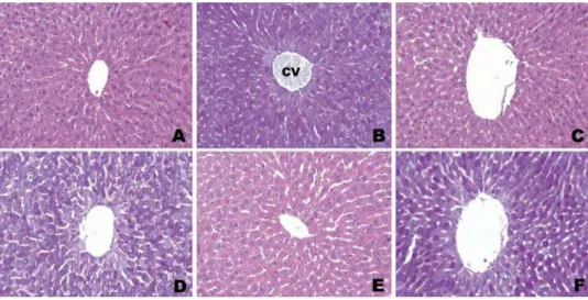
Sections of the ethanol, saline, and RSV groups did not present any histopathological alterations
(Figure 1). There was no significant difference
among these groups (p > 0.05). The liver tissues of the APAP group showed prominent portal and lobular changes including vascular congestion, necrosis, portal and focal inflammation, sinusoidal dilatation, and loss of glycogen content (Figure 2
(a–d)). The mean histopathological damage score
(MHDS) was 6.62 ± 0.37 in APAP group. There
Figure 1.Photomicrographs of liver tissue of control rats showing normal hepatic cells with central vein (CV). A. Group 1: Ethanol group. H-E; X20. B. Group 1: Ethanol group. PAS; X20. C. Group 2: Saline group. H-E; X20. D. Group 2: Saline group. PAS; X20. E. Group 3: RSV group. H-E; X20. F. Group 3: RSV group. PAS; X20.
were significant differences between APAP and ethanol, saline, RSV groups (p = 0.001, for all). In the APAP+RSV group, these changes were markedly reduced. Rarely, small necrotic hepato-cyte groups among normal-appearing hepatohepato-cytes were observed. Sinusoidal dilatation was not dis-tinct (Figure 2(e)). Loss of glycogen content was still prominent in PAS-stained sections (Figure 2
(f)). MHDS was 3.37 ± 0.32 in APAP+RSV group.
There was a significant difference between the APAP and APAP+RSV groups (p = 0.001).
MHDSs of the groups are shown in Table 1.
Immunohistochemical findings
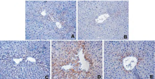
The livers of ethanol, saline, and RSV groups showed very weak iNOS expression. There were no significant differences between these groups (p > 0.05). However, livers of the APAP group showed intense iNOS expression than ethanol, saline and RSV groups (p < 0.05). In APAP group, iNOS immunostaining was most evident in pericentral hepatocytes. There were significant
differences between APAP group and ethanol, sal-ine, RSV groups (p < 0.0001, p < 0.0001, p = 0.001, respectively). In the same areas in APAP+RSV group, the intensity of iNOS immunostaining decreased with RSV administration, when com-pared to APAP group (p = 0.003) (Figure 3). The iNOS immunostaining scores are shown in
Table 2.
Electron microscopic findings
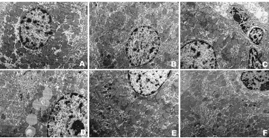
Liver sections of ethanol, saline, and RSV groups
showed normal ultrastructural appearance.
Conversely, severe changes were detected in APAP group including minimal intracytoplasmic edema, lipid droplets in hepatocytes, granular material accumulation, and closure in the space of Disse, sinusoidal endothelial degeneration, dense heterogeneous lysosomal accumulation in Kupffer cells, and locally degenerated Kupffer cells. Additionally, in this group, we also observed myelin figures within hydropic vacuoles in
peri-nuclear area and condensation in the
Figure 2.Photomicrographs of liver tissue of rats treated with acetaminophen. A. Group 4: APAP group showing severe necrosis (star) with disappearance of nuclei. H-E; X20. B. Group 4: APAP group showing inflammation areas (arrows). H-E; X20. C. Group 4: APAP group showing sinusoidal dilatation (arrows). H-E; X20. D. Group 4: APAP group showing loss of glycogen content. PAS; X20. E. Group 5: RSV administration ameliorates histopathological changes markedly in APAP-treated rat livers. H-E; X20. F. Group 5: APAP +RSV group showing loss of glycogen content, PAS; X20.
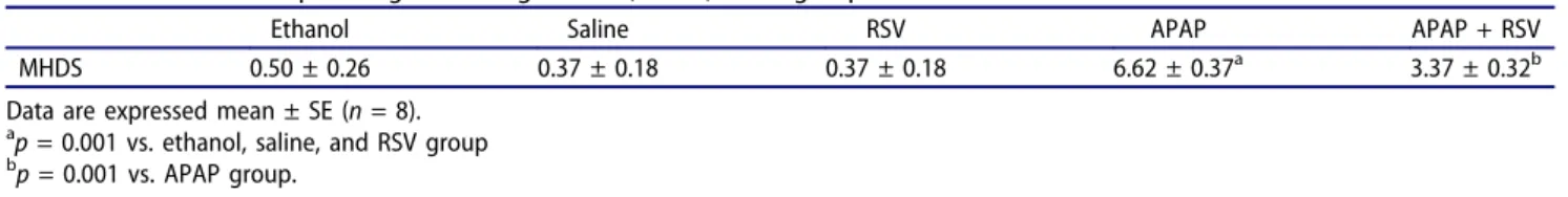
Table 1.The mean histopathological damage score (MHDS) of all groups.
Ethanol Saline RSV APAP APAP + RSV
MHDS 0.50 ± 0.26 0.37 ± 0.18 0.37 ± 0.18 6.62 ± 0.37a 3.37 ± 0.32b
Data are expressed mean ± SE (n = 8).
ap = 0.001 vs. ethanol, saline, and RSV group bp = 0.001 vs. APAP group.
mitochondrial matrix. Liver sections of APAP +RSV group were nearly normal in ultrastructural appearances. In this group, small lysosomes, rarely small myelin figures in hepatocyte cytoplasm, and locally minimal intracytoplasmic edema were observed (Figure 4).
Biochemical findings
The tissue MDA levels of APAP group were sig-nificantly increased in comparison with ethanol, saline, and RSV groups (p < 0.05, p < 0.05, p < 0.005, respectively). However, tissue MDA levels of APAP+RSV group was significantly lower than that of APAP group (p < 0.05). When compared with the ethanol, saline, and RSV groups, tissue GSH levels, SOD, and CAT activity in APAP group were significantly decreased. RSV administration resulted in a significant increase in mean GSH level, CAT, and SOD activities
(p < 0.05, p < 0.01, p < 0.005, respectively). The mean biochemical results of all groups are shown
in Table 3.
Discussion
Despite APAP being an antipyretic and pain-relieving anti-inflamatory drug, which is broadly used to treat cerebral pain, fever, and other pains, and promptly accessible without a prescription, when taken in heavy doses, it becomes an intense hepatotoxin and causes acute liver dysfunction in
humans and experimental animals.25 APAP is one
of the most common reasons of drug-induced hepatic toxicity around the world that leads to excessive treatment and hospitalization costs every year.26
In the present study, hepatic damage of APAP group was higher than other groups. Placke et al. reported that in mice given 600 mg/kg single dose
Figure 3.Immunohistochemical photomicrographs of liver tissue of rats for iNOS expression. The areas stained in brown indicated iNOS positive expression. The livers of ethanol, saline, and RSV groups showed very weak iNOS expression. A. Group 1: Ethanol group, X20. B. Group 2: Saline group, X20. C. Group 3: RSV group, X20. D. Group 4: APAP group showing most evident iNOS immunostaining in pericentral hepatocytes, X20. E. Group 5: APAP+RSV group showing that the intensity of iNOS immunostaining decreased with RSV administration, X20.
Table 2.The iNOS immunostaining scores of all groups.
Ethanol Saline RSV APAP APAP + RSV
Score 0.25 ± 0.16 0.12 ± 0.12 0.37 ± 0.18 2.50 ± 0.18a,b 1.37 ± 0.18c,d,e,f
Data are expressed mean ± SE (n = 8).
ap < 0.0001 vs. ethanol and saline group. bp = 0.001 vs. RSV group. cp = 0.002 vs. ethanol group. dp = 0.001 vs. saline group. ep = 0.005 vs. RSV group. fp = 0.003 vs. APAP group. ULTRASTRUCTURAL PATHOLOGY 43
of APAP at 60 minutes, alterations started for hydropic degeneration, followed by pyknosis, coa-gulative necrosis, and hepatocellular atrophy.27 Dash et al. reported that in paracetamol-treated group, severe hepatotoxicity was observed by severe necrosis with the disappearance of nuclei.28 Hussain et al. also observed severe congestion of blood vessels alongside vacuolization, hepatic cell necrosis, plasma cells infiltration, macrophages, eosinophils, degeneration of hepatocytes nuclei, and fibrosis.29 Khayyat et al. reported that liver specimens from mice in the APAP-administered group showed diffuse coagulative necrosis around
the vena centralis. Necrotic areas were
character-ized by eosinophilic, vacuolated, swollen
hepatocytes.26 Wu et al. reported that in the
APAP group, severe hemorrhage, sinusoidal con-gestion, and inflammatory cell infiltration dilated central vein and degenerated hepatocytes with perinuclear vacuolization.30 Polat et al. also seen similar histopathological alterations including intense necrotic foci, an increase of eosinophils, irregular Remark cordons, dilated sinusoids, intense erythrocyte aggregation, severe hyperchro-masia in the nucleus, and irregularity of the cell membrane in hepatocytes of the liver tissue, which
Table 3.The tissue oxidant–antioxidant parameters of all groups.
MDA (nmol/g wet tissue) GSH (nmol/g wet tissue) SOD (U/g protein) CAT (k/g protein)
Ethanol 362.00 ± 5.87 1.28 ± 0.14 115.87 ± 3.54 22.00 ± 0.32
Saline 374.12 ± 12.10 1.24 ± 0.09 119.50 ± 3.90 24.68 ± 0.52g
RSV 336.75 ± 4.29a,b 1.29 ± 0.04 152.75 ± 7.02g,h 30.42 ± 0.60b,g
APAP 464.25 ± 23.11c,d 0.89 ± 0.13c,d 104.37 ± 11.48i 19.97 ± 1.35d,j
APAP + RSV 384.12 ± 24.02e 1.47 ± 0.10f 161.75 ± 7.66e 27.78 ± 1.72k
Data are expressed mean ± SE (n = 8).
ap < 0.05 vs. ethanol group. bp < 0.005 vs. saline group.
cp < 0.05 vs. ethanol and saline group. dp < 0.005 vs. RSV group. ep < 0.05 vs. APAP group. fp < 0.005 vs. APAP group. gp < 0.005 vs. ethanol group. hp < 0.01 vs. saline group. ip < 0.01 vs. RSV group. jp < 0.05 vs. saline group. kp < 0.01 vs. APAP group
Figure 4.Transmission Electron Microscopic (TEM) photomicrographs of liver tissue of rats. Liver sections of ethanol, saline, and RSV groups showing normal ultrastructural appearance. A. Group 1: Ethanol group, X6300. B. Group 3: RSV group, X6300. C. Group 4: APAP group showing minimal intracytoplasmic edema, lipid droplets in hepatocytes, granular material accumulation, and myelin figures within hydropic vacuoles in perinuclear area, X6300. D. Group 4: APAP group showing intracytoplasmic edema and lipid droplets, X8000. E. F. Group 5: APAP + RSV group showing small lysosomes, rarely small myelin figures in hepatocyte cytoplasm and locally minimal intracytoplasmic edema X6300.
were given paracetamol.31 Our results are concor-dant with the literature.
The use of natural products in the treatment of in vivo and in vitro rodent models of liver injury
including APAP-induced hepatotoxicity is
popular.25,28,29,32,33 Among these products, RSV has been shown to be a powerful antioxidant and free radical scavenger.34 RSV provides protection against liver injury induced by ROS and inflam-matory cytokines and decreases hepatic lipid peroxidation.35 In previous studies, it was stated that RSV has beneficial effects on different liver injuries.35–43 In our study, RSV significantly improved light and electron microscopic altera-tions against detrimental effects of high-dose administration of APAP. It was suggested that RSV protected against APAP-induced liver toxicity due to its antioxidative and anti-inflammatory effects. McGill et al. reported that inflammation plays a significant role in APAP-induced liver toxicity and that RSV protected by decreasing
inflammation.34 Du et al. (2015) reported that
RSV prevents histopathological changes in
APAP-induced liver injury. Additionally, they emphasize that RSV may be a valid treatment option for APAP overdose.44 Wang et al. reported that centrilobular hepatocellular necrosis, intrahe-patic hemorrhage, and inflammatory cell
infiltra-tion were observed in APAP-induced
hepatotoxicity and RSV pretreatment for three days before APAP treatment prevents against liver injury.25
NO is a significant indication of the inflamma-tory process.45 In a variety of cells including neu-trophils, macrophages, and hepatocytes, iNOS is
induced by inflammatory cytokines.46,47 The
expression of iNOS leads to overproduction of NO, resulting in damage to neuronal, endothelial, and epithelial cells47 and it has been shown to be an important event in various models of liver injury.46,47 NO reacts with superoxide radicals, the formation of peroxynitrite by rapid reaction
of superoxide anion and NO mediates
nitrotyrosination.45,47 Peroxynitrite causes protein nitration and tissue injury.47During detoxification of peroxynitrite, GSH has an important role.45 In the current study, we observed a correlation between NO synthesis and APAP-induced liver damage as examined by the anti-iNOS intensity
in IHC-stained specimens. Previous studies have also reported that toxic doses of APAP lead to induction of the iNOS and nitrotyrosine expres-sion and increased tissue NO levels and serum nitrite/nitrate levels.45,46,48–50 Gardner et al. reported that cells isolated from APAP-treated (1 g/kg) rats produced more NO than cells from control animals. They concluded that NO is a significant indicator of APAP-induced hepatic toxicity.48
Ultrastructural studies related to APAP-induced hepatic injury are limited. Placke et al. reported that time- and dose-dependent (300 mg/kg and 600 mg/kg) ultrastructural changes were detected in APAP-induced hepatic damage in mice. The first ultrastructural changes (600 mg/kg of APAP single dose) on hepatocytes were seen at 60 min-utes. These cells had mild cytomegaly, decreased
density, and dispersion of organelles.
Mitochondria were compressed and were occa-sionally combined with little myelin figures. Those little intracristal calcium granules typically connected with mitochondria were not observed. Most of the endoplasmic reticulum was mildly elongated, round, or dilated. Centrilobular hepato-cytes from mice killed 4–6 hours following APAP were necrotic and degeneration had extended into mid zonal and periportal zones.27In current study, we used 1000 mg/kg single dose of APAP and we detected similar ultrastructural changes at 60 min-utes. We could not find any electron microscopic studies related with the role of RSV against APAP-induced hepatotoxicity in literature. Taken all together, we suggest that ultrastructural changes were commonly associated with higher APAP dose and RSV is excessively effective in improve-ment of ultrastructural damage. Although the pre-cise mechanism by which APAP causes cell injury is still unknown. When we consider all of these light and electron microscopic changes, it is sug-gested that mitochondria may play a significant role in the APAP-induced hepatic injury.
Oxidative stress has been hypothesized by many researchers to play a significant role in the etiology of APAP toxicity.26,31 APAP hepatotoxicity is defined as an imbalance between the production of ROS, antioxidants, and repair capacity.26MDA level is an important biomarker of lipid peroxidation and its increase in tissues, exposed to oxidative stress.26,31,45
MDA level elevation during APAP toxicity has been reported in the literature.26Several studies have indi-cated that APAP might involve free radicals and induce oxidative injury.5,26,28,30–33,45,49As a free radi-cal scavenger, GSH plays a significant role in the detoxification of ROS.31,51 Our study showed that RSV decreased levels of MDA and increased levels of hepatic antioxidant enzymes. We suggest that RSV decreases oxidative stress by directly scavenging free radicals and by upregulating antioxidant enzymes, including SOD and CAT. RSV acts as an antioxidant and it is effective in normalization of the antioxidant enzyme system. Wang et al. reported that APAP (400 mg/kg) treatment for 6 hours caused a signifi-cant decrease in liver mitochondrial GSH levels,
which was reversed by RSV pretreatment.25 Sener
et al. reported that a single dose of 900 mg/kg APAP hepatotoxicity was reduced by RSV in BALB-c mice. They determined that APAP caused a significant decrease in GSH levels while MDA levels were increased in liver. Conversely, when RSV adminis-tered following APAP, depletion of GSH and accu-mulation of MDA and neutrophil infiltration were reversed back to control.33 Our results are concor-dant with the literature.
Conclusion
We concluded that all of the histological and biochem-ical changes in APAP hepatotoxicity may be mediated by either lipid peroxidation or by peroxynitrite. We suggest that RSV has a potent effect against APAP-induced hepatic damage by providing the normaliza-tion of the antioxidant enzyme system. These findings emphasize the important role of dietary natural sub-stances and offer an innovative approach to coping with APAP-induced hepatotoxicity. Thus it may have potential beneficial effects in protection against the dose-dependent APAP administrations in patients. Further investigations should be conducted to untie the role of dietary compounds which are becoming more remarkable as alternative therapies.
Declaration of interest
The authors declare no conflicts of interest.
Funding
This study was financially supported by Scientific Research Fund of Inonu University, Turkey [Grant number: 2012/85].
References
1. Ghosh A, Sil PC. Antioxidative effect of a protein from Cajanus indicus L against acethaminophen-induced hepato-nephro toxicity. J Biochem Mol Biol. 2007;40 (6):1039–1049.
2. Galal RM, Zaki HF, Seif El-Nasr MM, Agha AM. Potential protective effect of honey against paraceta-mol-induced hepatotoxicity. Arch Iran Med. 2012;15 (11):674–680.
3. Chen X, Sun C-K, Han G-Z, et al. Protective effect of tea polyphenols against paracetamol-induced hepato-toxicity in mice is significantly correlated with cyto-chrome P450 suppression. World J Gastroenterol. 2009;15(15):1829–1835. .
4. Singh SK, Rajasekar N, Raj NAV, Paramaguru R.
Hepatoprotective and antioxidant effects of
Amorphophallus campanulatus against acetaminophen-induced hepatotoxicity in rats. Int J Pharm Pharm Sci. 2011;3(2):202–205.
5. Ilbey YO, Ozbek E, Cekmen M, et al. Melatonin pre-vents acetaminophen-induced nephrotoxicity in rats. Int Urol Nephrol. 2009;41:695–702.
6. Gamal El-Din AM, Mostafa AM, Shabanah OA, Al-Bekairi AM, Nagi MN. Protective effect of arabic gum against acetaminophen-induced hepatotoxicity in mice. Pharmacological Res. 2003;48:631–635.
7. Cao Y, Fu Z-D, Wang F, Lıu H-Y, Han R. Anti-angio-genic activity of resveratrol, a natural compound from medicinal plants. J Asian Nat Prod Res. 2005;7(3):205– 213.
8. Yar AS, Menevse S, Alp E, Helvacioglu F, Take G. The effects of resveratrol on cyclooxygenase-1 and cycloox-ygenase-2 mRNA and protein levels in diabetic rat kidneys. Mol Biol Rep. 2010;37:2323–2331.
9. Szkudelski T. Resveratrol-induced inhibition of insulin secretion from rat pancreatic islets: Evidence for pivo-tal role of metabolic disturbances. Am J Physiol Endocrinol Metab. 2007;293:901–907.
10. Hung L-M, Chen J-K, Huang -S-S, Lee R-S, Su M-J. Cardioprotective effect of resveratrol, a natural
antiox-idant derived from grapes. Cardiovasc Res.
2000;47:549–555.
11. Ignarro LJ, Buga GM, Wood KS, Byrns RE, Chaudhuri G. Endothelium-derived relaxing factor produced and released from artery and vein is nitric oxide. Proc Natl Acad Sci USA. 1987;84:9265–9269.
12. Nathan C. Nitric oxide as a secretory product of mam-malian cells. Faseb J. 1992;6:3051–3064.
13. Hibbs JBJ, Taintor RR, Vavrin Z. Macrophage cyto-toxicity: role for L-arginine deiminase and imino nitro-gen oxidation to nitrite. Sci. 1987;235:473–476. 14. Bonfoco E, Krainc D, Ankarcrona M, Nicotera P,
Lipton SA. Apoptosis and necrosis: Two distinct events induced, respectively, by mild and intense insults with N-methyl Daspartate or nitric oxide/superoxide in
cor-tical cell cultures. Proc Natl Acad Sci USA.
1995;92:7162–7166.
15. Dincel GC, Kul O. eNOS and iNOS trigger apoptosis in the brains of sheep and goats naturally infected with the border disease virus. Histol Histopathol. 2015;30 (10):1233–1242.
16. Brookes PS, Salinas EP, Darley-Usmar K, et al. Concentration-dependent effects of nitric oxide on mitochondrial permeability transition and cytochrome c release. J Biol Chem. 2000;275:20474–20479.
17. Moriya R, Uehara T, Nomura Y. Mechanism of nitric oxide-induced apoptosis in human neuroblastoma SH-SY5Y cells. FEBS Lett. 2000;484:253–260.
18. Özer MA, Polat N, Özen S, et al. Effects of
molsido-mine on retinopathy and oxidative stress ınduced by
radiotheraphy in rat eyes. Curr Eye Res. 2017;42 (5):803–809.
19. Gul M, Kayhan B, Elbe H, Dogan Z, Otlu A. Histological and biochemical effects of
dexmedetomi-dine on liver during an ınflammatory bowel disease.
Ultrastruct Pathol. 2015;39(1):6–12.
20. Uchiyama M, Mihara M. Determination of malonalde-hyde precursor in tissues by thiobarbituric acid test. Anal Biochem. 1978;86(1):271–278.
21. Ellman GL. Tissue sulphydryl groups. Arch Biochem Biophys. 1959;82:70–77.
22. Sun Y, Oberly LW, Li Y. A simple method for clinical assay of SOD. Clin Chem. 1988;34:479–500.
23. Lowry O, Rosenbraugh N, Farr L, Rondell R. Protein measurement with the folin-phenol reagent. J Biol Chem. 1951;183:265–275.
24. Casado A, De La Torre R, Lopéz-Fernández E. Antioxidant enzyme levels in red blood cells from cataract patients. Gerontol. 2001;47:186–188.
25. Wang Y, Jiang Y, Fan X, et al. Hepato-protective effect of resveratrol against acetaminophen-induced liver injury is associated with inhibition of CYP-mediated bioactivation and regulation of SIRT1– p53 signaling pathways. Toxicol Lett. 2015;236:82– 89.
26. Khayyat A, Tobwala S, Hart M, Ercal N. N-acetylcys-teine amide, a promising antidote for acetaminophen toxicity. Toxicol Lett. 2016;241:133–142.
27. Placke ME, Ginsberg GL, Wyand SD, Cohen SD. Ultrastructural changes during acute acetaminophen-induced hepatotoxicity in the mouse: a time and dose study. Toxicol Pathol. 1987;15:431–438.
28. Dash DK, Yeligar VC, Nayak SS, et al. Evaluation of
hepatoprotective and antioxidant activity of
Ichnocarpus frutescens (Linn.) R.Br. on paracetamol-induced hepatotoxicity in rats. Trop J Pharm Res. 2007 September;6(3):755–765.
29. Hussaın L, Ikram J, Rehman K, Tarıq M, Ibrahım M, Akash MSH. Hepatoprotective effects of Malva sylves-tris L. against paracetamol-induced hepatotoxicity. Turk J Biol. 2014;38:396–402.
30. Wu Y-L, Pıao D-M, Han X-H, Nan J-X. Protective effects of salidroside against acetaminophen-ınduced toxicity in mice. Biol Pharm Bull. 2008;31(8):1523– 1529.
31. Polat M, Cerrah S, Albayrak B, et al. Assessing the effect of leptin on liver damage in case of hepatic
injury associated with paracetamol poisoning.
Gastroenterol Res Pract. 2015;357360. DOI:10.1155/
2015/357360
32. Pang C, Zheng Z, Shi L, et al. Caffeic acid prevents acetaminophen-induced liver injury by activating the Keap1-Nrf2 antioxidative defense system. Free Radic Biol Med. 2016;91:236–246.
33. Sener G, Toklu HZ, Sehirli AO, Velioğlu-Oğünç A, Cetinel S, Gedik N. Protective effects of resveratrol against acetaminophen-induced toxicity in mice. Hepatol Res. 2006;35(1):62–68.
34. McGill MR, Du K, Weemhoff JL, Jaeschke H. Critical review of resveratrol in xenobiotic-induced hepatotoxi-city. Food Chem Toxicol. 2015;86:309–318.
35. Dalaklioglu S, Genc GE, Aksoy NH, Akcit F, Gumuslu S. Resveratrol ameliorates methotrexate-induced hepa-totoxicity in rats via inhibition of lipid peroxidation. Hum Exp Toxicol. 2013;32(6):662–671.
36. Chan CC, Cheng LY, Lin CL, Huang YH, Lin HC, Lee FY. The protective role of natural phytoalexin resvera-trol on inflammation, fibrosis and regeneration in cho-lestatic liver injury. Mol Nutr Food Res. 2011;55 (12):1841–1849.
37. Chan CC, Lee KC, Huang YH, Chou CK, Lin HC, Lee FY. Regulation by resveratrol of the cellular factors mediating liver damage and regeneration after acute toxic liver injury. J Gastroenterol Hepatol. 2014;29 (3):603–613.
38. Lee ES, Shin MO, Yoon S, Moon JO. Resveratrol inhi-bits dimethylnitrosamine-induced hepatic fibrosis in rats. Arch Pharm Res. 2010;33(6):925–932.
39. Oliva J, French BA, Li J, Bardag-Gorce F, Fu P, French SW. Sirt1 is involved in energy metabolism: The role of chronic ethanol feeding and resveratrol. Exp Mol Pathol. 2008;85(3):155–159.
40. Tunali-Akbay T, Sehirli O, Ercan F, Sener G. Resveratrol protects against methotrexate-induced hepatic injury in rats. J Pharm Pharm Sci. 2010;13 (2):303–310.
41. Nivet-Antoine V, Cottart CH, Lemaréchal H, et al. Trans-resveratrol downregulates Txnip overexpression occurring during liver ischemia-reperfusion. Biochimie. 2010;92(12):1766–1771.
42. Roy S, Sannigrahi S, Majumdar S, Ghosh B, Sarkar B. Resveratrol regulates antioxidant status, inhibits cyto-kine expression and restricts apoptosis in carbon tetra-chloride induced rat hepatic injury. Oxid Med Cell Longev. 2011;2011:703676.
43. Velioglu-Ogunc A, Sehirli O, Toklu H, et al. Resveratrol protects against irradiation-induced hepa-tic and ileal damage via its antioxidative activity. Free Rad Res. 2009;43:1060–1071.
44. Du K, McGill MR, Xie Y, Bajt ML, Jaeschke H. Resveratrol prevents protein nitration and release of endonucleases from mitochondria during acetamino-phen hepatotoxicity. Food Chem Toxicol. 2015;81:62–70. 45. Kuvandik G, Duru M, Nacar A, et al. Effects of erdos-teine on acetaminophen-induced hepatotoxicity in rats. Toxicol Pathol. 2008;36:714–719.
46. Gardner CR, Laskin JD, Dambach DM, et al. Reduced hepatotoxicity of acetaminophen in mice
lacking ınducible nitric oxide synthase: potential
role of tumor necrosis factor-α and interleukin-10. Toxicol Appl Pharmacol. 2002;184:27–36.
47. Kamanakaa Y, Kawabatab A, Matsuyaa H, Tagaa C, Sekiguchib F, Kawao N. Effect of a potent iNOS inhi-bitor (ONO-1714) on acetaminophen-induced hepato-toxicity in the rat. Life Sci. 2003;74:793–802.
48. Gardner CR, Heck DE, Yang CS, et al. Role of nitric oxide in acetaminophen-induced hepatotoxicity in the rat. Hepatol. 1998;26:748–754.
49. Hinson JA, Bucci TJ, Irwin LK, Michael SL, Mayeux PR. Effect of inhibitors of nitric oxide synthase on acetaminophen-induced hepatotoxicity in mice. Nitric Oxide: Biol Chem. 2002;6(2):160–167.
50. Zhao YL, Zhou GD, Yang HB, et al. Rhein protects against acetaminophen-induced hepatic and renal
toxicity. Food Chem Toxicol. 2011;49(8):1705–
1710.
51. Mutlib AE, Shockcor J, Espina R, et al. Disposition of glutathione conjugates in rats by a novel glutamic acid pathway: Characterization of unique peptide conju-gates by liquid chromatography/mass spectrometry and liquid chromatography/NMR. J Pharm Exp Ther. 2000;235:25–32.