35 PERINATAL LETHAL FORM OF HYPOPHOSPHATASIA
Hypophosphatasia is a rare and lethal metabolic bone disease characterised by low or absent serum and tissue-alkaline phosphatase activities resulting in insufficient bone mineralisation. Hypophosphatasia has been reported throughout the world and affects all races; however, it is especially prevalent in Manitoba, Canada, where about one in 25 individuals is a carrier and 1: 2.500 newborns manifest severe disease. Six forms of hypophosphatasia have been identified. The perinatal (lethal), infantile, childhood and adult types are distinguished from one another by the age at which clinical manifestations and bone
lesions are recognised. Patients with dental manifestations but no skeletal disease are considered to have odontohypophosphatasia. Pseudohypophosphatasia is a rare variant of hypophosphatasia characterised by normal serum alkalinephosphatase (ALP) activity (1). The severe infantile form of hypophosphatasia is autosomal-recessively inherited and is lethal in the neonatal period (2).
We report a neonate with congenital hypophosphatasia in which prenatal diagnosis could not be carried out, in spite of the termination of the mother’s former pregnancy because of abnormal skeletal development.
35 Beg m Atasay, Ayla G nlemez, Sevim nal K z late , Merih Berbero lu, Saadet Arslan
Begüm Atasay, Ayla Günlemez, Sevim Ünal Kızılateş, Merih Berberoğlu,
Saadet Arsan
35 Y.Ad, Y. Ad
PERINATAL LETHAL FORM OF
HYPOPHOSPHATASIA
–––––––––––––––––––––––––
* Department of Pediatrics, Ankara University, Ankara, Turkey
–––––––––––––––––––––––––––––––––––––––––––––––––––––––––––––––––––––––––––––––––––––––––––––––––––– Received: Feb. 23, 2001 Accepted: May 01, 2001
JOURNAL OF ANKARA MEDICAL SCHOOL Vol 55, No 1, 2002 35-38
SUMMARY
Hypophosphatasia is a rare bone disorder characterised by low or absent levels of the serum and tissue non-specific alkaline phosphatase necessary for normal bone mineralisation. The severe infantile form of the disease is autosomal-recessively inherited and is usually lethal in the neonatal period.
We report a neonate with congenital hypophosphatasia in which prenatal diagnosis could not be carried out, in spite of the termination of the mother s former pregnancy due to abnormal skeletal development. This report indicates the importance of the index case and the need for more careful prenatal evaluation of subsequent pregnancies.
Key Words: Hypophosphatasia, Neonate.
ÖZET
Perinatal Letal Hipofosfatazya
Hipofosfatazya, normal kemik mineralizasyonu i in gerekli olan serum ve doku alkalen fosfataz enzim aktivitesinin azalmas veya olmamas ile karekterli nadir bir kemik hastal d r. Yenido an d neminde l mle sonu lanan hastal n a r infantil formunda kal t m otozomal resesiftir.
Burada bir nceki gebeli i iskelet anomalisi nedeniyle sonland r lm ancak sonraki gebelikte prenatal tan yap lmam bir konjenital hipofosfatazya olgusu, prenatal tan n n vurgulanmas amac ile sunulmaktad r.
Patient report
A 1.97-kg female infant was born at 35 weeks of gestation by an emergency cesarian section due to fetal distress. The mother was a healthy 30-year-old whose prenatal care featured apparently normal sonographic evaluation. The infant’s first and fifth minute APGAR scores were 1 and 6 respectively, and she was resuscitated and immediately admitted to the neonatal intensive care unit.
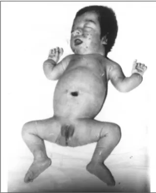
The infant was the result of the fourth pregnancy of consanguineous parents. Their first two children were healthy males; however, a third pregnancy was terminated in the second trimester due to abnormal skeletal development. There was no specific diagnosis of the deceased fetus, which had multiple skeletal malformations. Our patient had abnormalities apparent at birth, including short, bowed extremities, dimples in the extensor surfaces, generalised hypotonia, a small funnel chest, soft calvaria, very large fontanel, extremely wide cranial sutures, low-set ears and a depressed nasal bridge (Figure 1). Serum alkaline phosphatase activity was measured at birth and in the subsequent days. The measurements revealed very low levels of 15, 9 and 10 IU/L, respectively (Normal: 185-340). Serum calcium and phosphorus concentrations were 7.9 and 8.5 mg/dl, respectively.
Radiographs showed hypomineralisation of all bones, especially the calvarium, long bones and ribs; widening of sutures; and poor ossification of the calvarium, marked in the frontal and pariatal bones. Bowing was apparent in the distal portions of both upper and lower extremities. Bony spurs protruded laterally from the midshaft of the ulnae and fibulae. The major long bones had unmineralised osteoid protruding into the metaphysis, resulting in a moth-eaten appearance. (Figure 2).
The infant was in severe respiratory distress and was ventilated mechanically. She survived for six days, after which she suffered from increasing respiratory compromise due to her small chest and increased secretions.
36 PERINATAL LETHAL FORM OF HYPOPHOSPHATASIA
Figure 1. Physical appearance of the patient.
Figure 2. Radiograph of the patient. Note the moth-eaten appearance of the major long bones resulting from unmineralised osteoid protruding into the metaphysis.
Discussion
Generalised disruption of skeletal mineralisation in infants or children causes rickets, a result of subnormal levels of vitamin D, calcium and/or phosphorus. Hyphophosphatasia, on the other hand, is a rare bone disorder characterised by low levels of the tissue non-specific alkaline phosphatase (TNSALP) necessary for normal bone mineralisation. Blood and urine levels of ALP substrates, including phosphoetanolamine, inorganic pyrophosphate and pyridoxal phosphate, have been found to be increased in patients with hypophosphatasia (1).
The severity of hypophosphatasia correlates directly with the degree of deficiency of serum and tissue TNSALP activity. Perinatal hypophosphatasia, which manifests at birth, is lethal. The disease can be diagnosed by the typical appearance of short extremities, soft calvarium and respiratory distress. Characteristic radiological findings and low serum alkaline phosphatase levels confirm the diagnosis. Infants with this lethal form of hypophosphatasia usually die within a few days from respiratory insufficiency due to reduced thoracic volume and hypoplastic lungs (1).
The frequency of consanguinity and recurrence rates associated with the disease indicate an autosomal recessive mode of
inheritance in the neonatal and infantile forms (3). Prenatal diagnosis of perinatal and infantile hypophosphatasia is desirable, since the disease is lethal and there is no current treatment available (4). Methods of prenatal diagnosis include ultrasonic fetal examination and measurement of ALP activities in amniotic fluid, cultured amniotic fluid cells or chorionic villi (5). However, the reliability of these tests has not been definitively established. More reliable results have been noted with DNA analysis. The TNSALP locus maps to chromosome 1p34-p36 (4,6), and it has been shown recently that a mutation in the TNSALP gene resulted in the lethal form of hypophosphatasia. Prenatal diagnosis can be carried out using the ALP cDNA as a probe. Since impaired bone mineralisation can be observed using ultrasound, scanning seems to be another available method for prenatal diagnosis. Hypophosphatasia should be considered in the presence of polyhydramnios, low bone echogenity and signs of prominent falx cerebri (7).
Accurate prenatal ultrasonic diagnosis was not possible in our case, nor was there a precise diagnosis of the former deceased fetus or genetic counselling for the mother. This indicates the importance of diagnosis of the index case and the need for more careful prenatal evaluation of subsequent pregnancies.
37 Beg m Atasay, Ayla G nlemez, Sevim nal K z late , Merih Berbero lu, Saadet Arsan
38 PERINATAL LETHAL FORM OF HYPOPHOSPHATASIA
1. Whyte MP. Hypophosphatasia and the role of Alkaline phosphatase in skeletal mineralisation. Endocrine Reviews 1994; 15: 439-461. 2. Tekinalp G, Gürakan B, Yalçın S, et al.
Neonatal form of hypophosphatasia: Turkish J Pediatrics 1995; 37: 421-424.
3. Moore CA, Ward JC, Rivas ML, Magill HL, Whyte MP. Infantile Hypophosphatasia: autosomal recessive transmission to two related siblings. Am J Med Genet1990; 36: 15-22. 4. Kishi F, Matsuura S, Murano I, Akita A, Kajii.
Prenatal diagnosis of infantile hypophosphatasia. Prenat Diagn 1991; 11: 305-309.
5. Warren RC, Rodeck CH, Brock DJH, McKenzie
CF, Moscoso G, Barron L. First trimester diagnosis of hypophosphatasia with a monoclonal antibody to liver/bone/kidney isoenzyme of alkaline phosphatase. Lancet 1985; 5: 283-286.
6. Greenberg CR, Evans JA, McKendry-Smith S, et al. Infantile hypophosphatasia. Localisation from chromosome region 1p 36.1-34 and prenatal diagnosis using linked DNA markers. Am J Hum Genet 1990; 46: 286-292.
7. Von Dongen PWJ, Hamel BCJ, Nijhuis JG, et al. Prenatal follow up of hypophosphatasia by ultrasound: case report. Eur J Obstet Gynecol Reprod Biol 1990; 34: 283-288.