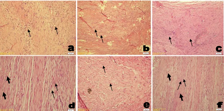
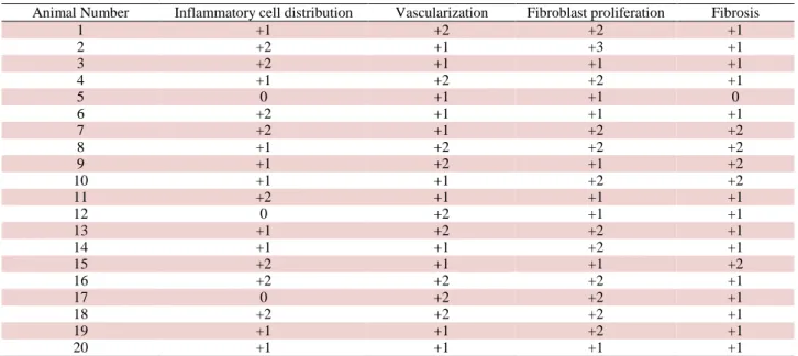
Başlık: Comparison of the effects of phonophoresis and ultrasound therapy on recovery of experimental tenorrhaphy in rabbits Yazar(lar):KARABAĞLI, Gamze; DÜZGÜN, Oktay; KARABAĞLI, Murat; HAKTANIR, Damla; GÜREL, Aydın; YETMEZ, Mehmet Cilt: 62 Sayı: 3 Sayf
Tam metin
Şekil
Benzer Belgeler
Yaptığımız çalışmada cerrahi rezeksiyon derecesi ile tümör nüksü arasında istatistiksel olarak anlamlı ilişki görülmese de, grade II ve IV cerrahi
The study group comprised patients with bipolar disorder who were recruited from outpatient clinics at Taipei City Psychiatric Center: 27 patients who received FLAI antipsychotics
According to the estimation results of SV model with dynamic leverage effect shown in Table 3, the φ coefficient indicating the permanence of Bitcoin volatility is
Yemek pişerken aşçı ekmek bulmak için oradan ayrılm ış, et kokusunu alan bir köpek yavaş yavaş sokularak, eti kapm ak istem iş, tencerenin kul bu boynuna
AB, decrease in accident and sickness absenteeism; EM, emergency measure; EP, employee participation; HS, health surveillance; IC, internal control; PM, protective measure; TI,
In the second month, statistically significant improvements con- tinued in walking VAS scores (p=0.007), flexion of the knee VAS scores (p=0.001), WOMAC pain scores (p=0.001), WOMAC
Düşük erime noktasına (32-42 °C) sahip parçalanmış veya hidrojene edilmiş bitkisel yağların yanı sıra 45-122 °C erime noktalarına sahip sıkı mono ve diasilgliseroller
1996 yılında bir hastanede yatarak tedavi gören uçucu madde kullanıcısı 78 olgunun 47 tanesinin madde kullandığı için veya hırsızlık, kavga nedeni ile polis
