A. U. Vet. Fak. DerI(.
36 (2) : 477 -48ı, ı989
BIFURCATIO TRACHEAE IN DOMESTIC AMMALS
Mehmet Kamil ÖcaII b'cil Hayvanlarda bifurcatio tracheae.
Özet: Eveil memeli hayvanlarm bilureaıio traeheae'sı makro-ana-tomik olarak incelendi. Buna göre, al, merkep ve slğIJ'da son Irachea halkası ile bronchus principalis sinisıer'in ilk halkas/ll//1 birbirine yapışmış olduğu gözlendi. Exıracartilaginous levlıalann al ve merkepıe Irachea üzerinde, sığlı'da ise bronchus principalis sinisler'in üzerinde bulunduğu görüldü. COI'ina Iracheae'l1In al, merkep ve slğıı'da her zaman yamnay şeklindeki kıkıı'dak ıara/indan desıeklendiği sapıandı.
Summar)': Gross analomical study was mQ(le on the bi(urcalio trac-heae in domeslic animals. lt was obserl'ed ıhat ıhe last traclıeal ring and the jirst bronchial canilage olıhe leji principal bmnclıus were fused in horse, donkey and OX. The extracartilaginous plates were seen 0/1the
trac-hea in horse and donkey and also on the lelı principal bronchus in ox. The carina tracheae is always supponed hy a crescent-shaped cartilage in horse, donkey and ox.
Introduction
The gross stmeture of the traehea is based mainıyon materials from the classic literature as in horse and ox by Sisson and Grossman (6), dog by Miller ct ai. (3). in addition to these, the biometrical studi-es of the trachea were reportcd in horsstudi-es and cattle by Lodgc (2) and in buffalo by Peshin and Prekash (5).
Although Getty (I) and Nickel et ai. (4) described the extracarti-laginous plates İn horse, there seems to be no enough evidence the car tilaginous structure of the bifurcatio tracheae. The purpose of this study is to present some additional anatomİcal observatİons on the bifurcatio tracheae in domestic animals.
4iıl MEHMET KAMiL ÖCAL
Materials and Methods
Six horses, sİx donkeys, six oxen, six dogs and six ca ts were used as animal materials İn this study. All the animal were adult but the breed and sex differences were not marked. The animals were lethally anesthetized and immediately af ter death the bifurcatio tracheae bet-ween the last fourth tracheal ring and at the level of the hilus pulmonis were removed and immersed in LO
1.,
formalin. The shape of the car-tilages was examined with the aid of a dissectİon microscope, then the photographs \-yere taken and siınplified drawings were made of the photographs.Observations and Discussion
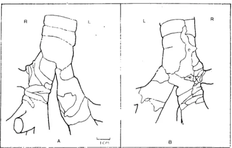
The last tracheal ring and the first bronchial ring of the kft prin-cipal bronchus are always fused in horsc, donkey and ox. AIso the last two tracheal rings showed a partİal fusion in horse and ox. Getty (I) and Nickel et aL.(4) recorded that the extracartilaginous plates are ta-ken place just cranİal to the bifurcatio tracheae İn horse. The present study confirms theİr findings. Furtherınore it was observed that the extracartilaginous plates were 4-6 in horse (Fig.
ı
e) and 3-5 İn donkeyA L.--...lıcm B
Figure ı. Schematic drawing of the bifurcatio tracheae of horse. Ventral vİew (A), dorsal view (B), left (L), right (R), extracartilaginous plates (e).
BrFURCATJO TRACHEAE rN DOMESTJC ANrMALS 1i9
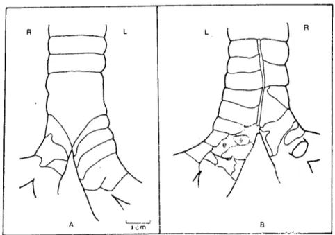
(Fig. 2e). The number of the cartilages varied according to the their size. The smaIler they were in size, the more they were in number. Most-Iy one or two smaIl, oval and thin extracartilaginous plates were seen on the left principal bronchus in ox (Fig. 3e).
i
L_._.
A_._._~_~_J
B
_
R
Figure 2. Schenıatic drawing of the bifurcatio tracheac of donkey. Ventral vicw (A), dorsal vicw (B), left (L), iight (R). cxtracarıilaginous plates (e).
The cranial cartilages of the left principal bronchus were almost the same shape of the tracheal rings in horse and ox. This has been sup-ported by the description of Getty (I).In donkey, they were broad and irregular in form Iike the right principal bronchial cartilages of ox. The cranial lobar bronchus leaves the right principal bronchus just caudal to the bifurcatio tracheae and therefore the number of the car-tilagines bronchales of the right principal bronchus was more than the left in horse and donkey. In dog (Fig. 4) and cat, the tracheal and the cranial bronchial cartilages which were almost cylindrical and the free ends did not meet dorsally had a same shape.
Getty (1) reported that the carina tracheae is usuaIly supported by a piece of cartilage which may be derived from the most distal trac-heal cartilage, from the proximal right or left bronchial cartilages or from a combination of both. But in the present study, it was found that the carina tracheae was supported by a crescent-shaped special
cartila-,ıBO MEHMET KAMiL ÖCAL
R L L R
B
---'---_._
.._,Figure 3. Schenıatic drawing of the bifurcatio tracheae of ox. Ventral vicw (A), dorsal view (B), lefı (L), right (R), cxıracartilaginoııs plaıes (e).
R
A
L
~
ıcm B
Figure 4. Schenıaıic drawing of the hifurcalİo tracheae of dog. Ventral view (A), dorsal view (B). left (L), rİght (R).
HIFURCATlO TRACHEAE .IN DOMFSTIC ANIMALS 1-RI
ge in all the specİmens of horse, donkey and ox. The dorsal end of the cartilage was thin and fiat, the ventral end was pointed. it is here pro-posed that the cartİlago carİna tracheae should be added to the Nomina Anatomica Veterİnarİa.
References
i. Getty, R. (1975): The Aııatom}' o/ the Domestic Aııimals. Fifth ed., W.B. Saunders Company, Philadelphia, London, Toronto.
2. Lodge, D. (1969): A sun'e)' o/ the tracheal dimeıısioııs in!ıorses and cattle in relatiaıı ta
the eııdotraclıeal tube size. Yet. Rec., 85: 300-303.
3. Millcr, M.E., Christensen, G.c. and Evans, H.E. (1964): Aııatomy o/ the Dog. W.B. Saundl.rs Company, Philadelphia, London.
4. Nickel, R., Schummer, A. and Seiferle, E. (1979): The Viscera o/the Domestic Mammals. Second ed., Yerlag Paul Parev, Berlin, Hamburg.
5. Peshin, P.K. and Prekash, P. (1975): A ııote011 the quaııtitatire anatomical study o/
the trachea iıı the Indiaıı Bu//alo (Bubalus bııbalis). Anat. Anı., 138: 463-467. 6. Sisson, S. and Grossman, J.D. (1959): The Aııatomy o/ the Domestic Animals,' Fourth