Summary
A case of congenital segmental spinal hypoplasia was described in a 40-day-old Holstein calf. The calf was submitted to our clinic (Istanbul University, Faculty of Veterinary Medicine, Education and Research Hospital, Surgery Clinic) with the complaint of not being able to rise to feet after birth. After clinical and radiologic inspection an anomaly was detected in spinal cord at the level of 3rd and 4th lumbar vertebrae. As the prognosis of the animal deteriorates, the calf was euthanized and systemic necropsy of the animal was performed. There was no malformation at the vertebrae around lumbar region, but a constriction was found at the third and fourth lumbar region of the spinal cord in macroscopy. It was detected that the sections prepared from the narrowest part of the medulla spinalis revealed the entire disappearance of the canalis centralis, microscopically. Furthermore syringomyelic cavities with different dimensions were observed in dorsal and ventral funiculi.
Keywords: Calf, Congenital malformation, Spinal cord, Hypoplasia
Holstein Irkı Bir Buzağıda Omurilikte
Kongenital Segmental Hipoplazi Olgusu
Özet
Bu olguda 40 günlük Holstein ırkı bir buzağıda segmental hipoplazi tanımlanmıştır. Buzağı doğum sonrası ayağa kalkamama şikâyeti ile kliniklerimize (İstanbul Üniversitesi, Veteriner Fakültesi, Eğitim ve Araştırma Hastanesi, Cerrahi Kliniği) getirildi. Klinik ve radyolojik muayene sonrasında 3. ve 4. lumbal vertebralar hizasında omurilikte anormallik saptandı. Prognozun kötüleşmesi üzerine buzağıya ötenazi ve sistemik nekropsi uygulandı. Makroskopide lumbal bölge vertebralarında herhangi bir malformasyon izlenmedi ancak 3. ve 4.lumbal segmentte omuriliğin çapında daralma tespit edildi. Omurilik çapının en dar kesiminden hazırlanan kesitlerde, mikroskopik olarak, kanalis sentralisin tamamen gözden silindiği izlendi. Ayrıca dorsal ve ventral funikuluslarda siringomiyelik kavitasyonlar şekillendiği belirlendi.
Anahtar sözcükler: Buzağı, Kongenital malformasyon, Omurilik, Hipoplazi
Congenital Segmental Hypoplasia of the Spinal Cord
in A Holstein Calf
Funda YILDIRIM
1
Aydin GUREL
1Yalcin DEVECIOGLU
2Ebru ERAVCI
2Kaan DONMEZ
31 2 3
Department of Pathology, Faculty of Veterinary Medicine, Istanbul University, TR-34320 Avcilar, Istanbul - TURKEY Department of Surgery, Faculty of Veterinary Medicine, Istanbul University, TR-34320 Avcilar, Istanbul - TURKEY Department of Surgery, Faculty of Veterinary Medicine, Selcuk University, TR-42003 Konya - TURKEY
Makale Kodu (Article Code): KVFD-2013-9166
The terms of myelodysplasia or dysraphic states describe congenital anomalies of the spinal cord. Spina bifida, neural tube closure defect and spinal duplication are most common congenital malformations occurred in cattle, concerning myelodysplasia [1]. Segmental hypoplasia of
the spinal cord is a rare form of myelodysplasia which is described as a developmental defect of one or more segments of the medulla spinalis [1]. Congenital malformations
of the spinal cord usually occur in calves and especially encountered in the thoraco-lumbar region [2]. In cattle,
those hypoplasia have been previously reported in Angus and other breeds [3,4]. Genetic and nutritional factors,
environmental factors like infections and toxins together with fertilization techniques have been shown as common reasons of congenital malformations [5,6]. Especially some
viral diseases, such as Akabane, Bovine viral diarrhea,
Blue-INTRODUCTION
İletişim (Correspondence)
+90 212 4737070 /17094
[email protected]Journal Home-Page: http://vetdergi.kafkas.edu.tr
online SubmiSSion: http://vetdergikafkas.org
CASE REPORT
Kafkas Univ Vet Fak Derg 19 (6): 1049-1052, 2013 DOI: 10.9775/kvfd.2013.9166
1050
Congenital Segmental Hypoplasia ...
tongue and Cache valley infections have been reported among the reasons of congenital anomalies [4,6]. It is strike
out that, such malformed animals cannot rise to their feet and their clinic aspects get worse after their birth. In this study, we aimed to describe a rare anomaly in a calf; congenital segmental hypoplasia of the spinal cord that was not associated with vertebral abnormalities.
CASE HISTORY
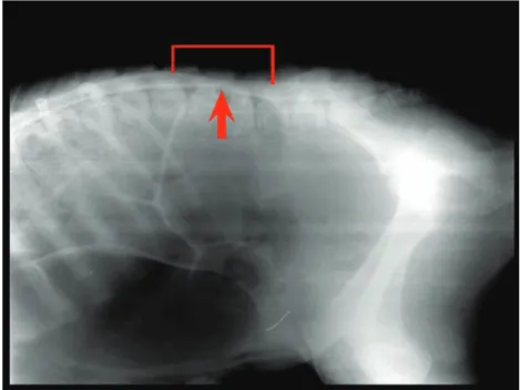
A 40-old-day Holstein calf with the complaint of not being able to rise to feet after birth was submitted to the Surgery Clinic of Istanbul University, Faculty of Veterinary Medicine, Education and Research Hospital. In clinical analyzes it is established that the animal had paraplegia. In neurologic inspection it is determined that the animal had neither tendon reflex nor deep pain sense in its rear legs. Because of ancylosis in the front legs which has occurred as a result of perpetual laying the front leg tendon reflex inspection was concluded as insignificant, however the animal had pain sense in the front legs. Although there was no pathologic alteration in the lumbar region radiography of the vertebrae, in cisternal myelography, a constriction was determined in spinal cord at the level of 3rd (L3) and 4th (L4) lumbar vertebras (Fig. 1).
In order to achieve decompression of spinal cord, hemil-aminectomie was performed to the calf. As the prognosis of the animal deteriorates after a week calf was euthanized with the approval of the animal’s owner and systemic necropsy of the animal was performed. Spinal canal of the calf was dissected transversally. After macroscopic examination tissue samples were collected into 10% formalin, processed routinely, stained with Heamatoxyline & Eosine and examined under light microscope.
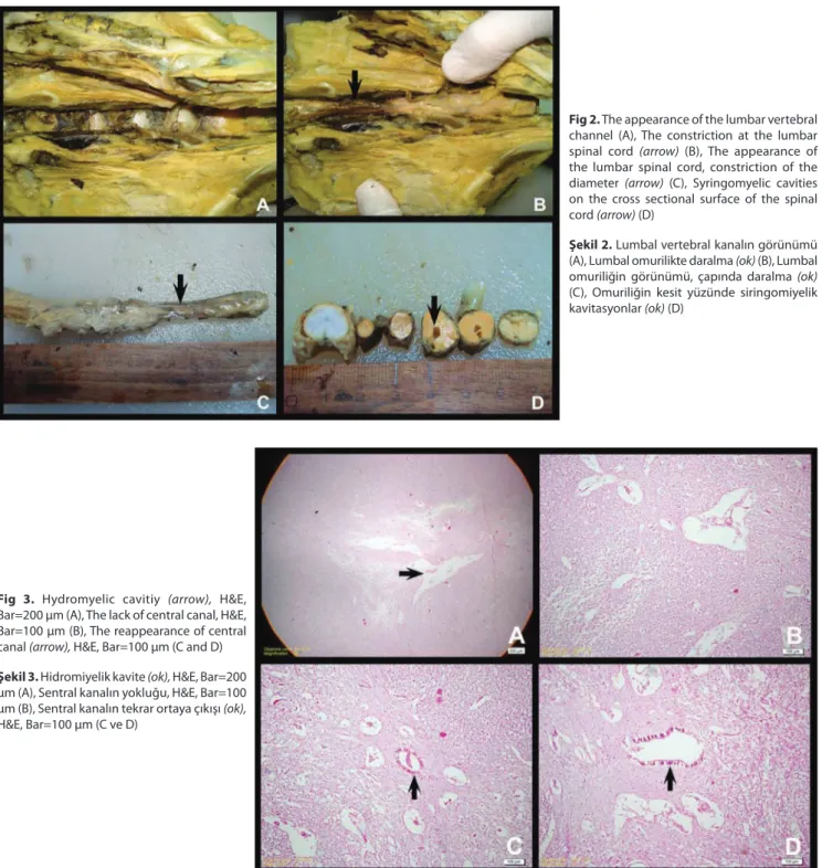
Macroscopically, there was no malformation at the vertebrae around lumbar region (Fig. 2A). But a constriction was found at the third and fourth lumbar region of the spinal cord. It was observed that the diameter of spinal cord was starting to get narrower at the cranial level of L3 and then it was starting to get broaden and reached its normal size at caudal level of L4 (Fig. 2B, 2C). It was determined that gray matter had an irregular and atrophic appearance in transversal sections. The anomaly observed at the lumbar spinal cord involved an area of 4 cm and at its narrowest point the diameter of medulla spinalis was 0.4 cm. The thickness of the cranial and the caudal parts of the narrowest point were 1.1 cm and 1.3 cm respectively. In the cross sections of the cranial part of the narrowest point, there were 1-2 mm dorsally located cavitation (Fig. 2D). Tissue samples prepared from cerebrum, cerebellum and spinal cord were evaluated microscopically. There was no inflammatory reaction in the any of tissue sections. Neuronal degeneration and atrophy in gray matter and evident demyelinisation in white matter were observed in sections prepared from hypoplastic part of medulla spinalis. Canalis centralis got broaden and became irregular. What is more, it was transformed to hydromyelic cavities in some areas (Fig. 3A). The section prepared from the narrowest part of the medulla spinalis revealed the entire disappearance of the canalis centralis (Fig. 3B). Furthermore, syringomyelic cavities with different dimensions were observed in dorsal and ventral funiculi. Then it was detected that the canalis centralis reappeared in the consecutive sections of lumbar spinal cord (Fig. 3C, 3D).
DISCUSSION
Generally vertebral defects are detected in cows with segmental hypoplasia defect of spinal cord [5]. In cases like
Fig 1. Myelography. The constriction at the 3rd and 4th
lumbar spinal cord
Şekil 1. Miyelografi. Omuriliğin 3. ve 4. lumbal
1051 YILDIRIM, GUREL, DEVECIOGLU ERAVCI, DONMEZ
Perosomus elumbis is characterized by the aplasia of the lumbosacral spinal cord and vertebrae[3,8]. However, in our
case there was neither defect nor anomaly in vertebras, but there was only segmental hypoplasia in the lumbar spinal cord.
Previously, cavitations of the spinal cord without vertebral defect were reported in spinal dysraphisms of cervical spinal cord [9,10] and in syringomyelia of thoracic
spinal cord [5,10]. Likewise in the cranial part of hypoplastic
medulla spinalis we detected a cavitation approximately 1-2 mm without any defect in vertebras. Besides, no links
between microscopic syringomyelic cavitations were observed. In spinal cord’s segmental hypoplasia of human, generally constriction of spinal channel and focal vertebral anomalies develops and it is acknowledged that they generate in relation with spinal dysgenesis [11,12]. Also in
our case the spinal cord was significantly narrower in the designated location, but the thicknesses of spinal cord in the cranial and caudal of that location were normal. As there were no vertebral neural arch or bone/skin lesions like the gashing of skin the segmental spinal cord hypoplasia in this case was not considered as neural channel defect. It is known that congenital anomalies were related with
Fig 2. The appearance of the lumbar vertebral
channel (A), The constriction at the lumbar spinal cord (arrow) (B), The appearance of the lumbar spinal cord, constriction of the diameter (arrow) (C), Syringomyelic cavities on the cross sectional surface of the spinal cord (arrow) (D)
Şekil 2. Lumbal vertebral kanalın görünümü
(A), Lumbal omurilikte daralma (ok) (B), Lumbal omuriliğin görünümü, çapında daralma (ok) (C), Omuriliğin kesit yüzünde siringomiyelik kavitasyonlar (ok) (D)
Fig 3. Hydromyelic cavitiy (arrow), H&E,
Bar=200 µm (A), The lack of central canal, H&E, Bar=100 µm (B), The reappearance of central canal (arrow), H&E, Bar=100 µm (C and D)
Şekil 3. Hidromiyelik kavite (ok), H&E, Bar=200
µm (A), Sentral kanalın yokluğu, H&E, Bar=100 µm (B), Sentral kanalın tekrar ortaya çıkışı (ok), H&E, Bar=100 µm (C ve D)
1052
Congenital Segmental Hypoplasia ...
infectious agents as well as genetic and nutritional factors. It has been reported that Fetal Akabane or Bluetongue virus infections were determined in some cases with hypoplasia and aplasia of spinal cord [4,6]. Because isolation
and identification for infectious agents was not performed, the aetiology of the present case could not be detected. On the other hand, as there was no inflammatory reaction in the microscopic evaluations of cerebrum, cerebellum and medulla spinalis, we considered that the determined hypoplasia might be related to an anomaly occurred during organogenesis depending on a genetic aberration.
The ancylosis observed in the front leg joints reminds ‘Arthrogryposis congenita’ which is also called as ‘Crooked Calf Syndrome’, known as a malformation characterized by arthrogryposis, torticollis and scoliosis and sometimes palatoschisis seen among cows[13]. Generally, some
deformities such as fracture, luxation, hyperextension, flexion or joint skew in the lower extremity joints are seen in the arthrogrypotic animals [14]. However, in our case, except
ancylosis, there was no finding mentioned above. Besides, the authors considered that ancylosis occurred secondarily as a result of perpetual laying after birth and immobility.
As the clinical findings were observed after birth and there were no pathological changes detected in the bone structure of the spinal channel and according to the histopathological findings we described the case as congenital segmental spinal cord hypoplasia. Since this was the first report in our country, the present case was considered to be worth of being presented due to its contributory value to veterinary literature.
REFERENCES
1. Leipold HW, Hiraga T, Dennis SM: Congenital defects of the bovine
central nervous system. Vet Clin North Am: Food Anim Pract, 9, 77-91, 1993.
2. Cho DY, Leopold HV: Spina bifida and spinal dysraphism in calves.
Zentalbl Vetinamed Med A, 24, 680, 1977.
3. Hiraga T, Abe M: Anatomical observation of six calves affected with
segmental aplasia of the spinal cord. Anat Rec, 219, 402-408, 1987.
4. Jubb KVF, Huxtable CR: Dysraphic states. In, Jubb KVF, Kennedy PC,
Palmer N (Eds): Pathology of Domestic Animals. 4th ed., 272-276, Academic
Press, Newyork, 1993.
5. Imai S, Moritomo Y: Segmental hypoplasia of the spinal cord in a
japanese black calf. J Vet Med Sci, 71, 337-340, 2009.
6. Noden DM, Lahunta A: Limb Development. The Embriyology of
Domestic Animals-Developmental Mechanisms and Malformations. pp.196-209 Williams&Wilkins, 428 East Preston Street Baltimore, 1985.
7. Jones CJ: Perosomus elumbis (vertebral agenesis and arthrogryposis)
in a stillborn Holstein calf. Vet Pathol, 36, 64-70, 1999.
8. Madarame H, Azuma K, Nozuki H, Konno S: Perocormus associated
with segmental aplasia of the cervical spinal cord in a Japanese Shorthorn calf. J Comp Pathol, 101, 225-230, 1989.
9. Gulbahar MY, Yuksel H, Soyguder Z, Ercin OF: Dicephalus,
arnold-chiari malformation, spinal dysraphism and other associated anomalies in a newborn holstein calf. Turk J Vet Anim Sci, 29, 565-570, 2005.
10. Ohfuji S: Spinal dysraphism in a newborn Holstein-Friesian calf. Vet
Pathol, 36, 607-609, 1999.
11. Scott RM, Wolpert SM, Barthoshesky LE, Zimbler S, Karlin L:
Segmental spinal dysgenesis. Neurosurgery, 22, 739-744, 1988.
12. Tortori-Donati P, Fondelli MP, Rossi A, Raybaud CA, Cama A, Capra V: Segmental spinal dysgenesis: Neuroradiologic findings with clinical
and embriyologic correlation. Am J Neuroradiol, 20, 445-456, 1999.
13. Samsar E, Akın F: Özel Cerrahi. pp.344-345, Medipres Matbaacılık
Yayincilik Ltd. Şti. Malatya, 2006.
14. Firat I, Yildiz F, Ozsoy S: A case of congenital flexure of fetlock in a