Original Paper
Pathophysiol Haemost Thromb 2009–10;37:72–76 DOI: 10.1159/000322916
Effects of Short-Term Streptozotocin-Induced
Diabetes and Vitamin C on Platelet Non-Enzymatic
Glycation
Saime Batırel
a
Ayşen Yarat
b
Nesrin Emekli
b
a
Medical Faculty, Istanbul Medipol University, and b Department of Biochemistry, Dentistry Faculty,
Marmara University, Istanbul , Turkey
tamin C levels were not statistically significant. Although the differences among the groups were not statistically signifi-cant, vitamin C administration increased NEG levels in the diabetic group. The results of this study demonstrate that 8 days of STZ-induced short-term diabetes did not cause a sig-nificant increase in NEG of platelets. However, the effect of vitamin C on platelet NEG needs to be further investigated.
Copyright © 2011 S. Karger AG, Basel
Introduction
Diabetes mellitus, one of the most prevalent metabol-ic syndromes worldwide, is characterized by hyperglyce-mia resulting in short-term metabolic changes in lipid and protein metabolism and in long-term irreversible vascular and connective tissue changes. These changes include diabetes-specific complications such as retinopa-thy, nephropathy and neuroparetinopa-thy, as well as macrovascu-lar complications such as atherosclerosis, potentially re-sulting in heart disease, stroke and peripheral vascular disease [1] . Links between chronic hyperglycemia and the development of long-term diabetes-specific complica-tions have been discovered but are not yet completely un-derstood [1, 2] .
Key Words
Diabetes ⴢ Streptozotocin ⴢ Vitamin C ⴢ Platelet ⴢ Glycation
Abstract
Diabetes mellitus is one of the most prevalent metabolic syndromes worldwide. Glycation, a chemical modification of proteins with reducing sugars, indicates a possible explana-tion for the associaexplana-tion between hyperglycemia and the wide variety of tissue pathologies. Non-enzymatic glycation (NEG) of platelet proteins is one of the key mechanisms in the pathogenesis of diabetic complications and may be signifi-cant in diabetic atherothrombosis. The aim of this study was to investigate the effects of streptozotocin (STZ)-induced short-term experimental diabetes on the glycation of plate-lets and to find out if vitamin C affected this glycation. A to-tal of 40 male Wistar albino rats, 200–250 g, were randomly divided into 4 groups (2 diabetic and 2 control groups). The diabetic groups were made diabetic by intraperitoneal injec-tion of STZ (65 mg/kg, citrate buffer pH 4.5). By daily intra-peritoneal injection, 80 mg/kg vitamin C (Roche, Turkey) was administered until the end of the experiment. Blood glucose levels of the diabetic groups were significantly higher than those at day 0 and also higher than those of the non-diabet-ic control groups. The changes in total protein, NEG and
Received: September 22, 2010
Accepted after revision: November 22, 2010 Published online: January 20, 2011
Glycation, a chemical modification of proteins with reducing sugars, indicates a possible explanation for the association [3, 4] between hyperglycemia and the wide variety of tissue pathologies. Research suggests that re-ducing sugars can react with the amino groups of long-lived proteins to produce non-enzymatic cross-links [5] . Formations of these cross-links occur as end stage prod-ucts of the Maillard reaction; they are known as advanced glycation end products (AGEs) [3, 6] .
AGEs are a class of complex, often unstable, reactive compounds formed in excess during aging and diabetes mellitus [5] . According to the ‘glycation hypothesis’, ac-cumulation of AGEs alters the structural properties of tissue proteins and reduces their susceptibility to catabo-lism. It has been shown that the process of AGE formation is accelerated by hyperglycemia. Some of the protein al-terations observed in diabetic patients resemble those in much older, non-diabetic patients, suggesting ‘diabetes-induced early aging’ [3–6] .
Growing evidence suggests that platelets of diabetic patients are larger and hyperreactive, showing increased adhesion and aggregation, as well as increased platelet-dependent thrombin generation [7] . Several mechanisms may account for the increased platelet reactivity in pa-tients with diabetes. Non-enzymatic glycation (NEG) of platelet proteins is one of the key mechanisms in the pathogenesis of diabetic complications and may be sig-nificant in diabetic atherothrombosis [8] .
Vitamin C, the antiscorbutic vitamin, is found in a large variety of foods but particularly in fruits and
vege-tables. The 2 major forms, L -ascorbic acid and
dehydro-L -ascorbic acid, are interconvertible through an
oxida-tion reducoxida-tion system which represents the fundamental basis for many of the biomedical roles of the vitamin [9] .
Vitamin C is actively taken up in high concentration by secretory cells of the islets of Langerhans where it is believed to play a role in antioxidant defense and as an electron donor in the post-translational enzymatic
pepti-dyl ␣ -amidation of islet peptides including amylin and
pancreatic polypeptide [10, 11] . In diabetes mellitus, the vitamin C metabolism is abnormal, and subjects have
been shown to have low vitamin C and high dehydro- L
-ascorbic acid concentrations in plasma [12] .
The pathogenesis of atherosclerosis in diabetes has several potential contributors, which include increased intravascular thrombin generation and reduced fibrino-lytic potential. Endothelial injury or plaque rupture with platelet adhesion and aggregation at the site of injury may be the critical event in producing morbidity and mortality from atherogenesis because most coronary
events occur with less than one-third narrowing of the vessel lumen. Therefore, platelets may assume an impor-tant role in the signal event in atherosclerosis in diabetes [13, 14] .
Consequently, the aim of this study was to investigate the effects of streptozotocin (STZ)-induced short-term experimental diabetes on the glycation of platelets and to find out if vitamin C affected this glycation.
Materials and Methods
A total of 40 male Wistar albino rats, 200–250 g, were ran-domly divided into 4 groups (2 diabetic and 2 control groups). They were kept at a constant temperature (22 8 1 ° C) with
12-hour light and dark cycles. The diabetic groups were rendered diabetic at day 0 by intraperitoneal injection of STZ (65 mg/kg, citrate buffer pH 4.5) [15] . Two days after the STZ injection, blood glucose levels were determined. Rats with a glucose level ! 150 mg/ dl were discarded from the experiment. After diabetes induction, 1 rat of the control and the diabetic group, respectively, was given 80 mg/kg vitamin C (Roche, Turkey) by daily intraperitoneal in-jection until the end of the experiment on day 8. Saline solution was given to the rest of the control and diabetic groups intraperi-toneally. On day 8, cardiac blood samples were taken from all rats under ether anesthesia. All experiments were carried out in ac-cordance with the guidelines of the Animal Care and Use Com-mittee of Istanbul University, Institute of Experimental Medicine. Blood glucose was measured by the Randox glucose kit (Ran-dox, GL3981, UK).
Blood samples were centrifuged for 5 min at 123 g, and plate-let-rich plasma was separated from the supernatant. Plateplate-let-rich plasma was further centrifuged for 10 min at 492 g for the pre-cipitation of the platelets, and the supernatant was used for the determination of vitamin C and glucose levels. The precipitate (platelet pellet) was washed 3 times with physiologic saline and then further diluted with 2 ml physiologic saline. This suspension was preserved for platelet count, glycosylation and protein deter-mination. Platelets were counted using phase contrast microscopy [16] .
NEG Assay
NEG of the platelet suspensions was assessed by the 2-thiobar-bituric acid method [17] . The latter involved hydrolysing each 0.5-ml homogenate with 0.5 0.5-ml of 0.5 M oxalic acid in an autoclave for 1 h at 124 8 1 ° C. To this, 0.5 ml 40% trichloracetic acid (w/v) was
added, mixed, centrifuged at 1,500 g for 10 min and filtered using filter paper. Absorbance at 443 nm was recorded. Then, 0.75 ml of supernatant was incubated in 0.25 ml of 0.05 M 2-thiobarbituric acid at 37 ° C for 30 min. After standing for 15 min at room
tem-perature, absorbance was again measured at 443 nm, and the dif-ferences between the first and second absorbances were calcu-lated. The protein glycation values were expressed as nmol of fructose/mg protein. NEG values were also divided by the platelet cell counts in order to obtain the NEG/platelet ratio. Commercial fructose (Sigma, St. Louis, Mo., USA) was used as a standard.
Protein Assay
Total protein levels of the platelet suspensions were deter-mined according to the method of Lowry et al. [18] . In alkaline medium, proteins are reacted with cupper ions and then are re-duced by folin reactive (phosphomolybdic-phosphotungstic acid). The absorbance of the blue-colored product at 500 nm was evalu-ated. Bovine serum albumin was used as a standard. The total protein level was expressed as % mg. Total protein levels were di-vided by the number of platelet cells in 1 mm 3 homogenate in or-der to obtain the protein/platelet ratio.
Determination of Vitamin C Levels
Vitamin C levels were determined by the dinitrophenylhydra-zine method based on the production of a derivative of dehydro-ascorbic acid (DHAA) with dinitrophenylhydrazine to measure the DHAA content, and indirectly, to measure total ascorbic acid after the native ascorbic acid has been oxidized to DHAA [19, 20] .
Statistical Analysis
Statistical analysis was carried out with the InStat statistics program using analysis of variance (ANOVA) and Student’s t test. p values ! 0.05 were regarded as significant.
Results
The mean levels of the blood glucose and body weight for the 6 groups at the end of 8 days are shown in table 1 . Prior to inducing diabetes (day 0), the groups were checked for differences in weight and blood glucose, but
none were found. After 8 days of diabetes, the blood glu-cose levels of the diabetic groups were significantly high-er than those at day 0 and also highhigh-er than those of the non-diabetic control groups. Vitamin C administration did not decrease the blood glucose level in the diabetic group ( table 1 ).
Also, after 8 days of diabetes, body weights of the rats in the diabetic groups decreased significantly compared with those at day 0 and also with those of the non-diabet-ic control groups ( table 1 ).
Total protein levels in the platelet suspensions were
di-vided by the number of platelets in 1 mm 3 homogenate:
they were found to be 0.144 8 0.06, 0.182 8 0.04, 0.202 8 0.12 and 0.179 8 0.11 mg/number of platelets in the control, control + vitamin C, diabetic and diabetic + vi-tamin C group, respectively. Differences between the groups were not statistically significant.
Vitamin C levels of the control, control + vitamin C, diabetic and diabetic + vitamin C group were 1.22 8 0.34, 1.35 8 0.38, 1.14 8 0.51 and 0.99 8 0.31 mg/dl, re-spectively. The mean vitamin C level of the diabetic + vitamin C group was lower than that of the control + vi-tamin C group; however, this difference was not statisti-cally significant.
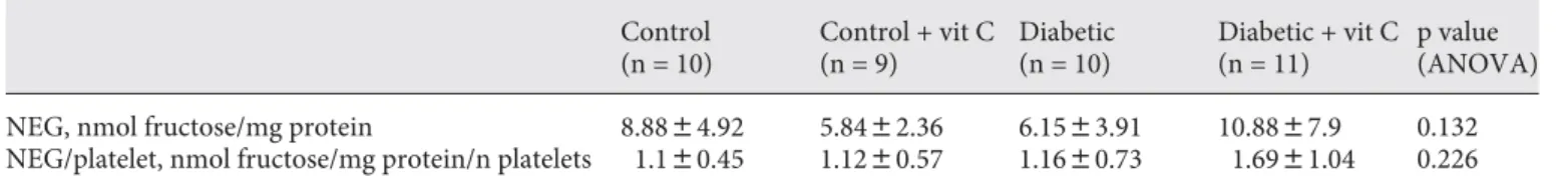
NEG levels of the platelet suspensions obtained from the 4 groups are given in table 2 . Although the differ-ences among the groups were not statistically significant,
Table 1. Mean levels of body weight and blood glucose at the end of 8 days Control (n = 10) Control + vit C (n = 9) Diabetic (n = 10) Diabetic + vit C (n = 11) p value (ANOVA) Weight, mg 243.9836.2 243.3838.3 202.8839.7* 194.7836.2* 0.0086 Blood glucose, mg/dl 149.7819.4 124.8828.7 254.18115.2* 247.28101.8* 0.0011
V alues are given as the mean 8 SD. vit C = Vitamin C. * p < 0.01, significantly different from the control and control + vitamin C groups.
Table 2. NEG values of the platelet suspensions obtained from the 4 groups Control (n = 10) Control + vit C (n = 9) Diabetic (n = 10) Diabetic + vit C (n = 11) p value (ANOVA)
NEG, nmol fructose/mg protein 8.8884.92 5.8482.36 6.1583.91 10.8887.9 0.132
NEG/platelet, nmol fructose/mg protein/n platelets 1.180.45 1.1280.57 1.1680.73 1.6981.04 0.226 V alues are given as the mean 8 SD. vit C = Vitamin C.
vitamin C administration increased NEG levels in the diabetic group. NEG/platelet ratios are also given in ta-ble 2 . Similarly, vitamin C administration insignificantly increased the NEG/platelet ratio in the diabetic group.
Discussion
The pathogenesis of diabetic complications continues to be a central issue in current diabetes research [2] . Type 2 diabetes mellitus increases atherothrombotic risk. Platelets in individuals with diabetes show increased ac-tivity at baseline and in response to agonists, ultimately leading to increased aggregation. The primary cause of mortality in the majority of patients with diabetes is atherothrombosis. Platelet hyperactivity (in combina-tion with abnormalities in coagulacombina-tion and fibrinolysis), which is characteristic in diabetes, undoubtedly is a con-tributing factor as platelets play a pivotal role in initiating and sustaining thrombi within vessels [21, 22] .
Acute in vivo hyperglycemia results in platelet activa-tion in type 2 diabetes. Furthermore, a link between hy-perglycemic spikes and the incidence of ischemic events has been demonstrated [23] . Hyperglycemia also causes NEG of platelet membrane proteins resulting in changes in protein structure and conformation, as well as in al-terations of membrane lipid dynamics [24] . This in turn can lead to enhanced expression of certain crucial platelet receptors, for instance, P-selectin and glycoprotein IIb/ IIIa, thus altering platelet activity [25] .
In the present study, we aimed to investigate the NEG of platelets in short-term acute hyperglycemia. Moreover, we aimed to investigate if vitamin C administration af-fected the level of this glycation in STZ-induced experi-mental diabetes. At the end of the 8-day experiexperi-mental period, no significant difference was found among the 4 groups; however, the vitamin-C-administered diabetic group presented the highest NEG value, as well as the lowest plasma vitamin C levels.
Several studies have demonstrated that diabetes is as-sociated with abnormalities in plasma vitamin C levels and ascorbate status [26, 27] . The loss of vitamin C in diabetes has been postulated to be due to the increased oxidative stress associated with the disease [28] . Benefits of vitamin C supplementation in humans with type 2 di-abetes have been described. Additionally, it has been pro-posed that dietary measures to increase plasma vitamin C may be an important strategy to counter diabetes [29] . Clearly, vitamin C has several important biological func-tions that may be of benefit in these situafunc-tions. It is
con-ceivable that one of these roles is the maintenance of the functional integrity of biologically active proteins, such as insulin by inhibition of glycation [30] .
However, in the literature, there are contradictory re-sults on the effect of vitamin C on glycohemoglobin lev-els. Weykamp et al. [31] suggested that supplementation of non-diabetics with 750 or 1,500 mg of vitamin C daily for 12 weeks does not cause any interference with glyco-hemoglobin determinations by HPLC, electrophoresis, affinity chromatography or immunoassay, and does not reduce in vivo hemoglobin glycation. On the other hand, Ely [32] reported antagonism of hemoglobin glycation by ascorbic acid in animals and humans. To our knowledge, the effect of vitamin C administration on platelet glyca-tion has not been studied before. Platelet hyperaggre-gability may be involved in the pathogenesis of diabetic micro- and macroangiopathy, and vitamin C has been suggested to reduce platelet aggregation [33] . Moreover, NEG of platelet membrane proteins has been reported to change platelet activity [25] .
Based on the results of this present study, we may sug-gest that diabetes induced by STZ for 8 days did not cause a significant difference in terms of platelet NEG. How-ever, among the 4 groups, the vitamin-C-administered diabetic group presented the highest NEG value. Accord-ingly, the oxidation of ascorbic acid has been reported to lead to the formation of several compounds which are capable of reacting with protein amino groups via a Mail-lard reaction [34] . The reaction by which ascorbic acid causes glycation and cross-linking of lens proteins dis-plays a rigid requirement for the presence of oxygen and is inhibited by the presence of glutathione. Oxygen is re-quired to oxidize ascorbic acid to DHAA and other prod-ucts, i.e. the active glycating species [35] .
In conclusion, although acute in vivo hyperglycemia results in platelet activation in type 2 diabetes, the results of this study demonstrate that 8-day STZ-induced short-term diabetes did not cause a significant increase in NEG of platelets. However, the effect of vitamin C on platelet NEG needs to be further investigated.
Acknowledgement
The authors thank Dr. Ebru Emekli-Alturfan for editing the manuscript.
References
1 Hudson BI, Hoffmann MA, Bucciarelli L, Wendt T, Moser B, Lu Y, Qu W, Stern DM, D’Agati V, Yan SD, Van SF, Grant PJ, Schmidt M: Glycation and diabetes: the RAGE
con-nection. Curr Sci 2002; 83: 1515–1521.
2 McCance DR, Dyer DG, Dunn JA, Bailie KE, Thorpe SR, Baynes JW, Lyons TJ: Maillard reaction products and their relation to com-plications in insulin-dependent diabetes
mellitus. J Clin Invest 1993; 91: 2470–2478.
3 Dyer DG, Dunn JA, Thorpe SR, Beilie KE, Lyons TJ, Mc Cance DR, Boynes JW: Accu-mulation of Maillard reaction products in skin collagen in diabetes and aging. J Clin
Invest 1993; 91: 2463–2469.
4 Kagami K, OndaK, Oka K, Hirano T: Sup-pression of blood lipid concentrations by vol-atile Maillard reaction products. Nutrition
2008; 24: 1159–1166.
5 Wautier JL, Guillausseau PJ: Advanced gly-cation end products, their receptors and
dia-betic angiopathy. Diabetes Metab 2001; 27:
535–542.
6 Ahmed N: Advanced glycation end prod-ucts: role in pathology of diabetic
complica-tions. Diabetes Res Clin Pract 2005; 67: 3–21.
7 Arjomand H, Roukoz B, Surabhi SK, Cohen M: Platelets and antiplatelet therapy in pa-tients with diabetes mellitus. J Invest Cardiol
2003; 15: 264–269.
8 Yamagishi S, Nakamura K, Imaizumi T: Ad-vanced glycation end products (AGEs) and diabetic vascular complications. Curr
Dia-betes Rev 2005; 1: 93–106.
9 Wardlow GM, Insel PM: The water soluble vitamins; in Wardlow GM, Insel PM: Per-spectives in Nutrition, ed 3. St Louis, Mosby, 1995, pp 437–479.
10 Zhou A, Thorn NA: High ascorbic acid con-tent in the rat endocrine pancreas.
Diabeto-logia 1991; 34: 839–842.
11 Zhou A, Nielsen JH, Farver O, Thorn NA: Transport of ascorbic acid and dehydro-ascorbic acid by pancreatic islets from
neo-natal rats. Biochem J 1991; 274: 739–744.
12 Will JC, Ford ES, Bowman BA: Serum vita-min C concentrations and diabetes: findings from the Third National Health and Nutri-tion ExaminaNutri-tion Survey. Am J Clin Nutr
1999; 70: 49–52.
13 Ross R: Atherosclerosis-inflammatory
dis-ease. N Engl J Med 1999; 340: 115–126.
14 Vinik AI, Erbas T, Park TS, Nolan R, Pit-tenger GL: Platelet dysfunction in type 2
dia-betes. Diabetes Care 2001; 24: 1476–1485.
15 Junod A, Lambert AE, Stauffacher W, Re-nold AE: Diabetogenic action of streptozoto-cin. Relationship of dose to metabolic
re-sponse. J Clin Invest 1969; 48: 2129–2139.
16 Staven P: Platelet count by phase contrast mi-croscopy – new diluting fluid for better
visu-alization. Scand J Lab Invest 1974; 33: 121–
123.
17 Parker KM, England JD, Hassel R, Goldstein PE: Improved colorimetric assay for
glyco-sylated hemoglobin. Clin Chem 1981; 27:
669–672.
18 Lowry OH, Rosebrough WI, Farr Al, Randal RJ: Protein measurement with the folin
phe-nol reagent. J Biol Chem 1951; 193: 265–275.
19 Brewster MA, Turley CP: Vitamin C; in Pesce AJ, Kaplan LA (eds): Methods in Clin-ical Chemistry. St Louis, Mosby, 1987, pp 574–581.
20 Tietz NW: Fundamentals of Clinical Chem-istry, ed 3. Philadelphia, Saunders, 1987, p 513.
21 Beckman JA, Creager MA, Libby P: Diabetes and atherosclerosis: epidemiology, patho-physiology and management. JAMA 2002;
287: 2570–2581.
22 Carr ME: Diabetes mellitus: a
hypercoagu-lable state. J Diabetes Complications 2001; 15:
44–54.
23 Mustard JF, Packham MA: Platelets and
dia-betes mellitus. N Engl J Med 1977; 297: 1345–
1347.
24 Winocour PD, Watala C, Kinlough-Rath-bone RL: Reduced membrane fluidity and increased glycation of membrane proteins of platelets from diabetic subjects are not asso-ciated with increased platelet adherence to
glycated collagen. J Lab Clin Med 1992; 120:
921–928.
25 Ferroni P, Basili S, Falco A, Davi G: Platelet activation in type 2 diabetes mellitus. J
Thromb Haemost 2004; 2: 1282–1291.
26 Sinclair AJ, Taylor PB, Girling AJ: Low plas-ma ascorbate levels in patients with type 2 diabetes mellitus consuming adequate
di-etary vitamin C. Diabet Med 1994; 11: 893–
901.
27 Will JC, Byers T: Does diabetes mellitus in-crease the requirement for vitamin C? Nutr
Rev 1996: 54: 193–202.
28 Noctor G, Foyer C: Ascorbate and glutathi-one: keeping active oxygen under control.
Annu Rev Plant Mol Biol 1998; 49: 249–279.
29 Sargeant LA, Wareham NJ, Bingham S, Day NE, Luben RN, Oakes S, Welch A, Khaw KT: Vitamin C and hyperglycemia in the Euro-pean Prospective Investigation into Cancer-Norfolk (EPIC-Cancer-Norfolk) study: a population
based study. Diabetes Care 2000; 23: 726–
732.
30 Yasser HA, Finbarr PM, Money MH, Barnett CR, Flatt PR: Vitamin C supplementation decreases insulin glycation and improves glucose homeostasis in obese hyperglycemic
(ob/ob) mice. Metabolism 2002; 51: 514–517.
31 Weykamp CW, Penders TJ, Baadenhuijsen FA, Muskiet WM, van der Slik W: Vitamin C
and glycohemoglobin. Clin Chem 1995; 41:
713–716.
32 Ely JTA: Unrecognized pandemic subclini-cal diabetes of the affluent nations: causes, cost and prevention. J Orthomolecular Med
1996; 11: 95–99.
33 Sarji KE, Kleinfelder J, Brewington P, Gon-zalez J, Hempling H, Colwell JA: Decreased platelet vitamin C in diabetes mellitus: pos-sible role in hyperaggregation. Thromb Res
1979; 15: 639–650.
34 Ortwerth BJ, Slighta SH, Prabhakarama M, Sunb Y, Smithb YB: Site-specific glycation of lens crystallins by ascorbic acid. Biochim
Biophys Acta 1992; 1117: 207–215.
35 Prabhakarama M, Ortwertha BJ: The glyca-tion-associated crosslinking of lens proteins by ascorbic acid is not mediated by oxygen
free radicals. Exp Eye Res 1991; 53: 261–268.