Objectif: L’objectif de cette étude était d’étudier l’anémie ferriprive et la carence en vitamine D parmi les enfants autistiques et d’évaluer l’importance des facteurs de risque (déterminants). Sujets et méthodes: il s’agissait d’une étude cas-témoins réalisée chez les enfants atteints d’autisme à la Hamad Medical Corporation au Qatar. Au total, 308 cas et un nombre égal de contrôles ont été inscrits. Le programme d’observation diagnostique de l’autisme générique (ADOS) était l’instrument utilisé pour diagnostiquer l’autisme. Résultats: L’âge moyen (± SD, en années) pour les enfants autistes versus témoins était de 5,39 ± 1,66 vs 5,62 ± 1,81. La valeur moyenne des taux de sérum dans les enfants autistes a été considérablement réduite et significativement plus faible que dans les enfants témoins (74,13 ± 21,61 ug/dL avec une médiane de 74 chez les enfants autistes 87,59 ± 23,36 ug/dL dans les témoins) (P = 0,003). De même, l’étude a révélé que la carence en vitamine D était considérablement plus fréquente chez les enfants autistes (18,79 ±
8,35 ng/mL) par rapport aux enfants en bonne santé (22,18 ± 9,00 ng/mL) (P = 0,004). Enfin, les valeurs moyennes de l’hémoglobine, de la ferritine, du magnésium; potassium calcium; phosphoreux; le glucose, le phosphate alcalin, l’hématocrite, le globule blanc (CMB) et le volume corpusculaire moyen [MCV] étaient statistiquement significativement plus élevés chez les enfants témoins sains que chez les enfants autistes (P < 0,001). L’analyse de régression logistique
Abstract
Aim: The aim of this study was to investigate iron deficiency anemia and Vitamin D deficiency among autism children and to assess the importance of risk factors (determinants). Subjects and Methods: This was a case–control study conducted among children suffering from autism at the Hamad Medical Corporation in Qatar. A total of 308 cases and equal number of controls were enrolled. The Autism Diagnostic Observation Schedule-Generic was the instrument used for diagnosis of Autism. Results: The mean age (±standard deviation, in years) for autistic versus control children was 5.39 ± 1.66 versus 5.62 ± 1.81, respectively. The mean value of serum iron levels in autistic children was severely reduced and significantly lower than in control children (74.13 ± 21.61 µg/dL with a median 74 in autistic children 87.59 ± 23.36 µg/dL in controls) (P = 0.003). Similarly, the study revealed that Vitamin D deficiency was considerably more common among autistic children (18.79 ± 8.35 ng/mL) as compared to healthy children (22.18 ± 9.00 ng/mL) (P = 0.004). Finally, mean values of hemoglobin, ferritin, magnesium; potassium, calcium; phosphorous; glucose, alkaline phosphate, hematocrit, white blood cell, and mean corpuscular volume were all statistically significantly higher in healthy control children as compared to autistic children (P < 0.001). Multivariate logistic regression analysis revealed that serum iron deficiency, serum calcium levels, serum Vitamin D levels; ferritin, reduced physical activity; child order, body mass index percentiles, and parental consanguinity can all be considered strong predictors and major factors associated with autism spectrum disorders. Conclusion: This study suggests that deficiency of iron and Vitamin D as well as anemia were more common in autistic compared to control children.
Keywords: Autism spectrum disorders, epidemiology, ferritin, iron deficiency, risk factors, Vitamin D
Address for correspondence: Prof. Abdulbari Bener, Department of Biostatistics and Medical Informatics, Cerrahpaşa Faculty of Medicine, Istanbul University and Istanbul Medipol University, International School of Medicine, 34098 Cerrahpasa, Istanbul, Turkey. E‑mail: [email protected]
Iron and Vitamin D Levels among Autism Spectrum Disorders
Children
Abdulbari Bener1,2, Azhar O. Khattab3,4, Dinesh Bhugra5, Georg F. Hoffmann6
1Department of Biostatistics and Medical Informatics, Cerrahpasa Faculty of Medicine, Istanbul University, Istanbul, Turkey, 2Department of Evidence for Population Health Unit, School of Epidemiology and Health Sciences, University of Manchester, Manchester, 5Institute of Psychiatry, Section of Cultural Psychiatry, King’s College London, London, England, UK, 3Department of Pediatrics, Rumailah and Hamad General Hospital, Hamad Medical Corporation, 4Department of Pediatrics,
Weill Cornell Medical College, Ar‑Rayyan, Qatar, 6Department of Pediatrics, University of Heidelberg, Baden‑Württemberg, Germany
Résumé
Access this article online
Quick Response Code:
Website:
www.annalsafrmed.org
DOI:
10.4103/aam.aam_17_17
This is an open access article distributed under the terms of the Creative Commons Attribution‑NonCommercial‑ShareAlike 3.0 License, which allows others to remix, tweak, and build upon the work non‑commercially, as long as the author is credited and the new creations are licensed under the identical terms.
For reprints contact: [email protected]
How to cite this article: Bener A, Khattab AO, Bhugra D, Hoffmann GF. Iron and vitamin D levels among autism spectrum disorders children. Ann Afr Med 2017;16:186-91.
I
ntroductIonAutism which has become a rather common disorder among 3–8 aged children may affect all aspects of a child’s life.[1,2] Genetic, nutritional, and environmental factors have
all been implicated as risk factors for autism.[3-8] Autism
is a neurodevelopmental disorder characterized by severe qualitative impairments in social interaction, and verbal and nonverbal communication, along with restricted, stereotyped interests, and behaviors.[3] The disorder is accompanied by
mental retardation in three out of four patients, and boys are four times more likely than girls to have the disorder. The underlying etiology is most likely multifactorial; and it is suggested that in most cases autism results from the interaction of multiple genetic and environmental factors.[1,2] As the risk of
autism spectrum disorder (ASD) is generally acknowledged to reflect both genetic and environmental factors,[3-6] the interplay
between genetic and environmental factors has become the subject of intensified research in the past several years.[3-9]
Iron deficiency is a major nutritional health concern among infants and young children resulting in insufficient iron to maintain normal cellular function. It affects 47% of children worldwide, 50% of children in developing countries,[3] and
6%–12% of children in developed countries.[9] An association
between iron deficiency and autism has been documented.[10-15]
Iron deficiency and iron deficiency anemia were more common in this clinical sample of children with global developmental delay and/or standard deviation (SD) than in the general population.[14,16] Vitamin D and iron have a unique role in
brain homeostasis, embryogenesis and neurodevelopment, immunological modulation (including the brain’s immune system), antioxidation, antiapoptosis, neural differentiation, and gene regulation.[17-23] Infants and children with ASDs
often have food selectivity and restricted diets, putting them at risk for nutritional deficiencies.[9] The previous studies have
demonstrated a high prevalence of iron deficiency in children with ASDs living in Wales, Canada, and Turkey.[7,11,12]
Although autism has a significant genetic component, it is primarily diagnosed through behavioral characteristics.[4-6]
Diagnosing autism has been formalized with instruments carefully designed to measure impairments indicative of autism in three developmental areas: language and communication, reciprocal social interactions, and restricted or stereotypical interests and activities. One of the most widely used instruments is the Autism Diagnostic Observation Schedule (ADOS)-Generic ADOS.[6] The ADOS consists of
a variety of semi-structured activities designed to measure social interaction, communication, play, and imaginative use of materials. The objective of this study was to investigate iron deficiency anemia and Vitamin D deficiency among autism children and to assess the importance of risk factors (determinants).
s
ubjectsandM
ethodsThis is a case–control study which was designed to determine the relationship between iron, anemia, Vitamin D, and autism in participants younger than 8-year-old at the Hamad Medical Corporation, Qatar. The survey was conducted from June 2011 to May 2014. This evaluation is based on 308 cases with ASDs and 308 control participants.
This study was approved by the Hamad General Hospital, Hamad Medical Corporation of Institutional Review Board Research Ethics Committee of and have been performed in accordance with the ethical standards laid down in the 1964 Declaration of Helsinki. All the persons who agreed to participate in this study gave their informed consent before their inclusion in the study.
Data collection
Study measurement‑clinical evaluation of autistic patients
This was based on clinical history taken from caregivers, clinical examination, and neuropsychiatric assessment. In addition, the degree of the disease severity was assessed using the ADOS; an instrument for diagnosing and assessing autism based on specific coded behaviors that are included in a scoring algorithm using the Diagnostic and Statistical Manual of Mental Disorders Version # IV diagnostic criteria, resulting in a Communication score, a Reciprocal Social Interaction score, and a Total score.[6,16,17] The ADOS is an observation measure designed
to assess reciprocal social interaction and communication, play, and use of imagination. The ADOS attempts to set a “social world” in which behaviors associated with ASDs can be observed through play, tasks, and conversation. The ADOS can be used to assist with educational planning.[6,16,17] The
ADOS was originally developed to be used in conjunction with the Autism Diagnostic Interview.[6,8,10] This combination
of instruments has been deemed the “gold standard” for the assessment of ASD.[6,8,10,16] The ADOS has been widely used in
the research and academic centers for approximately 15 years to classify children with an ASD diagnosis for research studies and to assist in making clinical diagnoses. Published validity studies suggest good predictive validity, with sensitivities
multivariée a révélé que la carence sérique en fer, les taux sériques de calcium, les taux sériques de vitamine D; ferritine, réduction de l’activité physique; l’ordre des enfants, les percentiles de l’IMC et la consanguinité parentale peuvent tous être considérés comme des prédicteurs forts et des facteurs majeurs associés aux troubles du spectre autistique. Conclusion: Cette étude suggère que la carence en fer et en vitamine D ainsi que l’anémie étaient plus fréquentes chez les enfants autistiques par rapport aux enfants témoins.
ranging from 90% to 97%, and specificities ranging from 87% to 94% for autism/ASDs versus other clinical diagnoses.[6,8,16]
The ADOS observation is run by a certified professional in a clinical environment and its duration can range from 30 to 60 min. Following the observation period, the administrator will then score the individual to determine their ADOS-based diagnosis, increasing the total time from observation through scoring to between 60 and 90 min in length.[6]
Autism participants aged younger than 8-year-old were identified from Pediatrics Clinics and School Health. As part of a cohort study, a random sample of 396 autism children was approached. Three hundred and eight participants gave consent and participated in the study; with a response rate of 77.8%. The study excluded the participants with following characteristics: calcium supplements or Vitamin D intake during the past 6 months before the study; history of epilepsy or antiepileptic drugs since they affect Vitamin D metabolism; any history of sun block use and the pubertal age around 10–11 since it is known that behavioral problems, and 25-hydroxyvitamin D2 (25OHD2) are affected by puberty and use of sun block.[8]
Selection of controls
Control participants younger than 8 years were identified from healthy participants who have not been diagnosed with ASD. This consisted of 405 participants who visited the primary health-care centers, of which only 308 participants were included with response rate of 76.0%. The healthy participants were selected after matching for the age, gender, and ethnicity of cases to give a good representative sample of the population studied.
Laboratory investigation
Blood collection and serum measurements of Vitamin D
Trained phlebotomists collected venous blood sample. The serum was separated and stored at 70°C until analysis. Serum 25OHD, a Vitamin D metabolite, was measured using a commercially available kit (DiaSorin Corporate Headquarter, Saluggia, Italy).[8] The samples were then assayed
using competitive binding radioimmunoassay technique. Participants were classified into four categories: (1) severe Vitamin D deficiency, 25OHD <10 ng/ml; (2) moderate deficiency, 25OHD 10–19 ng/ml; (3) mild deficiency, 25OHD 20–29 ng/ml; and normal/optimal level is between 30 and 80 ng/ml.[17-22] Additional baseline biochemical parameters
measured from the serum included Vitamin D in addition to calcium, phosphorus, magnesium, urea, parathyroid hormone, bilirubin, albumin, cholesterol, and triglycerides on the basis of the previous recommendations.[17-22]
We defined anemia as the state in which the hemoglobin concentration is 2 SDs lower than the mean hemoglobin concentration in the normal population of the same gender and age as defined by the WHO.[23,24] Based on this WHO
report, anemia was defined as hemoglobin concentration <11.0 g/dL in children. Iron deficiency was diagnosed if ferritin was <12 µg/L for children aged between 6 and 60 months. In
older children, iron deficiency was diagnosed by a ferritin level <15 µg/L or a ferritin level <30 µg/L in a child with C-reactive protein (CRP) (CRP blood test) ≥10 mg/L.[23,24]
Hematological Analysis was based on the instruction manual of the hematology analyzer (Sysmex Kx21). Total red blood cell (RBC) count, hemoglobin content (Hb; g/dL), hematocrit, total number of white blood cells (WBCs) lymphocyte (LYM) count LYM, LYM percentage, and platelet (PLT) count were assessed. In addition, mean corpuscular volume (MCV; fL), mean corpuscular hemoglobin (MCH; pg), MCH concentration, RBC distribution width (fL), PLT distribution width (fL), mean PLT volume (fL), and PLT larger cell ratio were also calculated.
The participants were interviewed by health professionals and nurses concerning their sociodemographic information such as age, gender, place of residence (urban and semi-urban), and family monthly income. Height and weight were measured using standardized methods. All participants wore light clothes and no shoes for this part of the examination.
Data are expressed as median, arithmetic mean, and standard deviation (SD) unless otherwise stated. Student’s t-test was used to ascertain the significance of differences between mean values of two continuous variables and test confirmed by nonparametric Mann–Whitney test. Fisher’s exact test (two-tailed) and Chi-square tests were performed to test for differences in proportions of categorical variables between two or more groups. Multivariate logistic regression analysis was used to assess the importance of risk factors (determinants) for autism. The level P < 0.05 was considered as the value for significance.
r
esultsTable 1 shows the sociodemographic characteristics of the studied autistic and healthy control children. The mean age (± SD, in years) of autistic versus control children was 5.39 ± 1.66 versus 5.62 ± 1.81. There were significant differences between autistic and control participants with respect to ethnicity (P = 0.023); higher educational level of the mother (P = 0.011); occupation of the mother (P = 0.011); higher monthly family income P = 0.019); higher consanguinity rate (P = 0.008); higher body mass index (BMI) (P < 0.001); less exposure to sun (P = 0.045); and less walking time per/day <60 min (P = 0.003).
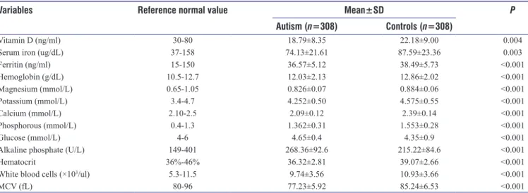
Table 2 presents baseline chemistry biomarkers of autistic and control children. The mean values of serum iron in autistic children (74.1 ± 21.61 ug/dL) was significantly much lower than the normal value in the control children (87.59 ± 23.36 ug/dL) (P = 0.003). Similarly, the study revealed that mean serum Vitamin D level was considerably lower among autistic (18.8 ± 8.3 ng/ml) compared to healthy children (22.2 ± 9.0 ng/ml) (P = 0.004). In addition, mean values of hemoglobin, ferritin, magnesium, potassium, calcium, phosphorous, glucose, alkaline phosphate, hematocrit,
WBC, and MCV were all statistically significantly higher in healthy controls compared to autistic children (P < 0.001). Table 3 identified potential risk factors for ASDs in children: serum iron deficiency (odds ratio [OR] = 2.83; confidence interval [CI] 1.81–4.72 P < 0.001); serum calcium level (OR = 2.74;CI 1.65–4.81, P < 0.001); serum Vitamin D level (OR = 2.36; CI 1.74–3.44, P = 0.002); ferritin (OR = 2.45;1.86–3.93;
P = 0.004); less physical activity (OR = 2.51;1.63–4.67; child
order (OR = 1.68;1.42–3.36; P = 0.019), BMI (OR = 2.39; CI 1.54–3.78, P = 0.028); and parental consanguinity (OR = 1.73; CI 1.48–2.65, P = 0.046) were considered as strong predictors and main factors associated with ASDs after adjusting for age, gender, and other variables.
d
IscussIonThis case–control study presents, to the best of our knowledge, the first report on an establishing level of iron deficiency in children with autism in Qatar and in Arabian Gulf Countries. Despite many studies, the epigenetics of iron deficiency are also still not well understood.[9,10,12,15] These results indicate a
significant low mean value of iron deficiency in children with ASDs. The mean values of iron deficiency and Vitamin D deficiency reported in this study are significantly lower in comparison with other reported studies.[3,6,10-15] Our findings
confirmed that iron deficiency and anemia are common in autism, in parallel with the previous reports.[4,8,12,15]
Iron deficiency is reported to be the most prevalent nutritional problem among children in the world today,[10-16,25] and there is
considerable evidence that iron is important for neurological functioning and development.[20-22] For the first time, this study
demonstrated abnormally low serum ferritin levels in children with autism in Arabian Gulf Countries. Serum ferritin levels were very low in children with autism compared to controls, this is consistent with the previous reported studies.[10-15] The
previous studies have revealed that iron deficiency was associated with autism and the current study revealed an increased risk of autism was noted among those with iron deficiency anemia, which was compatible with this report.[7,10-13]
As it is the case for most iron-deficient children in the general population,[12-15] iron deficiency in autism may be a result of
reduced dietary iron intake. Children with autism often have very restricted food preferences due to smell, taste, texture, or other characteristics of the foods.[7,12-13] Several studies[10-15] showed
Table 1: Sociodemographic characteristics of studied participants with autism versus controls
Variables Autism children (n=308), n (%) Control children (n=308), n (%) P Age (mean±SD) 5.39±1.66 5.62±1.81 Age group 3-4 years old 91 (29.5) 92 (29.9) 0.906 5-6 years old 113 (36.7) 117 (39.0) 7-8 years old 104 (33.8) 99 (32.1) Sex Male 153 (49.7) 137 (44.5) 0.170 Female 155 (50.3) 171 (55.5) Ethnicity of child Qatari 124 (40.3) 153 (49.7) 0.023 Non-Qatari/Arabs 184 (59.7) 155 (50.3) Educational level of father
Illiterate 12 (3.9) 12 (3.9) 0.495
Primary 34 (11.0) 24 (7.8)
Intermediate 46 (14.9) 45 (14.6) Secondary 109 (35.4) 102 (33.1) University and above 107 (34.7) 125 (40.6) Occupation of father Police/army/security 12 (3.9) 16 (5.2) 0.934 Sedentary/professional 109 (35.4) 110 (35.7) Clerk 32 (10.4) 33 (10.7) Businessman 77 (25.0) 71 (23.1) Government officer 78 (25.3) 78 (25.3) Educational level of mother
Illiterate 33 (10.7) 43 (14.0) 0.011
Primary 51 (16.6) 59 (19.2)
Intermediate 67 (21.8) 92 (29.9)
Secondary 72 (23.4) 48 (15.6)
University and above 85 (27.6) 66 (21.4) Occupation of mother
Homemaker 103 (33.4) 95 (30.8) 0.011
Sedentary/professional 177 (25.6) 80 (26.0)
Clerk 55 (17.9) 84 (27.3)
Business woman 73 (23.7) 49 (15.9)
Monthly income ($US dollars) 0.019
<$3000 77 (28.0) 102 (33.1) $3000-$6000 123 (39.4) 125 (40.6) >$6000 108 (32.7) 81 (26.3) BMI group <85th percentile 248 (89.4) 255 (80.3) <0.001 85th-95th percentile 39 (8.7) 34 (16.1) >95th percentile 21 (2.0) 15 (3.5) Consanguinity Yes 118 (38.3) 87 (28.2) 0.008 No 190 (61.7) 221 (71.8) Exposure to sun Yes 102 (33.1) 126 (40.9) 0.045 No 206 (66.9) 182 (59.1) Table 1: Contd... Variables Autism children (n=308), n (%) Control children (n=308), n (%) P
Walking time per/day <60 min
Yes 110 (35.7) 142 (46.1) 0.003
No 208 (67.5) 166 (53.9)
BMI=Body mass index, SD=Standard deviation
that nearly half of children with autism had an inadequate intake of dietary iron. In addition, Dosman et al.[7-13] reported
that twice as many preschoolers (69%) than school-aged children (35%) had an insufficient intake of dietary iron. Furthermore, Xia et al.[26] found that intake of iron increased
with age in 2–9-year-old children with autism. Because younger children with autism are more selective about what they eat, iron deficiency may be more prevalent in this age group.
A great deal of evidence has shown that iron is an important component in cognitive, sensori motor, and social-emotional development and functioning because the development of central nervous system processes is highly dependent on iron-containing enzymes and proteins.[27,28] Iron deficiency
increased the risk of psychiatric disorders, including mood disorders, ASD, attention deficit hyperactivity disorder, and developmental disorders.[21] Deficiency of iron in early life
may increase the risk of psychiatric morbidity. Our findings confirmed that iron deficiency and Vitamin D are common in autism, in parallel with the previous reports.[4,8,12,15,27,28]
Limitations of study
Although our study included a large sample of participants and is case–controlled, it has some limitations. Our study
was limited by the content of existing repositories that, for reasons related to the recruitment processes of those studies, contain very few individuals who did not meet the criteria for an autism classification. Data on the possible maternal iron deficiency and anemia before and after delivery are lacking. Another limitation is the iron source. This study did not include data on children kept on avoidance/restriction diets. It is known that avoidance or restriction diets are one of the modalities of therapy for some patients with autism. Data on duration of outdoor activity are lacking, another limitation in our study.
c
onclusIonThe current studies confirm that deficiencies of iron and Vitamin D and anemia were higher in autistic compared to control children. The results suggest that serum ferritin levels should be monitored in every case of autism as a part of baseline investigation.
What’s known on this
The studies strongly suggest that iron deficiency is linked to brain dysfunction. However, data are lacking with regards to the association between iron deficiency and autism.
What this study adds
The association between deficiencies of iron and Vitamin D and autism in young children and associated risk factors has never been reported in the literature. Perhaps, this is the first study to investigate an association between circulating levels of iron and autism among a highly endogamous population. The present study revealed that Vitamin D deficiency was also higher in autistic compared to healthy children. Supplementing infants with iron and Vitamin D might be a safe and effective strategy for reducing the risk of autism.
Acknowledgment
We are very grateful to Dr. Madeeha Kamal for help and support in data collection. This work was generously
Table 2: Clinical biochemistry baseline value of participants with autism versus controls
Variables Reference normal value Mean±SD P
Autism (n=308) Controls (n=308)
Vitamin D (ng/ml) 30-80 18.79±8.35 22.18±9.00 0.004
Serum iron (ug/dL) 37-158 74.13±21.61 87.59±23.36 0.003
Ferritin (ng/ml) 15-150 36.57±5.12 38.49±5.73 <0.001 Hemoglobin (g/dL) 10.5-12.7 12.03±2.13 12.86±2.02 <0.001 Magnesium (mmol/L) 0.65-1.05 0.826±0.07 0.884±0.06 <0.001 Potassium (mmol/L) 3.4-4.7 4.252±0.50 4.575±0.55 <0.001 Calcium (mmol/L) 2.10-2.5 2.09±0.12 2.39±0.14 <0.001 Phosphorous (mmol/L) 0.4-1.3 1.362±0.31 1.553±0.28 <0.001 Glucose (mmol/L) 4-6 4.65±0.4 4.35±0.9 <0.001
Alkaline phosphate (U/L) 149-401 268.36±92.6 215.22±84.6 <0.001
Hematocrit 36%-46% 36.32±2.81 39.07±2.66 <0.001
White blood cells (×103/ul) 5.3-11.5 9.74±3.56 10.93±3.66 <0.001
MCV (fL) 80-96 77.23±5.92 85.24±6.53 <0.001
MCV=Mean corpuscular volume, SD=Standard deviation
Table 3: Multivariate logistic regression analysis potential risk factors for autism disorder
Independent variables OR 95% CI P
Serum iron deficiency (ug/dL) 2.83 1.81-4.72 <0.001 Serum calcium level (mmol/L) 2.74 1.65-4.81 <0.001 Vitamin D deficiency (ng/ml) 2.36 1.74-3.44 0.002
Ferritin (ng/ml) 2.45 1.86-3.93 0.004
Less physical activity 2.51 1.63-4.67 0.005
Child order 1.68 1.42-3.36 0.019
BMI in percentiles 2.39 1.54-3.78 0.028
Consanguinity 1.73 1.48-2.65 0.034
supported and funded by the Qatar Foundation, grant NPRP 08-760-3-153. The project was partially supported and funded by the Qatar Diabetic Association, Qatar Foundation. The authors would like to thank the Hamad Medical Corporation for their support and ethical approval (HMC-MRC RC10226/10 RP# 12034/12-RC/70813/13 and RC# 71612/2012).
Financial support and sponsorship
Nil.
Conflicts of interest
There are no conflicts of interest.
r
eFerences1. Mefford HC, Batshaw ML, Hoffman EP. Genomics, intellectual disability, and autism. N Engl J Med 2012;366:733-43.
2. Landrigan PJ, Lambertini L, Birnbaum LS. A research strategy to discover the environmental causes of autism and neurodevelopmental disabilities. Environ Health Perspect 2012;120:a258-60.
3. American Psychiatric Association (APA). Diagnostic and Statistical Manual of Mental Disorders. 4th ed. Washington: American Psychiatric Association; 1994.
4. Deth R, Muratore C, Benzecry J, Power-Charnitsky VA, Waly M. How environmental and genetic factors combine to cause autism: A redox/methylation hypothesis. Neurotoxicology 2008;29:190-201.
5. Grabrucker AM. Environmental factors in autism. Front Psychiatry 2012;3:118.
6. Lord C, Risi S, Lambrecht L, Cook EH Jr., Leventhal BL, DiLavore PC,
et al. The autism diagnostic observation schedule-generic: A standard
measure of social and communication deficits associated with the spectrum of autism. J Autism Dev Disord 2000;30:205-23.
7. Dosman CF, Drmic IE, Brian JA, Senthilselvan A, Harford M, Smith R,
et al. Ferritin as an indicator of suspected iron deficiency in children
with autism spectrum disorder: Prevalence of low serum ferritin concentration. Dev Med Child Neurol 2006;48:1008-9.
8. Bener A, Khattab AO, Al-Dabbagh MM. Is high prevalence of Vitamin D deficiency evidence for autism disorder?: In a highly endogamous population. J Pediatr Neurosci 2014;9:227-33.
9. Moy RJ. Prevalence, consequences and prevention of childhood nutritional iron deficiency: A child public health perspective. Clin Lab Haematol 2006;28:291-8.
10. Shattuck PT, Durkin M, Maenner M, Newschaffer C, Mandell DS, Wiggins L, et al. Timing of identification among children with an autism spectrum disorder: Findings from a population-based surveillance study. J Am Acad Child Adolesc Psychiatry 2009;48:474-83.
11. Latif A, Heinz P, Cook R. Iron deficiency in autism and asperger syndrome. Autism 2002;6:103-14.
12. Hergüner S, Keleşoğlu FM, Tanıdır C, Cöpür M. Ferritin and iron levels
in children with autistic disorder. Eur J Pediatr 2012;171:143-6. 13. Chen MH, Su TP, Chen YS, Hsu JW, Huang KL, Chang WH, et al.
Association between psychiatric disorders and iron deficiency anemia among children and adolescents: A nationwide population-based study. BMC Psychiatry 2013;13:161.
14. Sidrak S, Yoong T, Woolfenden S. Iron deficiency in children with global developmental delay and autism spectrum disorder. J Paediatr Child Health 2014;50:356-61.
15. Bilgic A, Gurkan K, Turkoglu S, Faruk O, Kilic B, Uslu R. Iron deficiency in preschool children with autistic spectrum disorders. Res Autism Spectr Disord 2010;4:639-44.
16. Gotham K, Risi S, Dawson G, Tager-Flusberg H, Joseph R, Carter A, et al. A replication of the Autism Diagnostic Observation Schedule (ADOS) revised algorithms. J Am Acad Child Adolesc Psychiatry 2008;47:642-51.
17. Kamal M, Bener A, Ehlayel MS. Is high prevalence of Vitamin D deficiency a correlate for attention deficit hyperactivity disorder? Atten Defic Hyperact Disord 2014;6:73-8.
18. Bener A, Al-Hamaq AO, Saleh NM. Association between Vitamin D insufficiency and adverse pregnancy outcome: Global comparisons. Int J Womens Health 2013;5:523-31.
19. Bener A, Ehlayel MS, Tulic MK, Hamid Q. Vitamin D deficiency as a strong predictor of asthma in children. Int Arch Allergy Immunol 2012;157:168-75.
20. Bener A, Al-Ali M, Hoffmann GF. High prevalence of Vitamin D deficiency in young children in a highly sunny humid country: A global health problem. Minerva Pediatr 2009;61:15-22.
21. Bener A, Kamal M, Bener H, Bhugra D. Higher prevalence of iron deficiency as strong predictor of attention deficit hyperactivity disorder in children. Ann Med Health Sci Res 2014;4:S291-7.
22. Bener A, Hoffmann GF. Nutritional rickets among children in a sun rich country. Int J Pediatr Endocrinol 2010;2010:410502.
23. Sari M, de Pee S, Martini E, Herman S, Sugiatmi, Bloem MW, et al. Estimating the prevalence of anaemia: A comparison of three methods. Bull World Health Organ 2001;79:506-11.
24. World Health Organization. Iron Deficiency Anaemia: assessment, Prevention, and Control-A Guide for Program Managers. Geneva: WHO; 2001. p. 115. Available: from: http://www.who.int/nut/ documents/ida_assessment_prevention_control.pdf. [Last accessed on 2016 Aug 04].
25. Baker RD, Greer FR, Committee on Nutrition American Academy of Pediatrics. Diagnosis and prevention of iron deficiency and iron-deficiency anemia in infants and young children (0-3 years of age). Pediatrics 2010;126:1040-50.
26. Xia W, Zhou Y, Sun C, Wang J, Wu L. A preliminary study on nutritional status and intake in Chinese children with autism. Eur J Pediatr 2010;169:1201-6.
27. Johnson CP, Myers SM. Identification and evaluation of children with autism spectrum disorders. Pediatrics 2007;120:1183-215.
28. Walsh JA, Creighton SE, Rutherford MD. Emotion perception or social cognitive complexity: What drives face processing deficits in autism spectrum disorder? J Autism Dev Disord 2016;46:615-23.
![Table 3 identified potential risk factors for ASDs in children: serum iron deficiency (odds ratio [OR] = 2.83; confidence interval [CI]](https://thumb-eu.123doks.com/thumbv2/9libnet/5454352.105064/4.918.86.444.131.1078/table-identified-potential-factors-children-deficiency-confidence-interval.webp)