S E DA AR S L AN T H E E FFECT O F EARLY LI FE S T RE S S B il k en t U n iv er sity 2019
THE EFFECT OF EARLY LIFE STRESS ON BRAIN WHITE MATTER INTEGRITY AND WORKING MEMORY PERFORMANCE
A Master’s Thesis By SEDA ARSLAN Department of Psychology Bilkent University Ankara August 2019
THE EFFECT OF EARLY LIFE STRESS ON BRAIN WHITE MATTER INTEGRITY AND WORKING MEMORY PERFORMANCE
The Graduate School of Economics and Social Sciences Of
Ihsan Dogramacı Bilkent University
by
SEDA ARSLAN
In Partial Fulfillment of the Requirements for the Degree of MASTER OF ARTS IN PSYCHOLOGY
THE DEPARTMENT OF PSYCHOLOGY IHSAN DOGRAMACI BILKENT UNIVERSITY
ANKARA August 2019
iii
ABSTRACT
EFFECT OF THE EARLY LIFE STRESS ON THE BRAIN WHITE
MATTER INTEGRITY AND WORKING MEMORY PERFORMANCE
Arslan, Seda
M.A. in Psychology
Supervisor: Prof. Dr. Timothea Toulopoulou
August 2019
Former studies revealed that exposure to early life adversity is correlated
with alterations in the white matter structure, particularly, in the areas
associated with executive functioning and memory. Those alterations
include both volume and microstructural white matter integrity reductions in
the brain. A vast amount of the studies focused on volume reductions, and it
is not clear whether the alterations in the white matter integrity is associated
with cognitive functioning. The current study investigated the influence of
early life stress on white matter integrity in the anterior cingulate cortex
(ACC)and corpus callosum (CC) among the forty-six healthy participants.
Participants were split into two groups based on the Childhood Experience
of Care and Abuse Questionnaire (CECA.Q). Participants with relatively
low early life stress were compared with participants with relatively high
early life stress on fractional anisotropy (FA) and mean diffusivity (MD)
iv
memory performance of the participants in the n-back task. Findings
revealed that low-level early life stress did not significantly differ from
high-level of early life stress in terms of FA values. However, there were
significantly higher MD values in the high-level early life stress group
compared to low-level early life stress group. In terms of cognitive
performance, there were no performance differences between the two groups
on the n-back task. The findings suggest that the high level of early life
stress is associated with subtle white matter integrity changes in the brain
but does not affect the performance.
Keywords: Diffusion Tensor Imaging, Early Life Stress, Working Memory
v
ÖZET
BEYİN BEYAZ CEVHER MADDE BÜTÜNLÜĞÜ VE
ÇALIŞMA BELLEK PERFORMANSI ÜZERİNE ERKEN
YAŞAM STRESİNİN ETKİSİ
Arslan, SedaYüksek Lisans, Psikoloji
Tez Danışmanı: Prof. Dr. Timothea Toulopoulou Ağustos 2019
Önceki çalışmalar, erken dönemde yaşam sıkıntısına maruz kalmanın, özellikle yürütücü işlevsellik ve hafıza ile ilgili alanlarda, beyaz cevher dokusundaki değişikliklerle ilişkili olduğunu ortaya koydu. Bu değişiklikler beyindeki hem hacim hem de mikroyapısal beyaz cevher bütünlüğü
azalmasını içerir. Bununla birlikte, araştırmaların büyük bir kısmı hacim azalmasına odaklanmıştır ve beyaz cevher bütünlüğündeki değişikliklerin bilişsel işlevsellik ile ilişkili olup olmadığı açık değildir. Çalışma, kırk altı sağlıklı katılımcı arasında, erken yaşam stresinin, ön singulat korteks ve korpus kallosumdaki beyaz cevher bütünlüğü üzerindeki etkisini araştırdı. Katılımcılar Çocukluk Bakım Deneyimi ve Suistimal Anketi (CECA.Q) temelinde iki gruba ayrıldı. Göreceli olarak erken yaşam stresi düşük olan katılımcılarla göreceli olarak erken yaşam stresi yüksek olan katılımcılar anterior singulat korteks (ACC) ve korpus kallosumdaki (CC) fraksiyonel anizotropi (FA) ve ortalama difüzivite (MD) değerleri bakımından
vi
karşılaştırıldı. Başka bir analizde, katılımcıların n-back task üzerinde
çalışma belleği performansı araştırıldı. Bulgular, düşük seviyeli erken yaşam stresinin, FA değerleri açısından yüksek erken yaşam stresinden önemli ölçüde farklı olmadığını ortaya koydu. Bununla birlikte, yüksek seviye erken yaşam stres grubunda, düşük seviye erken yaşam stres grubuna göre anlamlı olarak daha yüksek MD değerleri bulundu. Bilişsel performans açısından, n-back taskta iki grup arasında performans farkı görülmedi. Mevcut çalışmanın bulguları, erken yaşam stresinin yüksek seviyesinin, beyindeki beyaz cevher bütünlüğü değişiklikleriyle ilişkili olduğunu, ancak performansı etkilemediğini göstermektedir.
Anahtar Sözcükler: Çalışma Belleği Performansı, Difüzyon Tensör
vii
TABLE OF CONTENTS
ABSTRACT . . . iii
ÖZET . . . v
TABLE OF CONTENTS . . . vii
LIST OF TABLES . . . x
LIST OF FIGURES . . . xi
CHAPTER 1: INTRODUCTION . . . 1
1.1 A Brief Overview on Early Life Stress . . . 1
1.2 The Effect of Early Life Stress on Working Memory Performance . . . 2
1.3 Relationship between Early Life Stress and Anterior Cingulate Cortex, Corpus Callosum . . . 3
viii
1.4 The Relationship between White Matter Integrity and Working
Memory Performance . . . 8
1.5 A Brief Overview on Fractional Anisotropy and Mean Diffusivity . . . 9
1.6 The Role of Anterior Cingulate Cortex and Corpus Callosum in Working Function . . . 10
CHAPTER 2: METHODS . . . 14
2.1 Participants . . . 14
2.2 Assessment of Early Life Stress . . . 14
2.3 Acquisition of Diffusion-Weighted Images . . . 16
2.4 Cognitive Paradigm . . . 16
2.5 Behavioral Data . . . 18
2.6 MRI Scanner . . . 19
2.7 Analysis of Diffusion-Weighted Images . . . 19
2.7.1 Preprocessing . . . 19
2.7.2 Statistical Analysis . . . 26
CHAPTER 3: RESULTS . . . 28
3.1 Analysis of Behavioral Data . . . 28
3.2 Tract-based Spatial Statistics Results . . . 31
ix
REFERENCES . . . 42 APPENDIX
x
LIST OF TABLES
3.1.1 The number and the percentage of the severity of the early life
adversity . . . 29 3.1.2 Descriptive statistics and results of working memory
accuracy . . . 30 3.2.1 The location of insignificant lower FA values . . . 32
3.2.2 Coordinates of the significant MD clusters . . . 34
3.2.3 The coordinates of the insignificant lower FA values while age is covaried . . . 35
xi
LIST OF FIGURES
1.3 Demonstration of hypothalamic-pituitary-axis adapted from the
study by Tapp et al. . . . 7
1.6.1 Regions of Cingulate Gyrus and Cingulum . . . 13
1.6.2 Regions of Corpus Callosum . . . 13
2.4 Demonstration of Working Memory Paradigm . . . 17
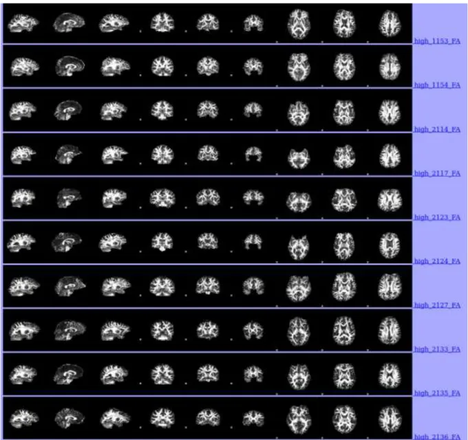
2.7.1.1 Pre-processed brain images of first 12 participants in the high-level of ELS groups . . . 22
2.7.1.2 Pre-processed brain images of last 10 participants in the high-level of ELS groups . . . 23
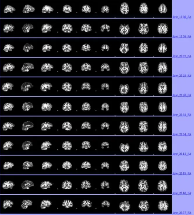
2.7.2.1 Pre-processed brain images of first 13 participants in low-level ELS group . . . 24
2.7.2.2 Pre-processed brain images of last 11 participants in low-level ELS group . . . 25
2.7.3 Skeletonized mean FA of the participants . . . 26
2.7.4 Skeletonized Mean Diffusivity image . . . 26
3.1.1 Comparative boxplot illustrating the CECA.Q scores between two age intervals . . . 30
3.2.1 Insignificant FA value differences between two groups . . . 32
3.2.2 Group difference depicting increment of MD values in the region of interest . . . 33
xii
3.2.3 Insignificant FA value differences between groups while age is
covaried . . . 35 A.1 Bar plots of average CECA.Q scores based on gender . . . 56 A.2 Average psychological, physical abuse and bullying assessment
scores based on gender . . . 57
A.3 Average n-back task performance scores between two
groups . . . 58 A.4 Average n-back task performance scores based on
1
CHAPTER 1
INTRODUCTION
1.1 A Brief Overview on Early Life Stress
Early life stress is also known as childhood trauma or maltreatment and stands for
stressful events that happen before the age of 18 involving psychological, physical or
sexual abuse, neglect, and bullying. Prevalence of early life adversity is high in the
population. Accordingly, 10 % of the population experienced emotional abuse, 26 %
experienced physical abuse, while 21 % of the population had sexual abuse in their
early life period (Dong et al., 2004). Various researches revealed that early life stress
often increases the risk to develop adult psychopathology and psychiatric disorders
such as major depressive disorder, post-traumatic stress disorder, bipolar disorder
(Calabrese, Molteni, Racagni & Riva, 2009; Pruessner et al., 2010; Heim, & Binder,
2012). Furthermore, early life adversity significantly has a deleterious effect on
individuals’ well-being. Accordingly, individuals exposed to early life stress were
significantly associated with an enhanced risk for cardiovascular disease and strokes,
especially in women population (Korkeila et al., 2010). Besides, it increases the risk
of premature death. Accordingly, the study of Brown et al. (2009) in which included
17.337 participants indicated that participants who had six or more than six stressors
in early life died approximately 20 years earlier than individuals without early life
2
1.2 The Effect of Early Life Stress on Working Memory Performance
The vast amount of literature indicates that early life stress has a debilitating effect
on cognitive functioning in adulthood (Irigaray et al., 2013; Lu et al., 2017; Masson,
Bussières, East-Richard, R-Mercier, & Cellard, 2015). There is a growing amount of
studies which investigates the relationship between early-life stress and working
memory. Working memory is part of the cognitive system that enables holding
information temporarily to process and manipulate the hold information (Miyake &
Priti, 1999). It is closely associated with complex cognitive abilities, such as
problem-solving and decision-making (Kyllonen & Christal, 1990). N-back task is
one of the main well-validated working memory tasks in which participants are
presented with multiple stimuli during the encoding phase, and then a response phase
in which participants are required to specify whether the stimuli presented is the
same as the stimuli presented in the encoding phase (Owen, McMillan, Laird &
Bullmore, 2005). Recent studies demonstrated the deleterious impact of early life
stress on working memory performance (Fuge et al., 2014; Majer, Nater, Lin,
Capuron & Reeves, 2010; Goodman, Freeman & Chalmers, 2019; Bos, 2009). For
instance, in the study of Majer et al. (2010), healthy adults who exposed to childhood
adversity had poorer working memory performance. Moreover, in the study of Bos
(2009), children with the early history of institutional care had impaired working
memory performance compared to their peers with no history of institutional care.
It is worth noting that diffusion tensor imaging (DTI) studies shed light on the
3
the myelination is produced in the first two years, and the critical process proceeds
through adolescence and adulthood (Yakovlev & Lecours, 1967). Development of
the microstructure of white matter is vulnerable to environmental effect, especially
during that period. During the development of the brain, remodeling of the synapse,
myelination and programmed cell death are crucial processes that affect both
organization of white matter and gray matter (de Graaf-Peters & Hadders-Algra,
2006). Thus, malicious experiences during this period create a possibility to disrupt
the neurodevelopmental process and cognitive development (Hart & Rubia, 2012).
According to both structural and functional neuroimaging research, prefrontal cortex
(PFC), cingulate gyrus, and corpus callosum are essential working memory-related
areas, and structural alterations in those areas are widely associated with
malfunctioning in working memory performance (Owen, McMillan, Laird &
Bullmore, 2005).
1.3 Relationship between Early Life Stress and Anterior Cingulate Cortex,
Corpus Callosum
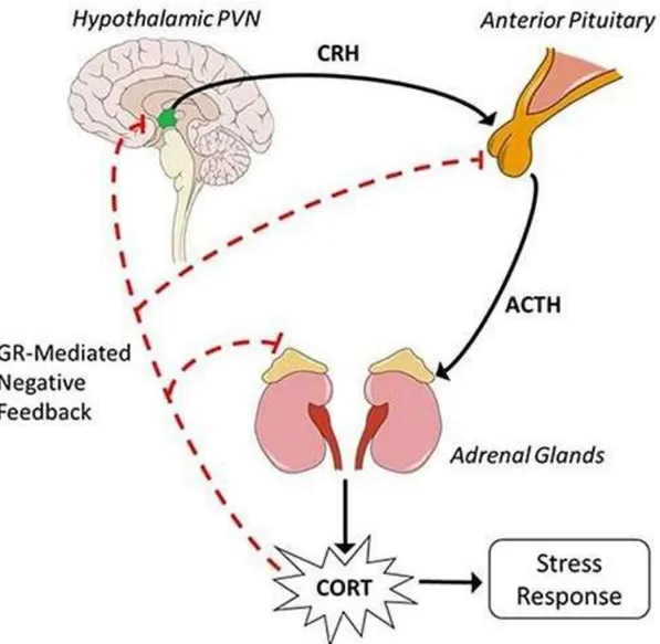
Early life stress considerably influences brain structure, function, and volume (Paul
et al., 2008). The hypothalamic-pituitary-adrenal axis (HPA) is considered as one of
the significant factors for the brain abnormalities in case of exposure to stress, and
early life adversity provokes persistent alterations in the HPA axis (Juruena, 2014;
Hunter, Minnis & Wilson, 2011). Briefly, the HPA axis regulates the hormonal
response system to stress, and glucocorticoid is known as the primary stress
4
corticotrophin-releasing factor (CRF) and arginine vasopressin (AVP) into vessels
bonding hypothalamus and pituitary gland. PVN and CRF affect pituitary gland to
generate and release adrenocorticotropic hormone (ACTH). ACTH leads to synthesis
and secretion of glucocorticoid from the adrenal glands. In humans, the primary
glucocorticoid is cortisol. Inhibitory and excitatory neurotransmitters on PVN
regulates the activation of the hypothalamus to organize the glucocorticoid activity.
In order to prevent extended activity, negative feedback loops regulate HPA axis to
preserve prearranged hormone degrees and homeostasis. For that purpose, cortisol
related negative feedback controls the release of CRF, ACTH, and AVP through the
anterior pituitary gland, PVN, and hippocampus. One of the cortisol receptors is
mineralocorticoid (MR), and in a stressful situation, cortisol density increases and
MR binds to glucocorticoids receptors (GR) to activate them. As a result, GR ends
the stress response (Stephens & Wand, 2012). Various studies on rats indicated that
as well as the hippocampus, medial prefrontal cortex involving anterior cingulate
have a significant role in the regulation of the glucocorticoid (Diorio, Viau &
Meaney, 1993; Sullivan & Gratton, 2002; Mizoguchi, Ishige, Aburada, Tabira, 2003).
For instance, according to the study of Diorio (1993), lesions on the cingulate cortex
significantly enhanced the level of ACTH and corticosterone after exposure to 20
minutes of a controlled stressor. Several human studies showed the significance of
the medial prefrontal cortex, including cingulate cortex on stress regulation. An
experiment with adolescents who gave salivary sample following a social stress test
and resting-state fMRI demonstrated a positive correlation between higher cortisol
5 Hamilton & Gotlib, 2011).
Furthermore, women with medial prefrontal cortex lesion displayed higher cortisol
response during the Trier social stress test (Buchanan et al., 2010). Accordingly, a
study with children indicated that girls with higher cortisol reactivity to stress had
disrupted white matter integrity in the right anterior cingulate cortex (Sheikh et al.,
2014). In another study, smaller left ACC volumes were associated with the
dysregulated HPA axis in healthy men participants (MacLullich et al., 2006). Those
human and animal studies shed light on the crucial role of the cingulate cortex on the
HPA axis.
Myelinated areas like cingulum, which constitutes the core part of cingulate gyrus
white matter, and corpus callosum, which is a nerve tract connecting the two
hemispheres of the brain, are vulnerable to effects of the early exposure to a high
degree of stress hormones since it suppresses glial cells which are essential for
myelination (Lauder, 1983). For instance, individuals with early life adversity
demonstrated a significant deterioration in the function of the oligodendrocytes in the
cingulate cortex, which is an indicator of disrupted myelination in that area.
However, depressed individuals with no history of abuse during childhood show no
such results. Altogether, these results show the significant influence of early life
stress on myelination in the cingulate cortex (Lutz et al., 2017). There are numerous
neuroimaging studies in humans to investigate the impact of early life stress on white
matter morphometry in ACC and CC. Accordingly, healthy individuals who had a
6
compared to healthy individuals with no history of early life stress (Cohen et al.,
2006; Baker et al., 2013). Similarly, high level of early life stress was associated with
decreased ACC white matter volume and with poorer spatial working memory
performance (Hanson et al., 2012). Moreover, early childhood adversity rather than
psychiatric disease was correlated with decreased CC size in children with
post-traumatic stress disorder (Teicher et al., 2004; Teicher et al., 1997).
In opposition to morphometry studies, few studies examined the microstructural
integrity of brain white matter in case of exposure to early life stress. Accordingly,
young adults who had exposure to verbal abuse in early life had reduced white matter
integrity in the cingulum, temporal gyrus, and left body of the fornix (Choi, Jeong,
Rohan, Polcari & Teicher, 2009). Similarly, children exposed to early neglect had
decreased white matter integrity in cingulum (Hanson et al., 2013). Moreover,
adolescents who experienced childhood adversity had lower white matter integrity in
CC and cingulum bundle (Huang, Gundapuneedi & Rao, 2012). Children and older
adults who had early life stress demonstrated lower white matter integrity in the genu
of the corpus callosum (Seckfort et al., 2008; McCarthy-Jones et al., 2018; Lu et al.,
7
Figure 1.3. Demonstration of hypothalamic-pituitary-axis adapted from the study by Tapp et al. (2019)
8
1.4 The Relationship between White Matter Integrity and Working Memory Performance
White matter is responsible for connecting individual neurons in different brain
areas, and it fills approximately half of the brain, and it plays a vital role in action
potentials. Myelinated axons primarily constitute the main structure of white matter
(Charlton et al., 2010). Several studies on aging suggested that there is a significant
relationship between white matter integrity and working memory performance
(Charlton et al., 2010; Zahr, Rohlfing, Pfefferbaum & Sullivan, 2009; Charlton et al.,
2008; Kennedy & Raz, 2009). Similarly, children who had decreased white matter
integrity had reduced performance on working memory performance (Hanson et al.,
2013).
Studies with patients also have a contribution to investigate the relationship between
working memory and white matter integrity. Notably, patients who are at the very
early stage of multiple sclerosis had altered functional connectivity in working
memory-related areas, and the alteration was related to changes of the white matter
diffusivity (Au Duong et al., 2005). Similarly, multiple sclerosis patients with
impaired cognition had severe reductions in white matter integrity compared to
patients with preserved cognition (Hulst et al., 2013). In case of schizophrenia,
severe white matter integrity reductions in the cingulum bundle and CC is
demonstrated and those white matter reductions were associated with poorer working
memory performance and increased reaction time (Kubicki et al., 2003; Wang et al.,
9
1.5 A Brief Overview on Fractional Anisotropy and Mean Diffusivity
The common white matter microstructure measures are fractional anisotropy (FA)
and mean diffusivity (MD) and they are measured by using Diffusion Tensor
Imaging (DTI). Briefly, diffusion tensor imaging has diffusion tensors, which
involves eigenvectors (ê1, ê2, ê3) and eigenvalues (λ1, λ2, λ3). In terms of FA,
diffusion of the water molecules varies from 0 to 1. Value of 1 means the occurrence
of the diffusion is along one axis (anisotropic diffusion), and the value of 0 means
diffusion occurs along with all directions (isotropic diffusion). Axons limits the travel
of the water molecules, hence, create an anisotropic diffusion. In the case of
anisotropic diffusion, eigenvalues are significantly different than each other
(λ1 > λ2 > λ3) while in case of isotropic diffusion, they are nearly equal (λ1 ~ λ2 ~ λ3).
FA indicates axonal diameter, axonal density, and complexity of the fiber tracts.
Mainly, reduced FA can be ascribed to deterioration of the myelin sheaths and
membranes of axons (O’Donnell & Westin, 2011).
Another common scalar to investigate white matter integrity is mean diffusivity. It is
calculated by averaging the eigenvalues of tensors ((λ1+λ2+λ3)/3). It calculates total
diffusivity in a specific tissue and depicts the average movement of the water
molecules as independent of tissue directivity (Fushimi et al., 2007). Particularly,
10
1.6 The Role of Anterior Cingulate Cortex and Corpus Callosum in Working
Memory Function
Neuroimaging techniques, which comprise DTI, provided crucial information
related to roles of anterior cingulate cortex and corpus callosum on cognitive
processing.
The development of diffusion tensor imaging enabled scientists to examine the
white matter integrity more elaborately. According to DTI studies, cingulate gyrus
and corpus callosum (CC) are significant working memory-related white matter
pathways (Charlton, Barrick, Lawes, Markus & Morris, 2010). Also, white matter
integrity in both cingulum and CC significantly associated with working memory
performance in the early adult population (Privado et al., 2014). Accordingly, healthy
subjects who have lower white matter integrity in anterior cingulate demonstrated
lower accuracy in 2-back working memory task (Takahashi et al., 2010). An analysis
of white matter tracts with young and old adults indicated white matter integrity in
the genu of CC and working memory correlations (Zahr, Rohlfing, Pfefferbaum &
Sullivan, 2009). Accordingly, white matter integrity in CC and working memory
performance demonstrated a significant association in children population, although
the effect of age was removed (Nagy, Westerberg & Klingberg, 2004). Researches
with patient groups involving subjects with multiple sclerosis, alcoholism
demonstrated working memory impairments and white matter damage in the
cingulum and CC (Harris et al., 2008; Dineen et al., 2009). Furthermore, children
11
white matter integrity in CC predicted imperfect verbal working memory (Treble et
al., 2013). According to fMRI studies, the anterior cingulate cortex is one of the
significant activated areas during the working memory tasks in both children and
adults (Botvinick, Cohen, Carter, 2004; Kerns et al., 2004; Lenartowicz, McIntosh,
2005; McCarthy et al., 1994; Casey et al., 1995; Nelson et al., 2000; Glabus et al.,
2003). Moreover, a study combined both fMRI and DTI analysis indicated white
matter integrity in the ACC and CC were correlated with fMRI activation in those
areas during the working memory task (Olesen, Nagy, Westerberg & Klingberg,
2003).
Lesion studies provide insights on the contribution of the ACC to working memory.
Lesions, which involves orbitofrontal tissue together with the cingulate cortex,
damaged spatial working memory performance in monkeys (Bachevalier & Mishkin,
1986).
The diagnosis of the participants exposed to early life stress is based on assessment
tools, which include assessments on abuse, neglect, and household dysfunction.
Childhood Abuse and Care Questionnaire (CECA-Q) is one of the extensively used
tools for measuring the early life adversity within participants. It was produced and
tested in UK women population with a history of abuse, depression, and neglect. Its
reliability and validity are proven by several studies (Li et al., 2014; Bifulco,
Bernazzani, Moran & Jacobs, 2005).
Prevailing literature suggests that white matter integrity in ACC and CC is
12
important for working memory function. There is a gap in the literature in terms of
observing the interactive relationship between the effect of early life stress on the white
matter integrity in ACC, CC, and working memory performance. Besides, the
researches which investigated the effect of early life stress on white matter integrity
mostly focused on observing the changes in the diffusivity of the water molecules in
the brain (FA). Thus, this study investigates the underlying neuronal effects of early
life stress on ACC and CC in a broader way by also analyzing the average movements
of the water molecules in the region of interest in order to investigate more elaborately
the microstructure of the tissue, and examines their relationship on the working
memory performance at the exploratory level.
Based on the literature, the primary hypothesis of the current study is that early life
stress is associated with decreased FA values and increased MD values in cingulate
cortex and corpus callosum and since the altered white matter integrity in ACC and
CC is associated with altered working memory performance as indicated above,
secondary hypothesis is that early life stress is associated with a poorer working
memory performance as a result of altered white matter integrity in the cingulate cortex
13
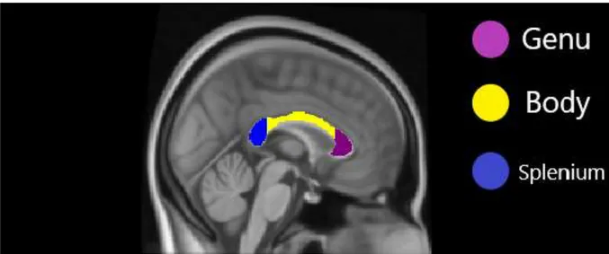
Note. Cingulate cortex was generated via Harvard-Oxford Cortical Structural Atlas in
FSL. Cingulum was generated via JHU (John Hopkins University) white matter tractography atlas in FSL.
Figure 1.6.1. Regions of Cingulate Gyrus and Cingulum on MNI152 Standard Image
Note. Generated via JHU (John Hopkins University) white matter tractography atlas in FSL
14
CHAPTER 2
METHOD
2.1 Participants
The current study involved 46 Turkish participants. The age of the participants was
between 17 and 24 (M= 20.15, SD= 2.29). 22 of the participants were male, and 24
of them were female. Participants were recruited via posters and advertisements
across schools, universities, and cafes in Ankara. The prerequisite for participating in
the study was a normal visual perception to be able to perceive the working memory
task accurately on the screen. Before joining the study, participants had to sign a
written informed consent. Legal guardians were required to sign the informed
consent for the participants whose ages were under 18. Participants who completed
the experiment received 50 liras. Participants with neurological or psychiatric
disorders were excluded from the study.
2.2 Assessment of Early Life Stress
Childhood Care and Abuse Questionnaire (CECA.Q) assess the level of stress
exposure before the age of 17. It is a comprehensive and reliable assessment of early
life adversity. The instrument screens both early life adversity (ages of 0-12) and
adversity during the later stages of childhood (ages of 12-16). The questionnaire
consists of 3 sections which examine family arrangement (e.g., parental loss and
15 abuse).
In terms of scoring, each item includes a two-point Likert scale either for yes/no
questions or for three multiple-choice questions. (0 point for no, 1 point for yes, 2
point for refused to answer). ‘’Yes’’ indicates the presence of related stress factor
while no indicates the opposite. For instance, ‘‘Ever had any unwanted sexual
experiences’’ is a sexual abuse-related question whose answer options are yes or no.
After summing the total points of each participant, the total score of the participants
whose score fell within and below the mean (M= 5.46) was classified as low early
life stress exposure (n= 24) while scores above mean were classified as high early
life stress exposure (n= 22).
In addition to CECA.Q assessment, psychological, physical abuse and bullying
assessments were used to assess the severity and type of the early life adversity. In
the psychological abuse assessment, participants are asked whether they had
experienced humiliation, rejection before the age of 17, and if they had, they were
required to provide information on the severity of the abuse (e.g. ‘0= None, 1=
Some, 2=Moderate, 3= Marked’’). In the physical abuse assessment, participants are
required to answer whether they experienced an attack or hitting before the age of 17
and again, the severity of the adversity is scored as alike to psychological abuse
assessment. In the bullying assessment, participants are asked whether people who
were similar age said hurtful things or hurtful names, ignored or hit them before the
age of 17, and the same severity scores were used. Adversities were scored in a
16
individual, and the maximum score was three, which indicates participants were
exposed to all three of early life adversities.
2.3 Acquisition of Diffusion-Weighted Images
Diffusion-weighted images of the entire brain were acquired by using an
echo-planar imaging sequence lasting 7 minutes 22 seconds to acquire slices in sagittal
(R>> L), coronal (A>>P) and transversal (F>>H) planes [repetition time (TR), 10740
ms; echo time (TE), 102 ms; slice thickness, 2 mm; b-values, 1000 s/mm²; diffusion
gradient directions, 33; field of view (FoV), 256 mm; acquisition matrix, 256 x 256;
voxel size 2x2x2 mm].
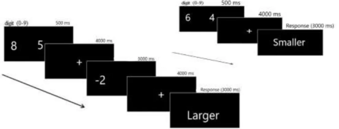
2.4 Cognitive Paradigm
In the current study, working memory performance was assessed via a
well-validated arithmetic n-back task. In the working memory paradigm, participants were
applied numerical size tasks or more complex tasks, which involves numerical
computations as well as numerical size judgments (Tan et al., 2012). Participants
were given a brief presentment of the task, which informs the participants about the
significant points that are required to pay attention before performing the working
memory paradigm in the MRI scanner. The response phase was presented for 3
seconds, and participants were required to decide by pressing the either right or left
button.
The events included a judgment task where participants were presented two-digit
17
and in the response phase, they were expected to choose the larger or smaller number
based on the instruction which says choose larger or smaller remembered number ,
and computational judgement in which participants were required to make a
numerical subtraction of 2 or 3 from the remembered number on the left or right side
in the encoding phase and then, choose larger or smaller number in the response
phase based on the instruction screen. Encoding phase was shown on the screen for
0.5 seconds, and a fixation point appeared for 4 seconds after it. The numbers of the
correct and incorrect answers were equally spread either on the left or the right side.
The total numbers of the events included ten trials.
18
2.5 Behavioral Data
Working memory performance was measured by using the percentage of the correct
responses and analysis of the behavioral data was run in IBM SPSS Statistics version
22. Accordingly, an independent samples t-test was applied in order to check whether
two stress groups (low vs. high) significantly differ from each other in terms of age
and gender. Independent samples t-test was applied to check whether total CECA.Q
scores of participants in both groups significantly differed. Also, a correlational
analysis was run in order to investigate whether the total scores of each participant in
CECA.Q assessment were significantly correlated with the score of the
psychological, physical abuse and bullying scores to observe the consistency of the
early life adversity reports of the participants between two assessments. Additionally,
we used a frequency analysis in order to investigate the overall severity levels of
early life stress. Moreover, early life adversity between 0-12 and 12-16 ages were
compared via paired samples t-test in order to find whether early life stress differed
for the same participants within those different age intervals. In order to investigate
whether working memory performance significantly differs between the two stress
groups (high vs. low), an independent samples t-test is applied. The dependent
variable was working memory performance while the independent variable was the
level of exposure to early life stress.
A correlation analysis was conducted to determine whether the total score of early
life stress was covaried with working memory performance on the n-back task. The
19
2.6 MRI Scanner
3 Tesla Siemens Magnetom scanner was used at the Bilkent University National
Magnetic Resonance Research Center (UMRAM), Ankara, Turkey to produce
diffusion-weighted images and to enable the whole brain coverage; radio-frequency
pulses were utilized via a 32-channel head coil. In order to protect subjects from the
noise of the scanner, flexile earplugs were used and prevent the head motion of the
participants, and head stabilizer cushions were applied both sides of the head. 31.5’’
telemedicine LCD 59 Hz refresh rate and 1920x1080 pixel resolution monitor was
utilized to present the stimuli to the participants who were lying in the scanner. To be
able to bring the monitor into the visual field of the participants, a mirror was put
onto the head coil. Generation of the stimulus and accuracy of the responses were
provided via Neurobehavioral Systems program. Moreover, participants’ responses
were recorded and transferred to an excel file during the working memory task via
the fiber-optic response box.
2.7 Analysis of Diffusion-Weighted Images
2.7.1 Preprocessing
The raw diffusion-weighted images with 33 volumes, which includes 128x128x64
voxels with 2x2x2mm resolution were stored by Snygo MR B17 software system in
the computer of magnetic resonance scanner. DICOM2NII software program was
used to convert to raw Dicom image files (.ima extension) into nifti (.nii extension)
20
FMRIB’s Diffusion Toolbox in FSL 5.0 (Functional MRI of the Brain Software
Library), which uses the Linux operating system. For the preprocessing step, all the
diffusion-weighted images were aligned with each other to correct the head motion
of the subjects and eddy current distortions by using Eddy tool. Then, a brain mask
was created and non-brain tissue, such as the skull, was removed based on the binary
brain mask created by BET tool in FSL. After the preprocessing steps, diffusion
tensors were calculated by using b values, and finally, maps of diffusion anisotropy
(FA) and mean diffusivity (MD) were generated.
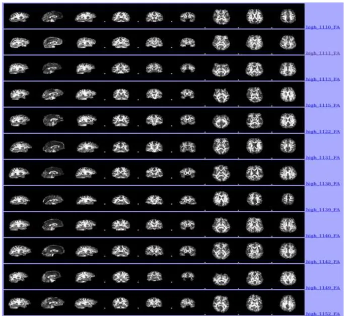
Each step was individually repeated for every 46 participants in the study. In order to
make group comparison, tract-based spatial statistics (TBSS) was performed. In the
first step of TBSS, all the processed FA images were placed into a new subdirectory
called as FA and generated a file called as slicedir which involved each input images
respectively (see Figure 2.7.1.1 and Figure 2.7.2.1). In the second step of TBSS,
individual FA images were aligned into a common 1x1x1 mm standard space
(FMRIB58_FA). In the third step of TBSS, the whole aligned data were transformed
into common space MNI152 (Montreal Neuroimaging Institute). The mean of
affine-transformed data was averaged in order to generate mean FA, and then, skeletonized
mean FA was derived from the mean FA. In the fourth step, the individual FA image
values were projected onto each voxel of the skeleton for the fine alignment. The
threshold value of 0.2 for the FA levels was chosen in order to prevent low mean FA
regions and variability of the inter-subject (see Figure 2.7.3). Group comparison for
21
and original non-linear registration was implemented to MD data. After unifying the
warped MD data of entire participants with 4D file, the revealed images were
projected onto original skeletonised mean FA. As a result, we had MD images of
22
Note. It enables the comparison of each participants’ brain images to see whether
there is a problem with the alignment and shape of the brain images and allows checking the place of each participant within the group
Figure 2.7.1.1. Automatically produced pre-processed brain images of first 12 participants in the high-level of ELS groups.
23
Figure 2.7.1.2. Automatically produced pre-processed brain images of last 10 participants in the high-level of ELS group
24
Figure 2.7.2.1. Automatically produced pre-processed brain images of first 13 participants in low-level ELS group
25
Figure 2.7.2.2. Automatically produced pre-processed brain images of last 11 participants in low-level ELS group
26
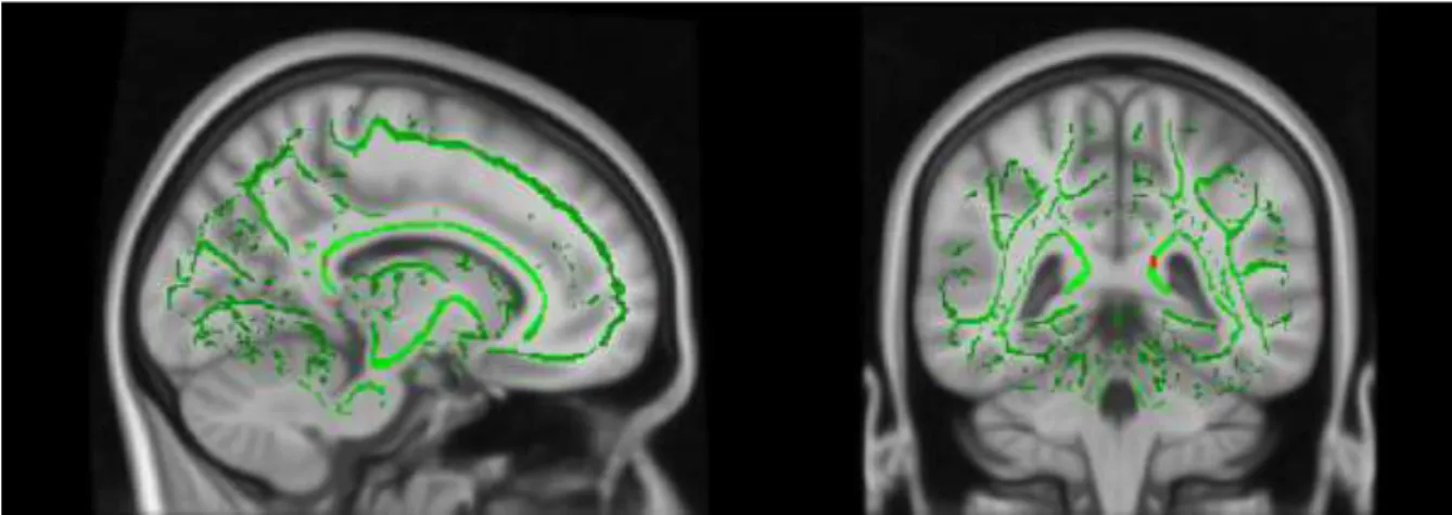
Note. The image depicted on standard MNI152 structural template Figure 2.7.3. Skeletonized mean FA of the participants
Note. The image depicted on FSL_HCP1065_MD standard image Figure 2.7.4. Skeletonised Mean Diffusivity image
2.7.2 Statistical Analysis
The skeletonised diffusion-weighted data underwent a higher-level analysis by
using GLM Setup tool in FSL. It enabled to generate voxel-wise contrasts for the
entire skeleton to carry out the group comparison. For the group comparison, two
high-27
early life stress group, while the second column included a relatively low-early life
stress group in the statistical matrix design. Moreover, a univariate GLM analysis
was used to investigate whether age affects the group differences. Mean age was
subtracted from each participant’s age and added in the third column as a covariate in
order to interpret the differences between the groups. The voxel-wise comparison
was finished by using Randomise command in order to investigate where the
significantly reduced FA and MD values were between two groups by using 5000
permutations. Family-wise error correction was carried out in the randomise
command, which enabled correction for false positives. The significant clusters
(p<0.05) were displayed by tbss_fill command, which shows the significant results
thicker as an overlay on the white matter skeleton. Anatomic location of the region of
interest was produced by using MNI Atlas, JHU ICBM-DTI-81 White Matter Labels
28
CHAPTER 3
RESULTS
3.1 Analysis of Behavioral Data
No significant age difference was found between relatively lower level of ELS
group (M= 19.88, SD= 2.32) and relatively higher level of ELS group (M= 20.45,
SD= 2.28) as a result of independent sample t-test analysis (t(44) = -0.851, p =
0.399). No significant gender differences between two groups (relatively lower ELS
vs. relatively higher ELS) were found as a result of independent sample t-test
analysis (t(44) = 0.86, p = 0.394). Independent samples t-test also indicated that the
total CECA-Q assessment scores of participants in low-level early life stress group
(M = 2.42, SD = 1.58) were significantly lower than the total CECA-Q assessment
scores of the participants in the high-level early life stress group (M = 8.77, SD =
2.11; t(44)= -11,59 p = 0.00). Moreover, Pearson correlation analysis revealed a
strong positive correlation between total score in CECA.Q assessment (M= 5.46,
SD= 3.69) and total score in psychological, physical abuse and bullying assessment
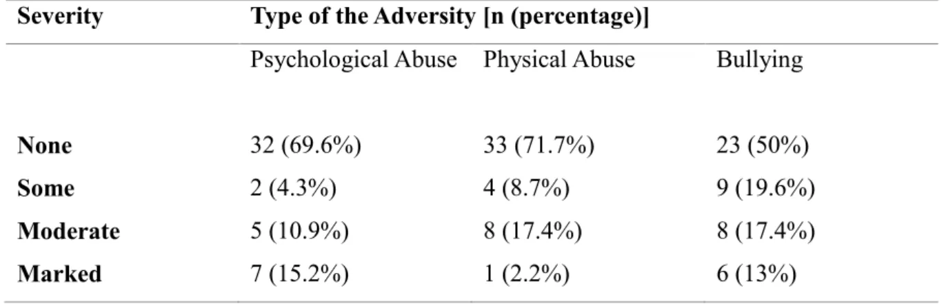
(M= 1.09, SD= 0.93; r(44)= 0.54, p< 0.01). According to frequency analysis, 13% of
the participants had marked level psychological abuse, 2% of the participants had
marked level physical abuse and 13% of the participants experienced marked level
bullying (see Table 3.1.1). Moreover, early life adversity between 0 to 12 years old
29
SD=1.68) were compared via paired samples t-test. We found a significant positive
correlation between two age intervals (r(44) = 0.76, p< 0.001), and no significant
mean difference between two groups (t(43) = 0.95, p = 0.34; see Figure 3.1.1).
We found no significant difference in working memory performance between higher
level early life stress group (M= 90.87, SD= 3.35) and lower level early life stress
group (M= 92.13, SD= 1.72) as a result of independent samples t-test ( t(44)= 1.62,
p= 0.11). Moreover, Pearson correlation analysis showed no correlation between
working memory performance (M=91.52, SD=2.67) and total score of each
participant in CECA.Q assessment (M = 5.28, SD = 3.89; r(44) = -0.13, p = 0.36).
Table 3.1.1. The number and the percentage of the severity of the early life adversity types in the subject population
Severity Type of the Adversity [n (percentage)]
Psychological Abuse Physical Abuse Bullying
None 32 (69.6%) 33 (71.7%) 23 (50%)
Some 2 (4.3%) 4 (8.7%) 9 (19.6%)
Moderate 5 (10.9%) 8 (17.4%) 8 (17.4%)
30
Figure 3.1.1. The comparative boxplot illustrates the CECA.Q scores between two age intervals
Table 3.1.2. Descriptive statistics and results of working memory accuracy
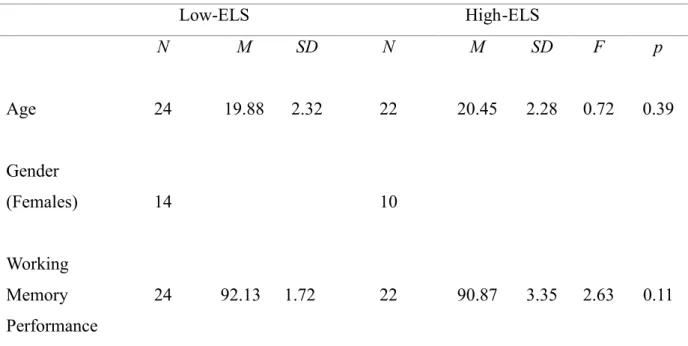
Low-ELS High-ELS N M SD N M SD F p Age 24 19.88 2.32 22 20.45 2.28 0.72 0.39 Gender (Females) 14 10 Working Memory Performance 24 92.13 1.72 22 90.87 3.35 2.63 0.11
31
3.2 Tract-based Spatial Statistics Results
In order to investigate the white matter integrity differences in anterior cingulate
cortex and corpus callosum between two groups (low level of ELS vs. high level of
ELS), an unpaired two-sample t-test was applied in GLM analysis. We found no
significant differences between high level of ELS (M= 0.385, SD= 0.14) compared to
low level of ELS group in terms of FA values (M= 0.387, SD= 0.28; (t(44)= 0.32, p=
0.74); see Figure 3.2.1 and Table 3.2.1 for coordinates). As a result of Univariate
GLM Analysis, age did not affect the group difference in terms of FA values in the
region of interests. In other words, we found no significant difference between two
groups whilst adjusting age as covariate (F(1,44)= 0.048, p= 0.82, see Figure 3.2.3
and Table 3.2.3 for coordinates).
Correlation analysis indicated a non-significant correlation between FA values of
each participant (M= 0.38, SD = 0.022) and working memory performance (M =
91.52, SD = 2.67; r(44) = 0.13, p = 0.38).
High-level ELS group significantly differed from the low-level ELS group in terms of MD values as a result of unpaired two-sample t-test. MD values in high level ELS group (MD=0.00069, SD=0.00008) was significantly higher than low level ELS group (M= 0.00064, SD= 0.00005, p< 0.031; see Figure 3.2.2, see Table 3.2.2).
Moreover, correlation analysis showed no significant correlation between MD values of each participants (M= 0.00067, SD= 0.00007) and working memory performance (M= 91.52, SD= 2.67; r(44)=0.72, p=0.64).
32
Note. Green indicates the mean FA skeleton. FA reduction clusters depicted on
standard MNI152 structural template. Red-orange demonstrates clusters of voxels (FWE corrected for multiple comparisons) with reduced values in high-ELS compared to low-ELS. Abbreviations: FA= fractional anisotropy; ELS= Early life stress; FWE= Family-wise errors
Figure 3.2.1. Insignificant FA value differences in the splenium of corpus callosum between high-level of ELS and low-level of ELS groups
Table 3.2.1. The location of insignificant lower FA values
Lower FA Values x y z
33
Note. Green demonstrates mean FA skeleton superimposed on mean FA image.
Red-orange indicates significant clusters of voxels (FWE corrected for multiple comparisons at p< .05) with higher values in high-ELS compared to the low-ELS group.
Figure 3.2.2. Group difference depicting increment of MD values in the region of interest
34
Table 3.2.2. Coordinates of the significant MD values
MD Clusters Anatomical Definition of Location of the
Cluster x y z
7 Right Cingulum (Cingulate Gyrus) 11 -31 34
8 Right Superior Corona Radiata 17 14 30
9 Right Cingulum (Cingulate Gyrus) 9 12 30
10 Body of Corpus Callosum 18 12 32
11 Genu of Corpus Callosum 1 17 0
12 Left Posterior Corona Radiata -17 -37 38
16 Body of Corpus Callosum 1 11 23
18 Left Cingulum (Cingulate Gyrus) -10 -48 25
19 Body of Corpus Callosum 18 -18 36
20 Body of Corpus Callosum 17 19 26
21 Splenium of Corpus Callosum -14 -50 20
30 Genu of Corpus Callosum 2 21 17
35
Note. Green indicates the mean FA skeleton. FA reduction clusters depicted on
standard MNI152 structural template. Red-orange demonstrates clusters of voxels (FWE corrected for multiple comparisons)
Figure 3.2.3. Insignificant FA value differences between groups while age is covaried
Table 3.2.3. The coordinates of the insignificant lower FA values while age is covaried
Lower FA Values x y z
36
CHAPTER 4
DISCUSSION AND CONCLUSION
The current thesis primarily aims to assess whether participants with a higher level
of early life stress had altered white matter integrity in the anterior cingulate cortex
and corpus callosum. A secondary aim was to assess whether decreased white matter
integrity in anterior cingulate cortex and corpus callosum was associated with
differences in the working memory performance between two groups. Although there
are studies which demonstrated the decreased white matter in the related areas, those
studies mostly focused on the macrostructural alterations in the white matter, and
there are few studies which focused on microstructural white matter alterations in the
region of interest and its effect on the cognitive performance. As a result, the current
study indicated that exposure to the high level of early life stress before the age of
17, did not significantly affect the FA values but revealed significantly increased MD
values in the cingulum, the corpus callosum, and superior corona radiata. The level
of the ELS was not associated with working memory performance.
In terms of mean diffusivity alterations, high level of ELS group significantly had
higher MD values compared to low-level ELS group in the corpus callosum, anterior
corona radiata, and cingulum. Previous DTI studies related to the development of the
white matter integrity during childhood and adolescence indicated that MD continues
37
Lebel, 2018). Alterations in MD might indicate a variance inside of intra and
extracellular space and decrease in neuropil (Selemon & Goldman-Rakic, 1999) or
altered boundaries which limit the movement of the water molecules such as cell
membranes (Bosch et al., 2012). Thus, increased MD values in the current study
might reflect an abnormal white matter integrity development as a consequence of
exposure to stressful events in early life. The results of the current study were
consistent with the study by Teicher et al. (2010). According to their research,
subjects exposed to verbal abuse in school years had higher MD values in the corpus
callosum and the corona radiata. Moreover, the level of verbal abuse exposure was
correlated with elevated MD values in those areas (Teicher, Samson, Sheu, Polcari &
McGreenery, 2010).
The result might be explained via the exacerbation of the inflammation in the brain.
Precisely, stress causes glucocorticoid release through activating the HPA axis.
Glucocorticoids can influence the neuro-inflammation by increasing the circulation
of the pro-inflammatory molecules (Marsland, Walsh, Lockwood & John-Henderson,
2017), and stress seems to increase the inflammatory state by increasing the secretion
of the pro-inflammatory molecule and then, inflammation engenders water increases
in the tissue, hence, causes increased mean diffusivity. A recent study indicated that
children who had early life stress showed continued elevated inflammatory levels in
adulthood (Danese, Pariante, Caspi, Taylor & Poulton, 2007). Consequently, findings
of the present study might suggest that the stress level in the high-ELS group was
38
as a response to stress. Unlike MD values, a weak association between FA values and
circulation degrees of inflammatory cytokine interleukin-6 was revealed in the study
by Molesworth et al. (2014).
In terms of the fractional anisotropy alterations in the white matter integrity, the data
may suggest that axonal or myelination outcomes of early life stress may not be
apparent in healthy participants until a threshold of the severeness is attained. In the
study by Kim et al. (2005), patients with post-traumatic stress disorders (PTSD) had
lower FA values in the anterior cingulate cortex, and the decrease level of the white
matter integrity was related to the severity level of the PTSD symptoms.
Furthermore, research by Sara et al. (2018) revealed that major depressive disorder
patients who experienced early life stress had reduced FA values in the cingulum and
corpus callosum compared to healthy patients exposed to early life stress. As
indicated above, while the participants in this study experienced ELS, none of them
were currently having any psychological problem at the clinical level as a result of
ELS, this may also indicate that participants might be moderately resilient to stress.
Besides, different stressors have different structural and functional consequences.
Especially, emotional abuse and sexual abuse are early life stress events that are
considered to have more harmful impacts on the brain development. In the study of
Hanson et al. (2013), children who experienced early life adversity had lower FA
values in anterior cingulate cortex and disruptions performance in the working
memory, however, the population in the study included children who grew up in the
39
care-provider, inadequate amount of toy, inadequate linguistic stimulant, and
nutrition. Additionally, children exposed to early deprivation had reduced FA values
and increased MD values in cingulum. Also, the time of the exposure to early
deprivation shown a reverse association with FA values, but independent from MD
values (Kumar et al., 2014). Each of these severe experiences may significantly
influence the brain structure, especially in the developmental period. Thus, the
severity of the adverse experiences in early life might play a key role in fractional
anisotropy alteration, which, as discussed before, is an index of myelination. In the
present study, overall ELS severity was low, specifically, 15.2% of the participants
experienced marked level psychological abuse, and 13% of the participants had
marked level bullying while 2.2% of the participants had marked level physical
abuse experience. Thus, because of the small sample size, the effect of the severity of
the early life adversity could not be analyzed; thus, it requires further analysis.
Another essential factor for changes in fractional anisotropy values might be social
support. It is considered as the central preservative agent against the neurobiological
and mental influence of early life adversity. In the study of Leicht-Deobald et al.
(2018), participants with ELS who have high social support from work environment
showed lower stress reactivity to psychosocial stress test. Moreover, there are studies
which indicated that social support was significantly correlated with decreased
cortisol levels in saliva as the reaction to acute stress (Ditzen et al., 2008; Heinrichs,
Baumgartner, Kirschbaum & Ehlert, 2003; Eisenberger, Taylor, Hilmert &
40
support in daily life can have a favorable influence on the HPA axis. Accordingly,
81.39% of our participants had social support either from adults or peers. Thus, it
could have protected the participants from having altered white matter integrity as
assessed by FA values.
In terms of working memory performance, the results of the present study are
consistent with the study by Seckfort et al. (2008) which examined working memory
performance in a population with early life stress whose ages varied between 8 and
73 years old. Their results showed no difference in the memory performance when
compared to the control group with no childhood adversity, although they found
decreased white matter integrity in the corpus callosum. Thus, early life stress might
create subtle alterations on the brain structure but not on the function.
In the current thesis, grouping method was double-checked by using psychological,
physical abuse, and bullying assessment. Total scores in CECA.Q assessment were
correlated strongly (0.54) with scores in psychological, physical abuse, and bullying
assessments. It means that when the rating of the participants increased in CECA.Q
assessment, their score increased in another assessment accordingly. Thus, it shows
the reliability of the grouping method in the current experiment.
The CECA.Q assessment enabled us to investigate the early life stress experiences
of the participants between age 0-12 and 12-16 in order to investigate whether early
life stress differed for the same participants within those different age intervals and
results indicated that life experiences in age interval of 0-12 are approximately
41
The first limitation of the current study was small sample size. We found
insignificant lower FA values in the splenium of the corpus callosum area of the
high-level ELS group. That area might not reach a significant level because of the
small sample size, which may cause the reflection of the type-II error. Thus, the
small sample size should be considered as an essential factor while interpreting the
results. Another limitation was that early life experiences in this study were based on
the retrospective reports. Hence, it is vulnerable to subjectivity bias in the
comprehension of early life experiences. Objective measurements like medical
reports and saliva analysis would be involved in the future study.
As a conclusion, the high level of early life stress was associated with higher mean
diffusivity in the left and right cingulum and corpus callosum. However, high-level
early life stress had no significant influence on fractional anisotropy values in those
areas and did not cause the distortions in the working memory performance.
Psychiatric disorders and cognitive abnormalities presumably occur because of the
convergence of the genetic vulnerability and early-life adversity in the critical
42
REFERENCES
Andersen, S. L., & Teicher, M. H. (2008). Stress, sensitive periods and maturational events in adolescent depression. Trends in Neurosciences, 31(4), 183-91.
Au Duong, M. Van, Audoin, B., Boulanouar, K., Ibarrola, D., Malikova, I., Confort- Gouny, S., … Ranjeva, J. P. (2005). Altered functional connectivity related to white matter changes inside the working memory network at the very early stage of MS. Journal of Cerebral Blood Flow and Metabolism, 25(10), 1245-53.
Baker, L. M., Williams, L. M., Korgaonkar, M. S., Cohen, R. A., Heaps, J. M., & Paul, R. H. (2013). Impact of early vs. late childhood early life stress on brain morphometrics. Brain Imaging and Behavior, 7(2), 196-203.
Bachevalier, J., & Mishkin, M. (1986). Visual recognition impairment follows ventromedial but not dorsolateral prefrontal lesions in monkeys. Behavioural
Brain Research, 20(3), 249-61.
Bifulco, A., Bernazzani, O., Moran, P. M., & Jacobs, C. (2005). The childhood experience of care and abuse questionnaire (CECA.Q): Validation in a community series. British Journal of Clinical Psychology, 44(4), 563-81.
Bos, K. J. (2009). Effects of early psychosocial deprivation on the development of memory and executive function. Frontiers in Behavioral Neuroscience, 3(16).
Bosch, B., Arenaza-Urquijo, E. M., Rami, L., Sala-Llonch, R., Junqué, C., Solé- Padullés, C., … Bartrés-Faz, D. (2012). Multiple DTI index analysis in normal aging, amnestic MCI and AD. Relationship with neuropsychological
performance. Neurobiology of Aging, 33(1), 61-74.
Botvinick M.M., Cohen J.D., Carter C.S. (2004). Conflict monitoring and anterior cingulate cortex: an update. Trends in Cogn. Sci., 8(12), 539-46.
43
Brown, D. W., Anda, R. F., Tiemeier, H., Felitti, V. J., Edwards, V. J., Croft, J. B., & Giles, W. H. (2009). Adverse Childhood Experiences and the Risk of Premature Mortality. American Journal of Preventive Medicine, 37(5), 389-396.
Buchanan, T. W., Driscoll, D., Mowrer, S. M., Sollers, J. J., Thayer, J. F.,
Kirschbaum, C., & Tranel, D. (2010). Medial prefrontal cortex damage affects physiological and psychological stress responses differently in men and women.
Psychoneuroendocrinology, 35(1), 56-66.
Calabrese, F., Molteni, R., Racagni, G., & Riva, M. A. (2009). Neuronal plasticity: A link between stress and mood disorders. Psychoneuroendocrinology, 34, 208-216.
Casey, B. J., Cohen, J. D., Jezzard, P., Turner, R., Noll, D. C., Trainor, R. J., Rapoport, J. L. (1995). Activation of prefrontal cortex in children during a nonspatial working memory task with functional mri. Neuroimage, 2(3), 221-9.
Charlton, R. A., Barrick, T. R., Lawes, I. N. C., Markus, H. S., & Morris, R. G. (2010). White matter pathways associated with working memory in normal aging. Cortex, 46(4), 474-89.
Charlton, R. A., Landau, S., Schiavone, F., Barrick, T. R., Clark, C. A., Markus, H. S., & Morris, R. G. (2008). A structural equation modeling investigation of age-related variance in executive function and DTI measured white matter damage.
Neurobiology of Aging, 29(10), 1547-55.
Choi, J., Jeong, B., Rohan, M. L., Polcari, A. M., & Teicher, M. H. (2009). Preliminary evidence for white matter tract abnormalities in young adults exposed to parental verbal abuse. Biological Psychiatry, 65(3), 227-34.
Cohen, R. A., Grieve, S., Hoth, K. F., Paul, R. H., Sweet, L., Tate, D., … Williams, L. M. (2006). Early Life Stress and Morphometry of the Adult Anterior
44
Danese, A., Pariante, C. M., Caspi, A., Taylor, A., & Poulton, R. (2007). Childhood maltreatment predicts adult inflammation in a life-course study. Proceedings of
the National Academy of Sciences, 104(4), 1319-1324.
Dineen, R. A., Vilisaar, J., Hlinka, J., Bradshaw, C. M., Morgan, P. S.,
Constantinescu, C. S., & Auer, D. P. (2009). Disconnection as a mechanism for cognitive dysfunction in multiple sclerosis. Brain, 132(1), 239-49.
Diorio, D., Viau, V., & Meaney, M. J. (1993). The role of the medial prefrontal cortex (cingulate gyrus) in the regulation of hypothalamic-pituitary-adrenal responses to stress. The Journal of Neuroscience : The Official Journal of the Society for Neuroscience, 13(9), 3839-47.
Ditzen, B., Schmidt, S., Strauss, B., Nater, U. M., Ehlert, U., & Heinrichs, M. (2008). Adult attachment and social support interact to reduce psychological but not cortisol responses to stress. Journal of Psychosomatic Research, 64(5), 479-86.
Dong, M., Anda, R. F., Felitti, V. J., Dube, S. R., Williamson, D. F., Thompson, T. J., Giles, W. H. (2004). The interrelatedness of multiple forms of childhood abuse, neglect, and household dysfunction. Child Abuse and Neglect, 28(7), 771- 784.
Eisenberger, N. I., Taylor, S. E., Gable, S. L., Hilmert, C. J., & Lieberman, M. D. (2007). Neural pathways link social support to attenuated neuroendocrine stress responses. NeuroImage, 35(4), 1601-12.
Fuge, P., Aust, S., Fan, Y., Weigand, A., Gärtner, M., Feeser, M., … Grimm, S. (2014). Interaction of early life stress and corticotropin-releasing hormone receptor gene: Effects on working memory. Biological Psychiatry, 76(11), 888-894.
Fushimi, Y., Miki, Y., Okada, T., Yamamoto, A., Mori, N., Hanakawa, T., Togashi, K. (2007). Fractional anisotropy and mean diffusivity: Comparison between 3.0-T and 1.5-T diffusion tensor imaging with parallel imaging using histogram and
45
region of interest analysis. NMR in Biomedicine, 20(8), 743-8.
Glabus, M. F., Horwitz, B., Holt, J. L., Kohn, P. D., Gerton, B. K., Callicott, J. H., … Berman, K. F. (2003). Interindividual Differences in Functional Interactions among Prefrontal, Parietal and Parahippocampal Regions during Working Memory. Cerebral Cortex, 13(12), 1352-61.
Goodman, J. B., Freeman, E. E., & Chalmers, K. A. (2019). The relationship between early life stress and working memory in adulthood: A systematic review and meta-analysis. Memory, 27(6), 868-880
de Graaf-Peters, V. B., & Hadders-Algra, M. (2006). Ontogeny of the human central nervous system: What is happening when? Early Human Development, 82(4), 257-266.
Hanson, J. L., Chung, M. K., Avants, B. B., Rudolph, K. D., Shirtcliff, E. A., Gee, J. C., … Pollak, S. D. (2012). Structural Variations in Prefrontal Cortex Mediate the Relationship between Early Childhood Stress and Spatial Working Memory.
Journal of Neuroscience, 32(23), 7917-25.
Hanson, J. L., Adluru, N., Chung, M. K., Alexander, A. L., Davidson, R. J., & Pollak, S. D. (2013). Early neglect is associated with alterations in white matter integrity and cognitive functioning. Child Development, 84(5), 1566-78.
Harris, G. J., Jaffin, S. K., Hodge, S. M., Kennedy, D., Caviness, V. S., Marinkovic, K., … Oscar-Berman, M. (2008). Frontal white matter and cingulum diffusion tensor imaging deficits in alcoholism. Alcoholism: Clinical and Experimental
Research, 32(6), 1001-13.
Hart, H., & Rubia, K. (2012). Neuroimaging of child abuse: a critical review.
Frontiers in Human Neuroscience, 6(6).
Heim, C., & Binder, E. B. (2012). Current research trends in early life stress and depression: Review of human studies on sensitive periods, gene-environment
46
interactions, and epigenetics. Experimental Neurology, 223(1), 102-111.
Heinrichs, M., Baumgartner, T., Kirschbaum, C., & Ehlert, U. (2003). Social support and oxytocin interact to suppress cortisol and subjective responses to
psychosocial stress. Biological Psychiatry, 15(12), 1389-98.
Huang, H., Gundapuneedi, T., & Rao, U. (2012). White matter disruptions in adolescents exposed to childhood maltreatment and vulnerability to psychopathology. Neuropsychopharmacology, 37(12), 2693-2701
Hulst, H. E., Steenwijk, M. D., Versteeg, A., Pouwels, P. J. W., Vrenken, H.,
Uitdehaag, B. M. J., … Barkhof, F. (2013). Cognitive impairment in MS: Impact of white matter integrity, gray matter volume, and lesions. Neurology, 80(11), 1025-32.
Hunter, A. L., Minnis, H., & Wilson, P. (2011). Altered stress responses in children exposed to early adversity: A systematic review of salivary cortisol studies.
Stress, 14(6), 614-626.
Irigaray, T. Q., Pacheco, J. B., Grassi-Oliveira, R., Fonseca, R. P., Leite, J. C. de C., & Kristensen, C. H. (2013). Child maltreatment and later cognitive functioning: a systematic review. Psicologia: Reflexão e Crítica, 26(2), 376- 387.
Juruena, M. F. (2014). Early-life stress and HPA axis trigger recurrent adulthood depression. Epilepsy and Behavior, 38, 148-59.
Kennedy, K. M., & Raz, N. (2009). Aging white matter and cognition: Differential effects of regional variations in diffusion properties on memory, executive functions, and speed. Neuropsychologia, 47(3), 916-27.
Kerns JG, Cohen JD, MacDonald AW, Cho RY, Stenger VA, Carter C.S (2004). Anterior cingulate conflict monitoring and adjustments in control. Science,
47
Kim, M. J., Lyoo, I. K., Kim, S. J., Sim, M., Kim, N., Choi, N., Renshaw, P. F. (2005). Disrupted white matter tract integrity of anterior cingulate in traumasurvivors. NeuroReport, 16(10), 1049-53.
Korkeila, J., Vahtera, J., Korkeila, K., Kivimäki, M., Sumanen, M., Koskenvuo, K., & Koskenvuo, M. (2010). Childhood adversities as predictors of incident coronary heart disease and cerebrovascular disease. Heart, 36(4), 298-303.
Kubicki, M., Westin, C. F., Nestor, P. G., Wible, C. G., Frumin, M., Maier, S. E., … Shenton, M. E. (2003). Cingulate fasciculus integrity disruption in
schizophrenia: A magnetic resonance diffusion tensor imaging study. Biological
Psychiatry, 54(11), 1171-80.
Kubicki, M., Niznikiewicz, M., Connor, E., Ungar, L., Nestor, P. G., Bouix, S., Shenton, M. E. (2009). Relationship between white matter integrity, attention, and memory in schizophrenia: A diffusion tensor imaging study. Brain Imaging
and Behavior, 3(2), 191-201.
Kumar, A., Behen, M. E., Singsoonsud, P., Veenstra, A. L., Wolfe-Christensen, C., Helder, E., & Chugani, H. T. (2014). Microstructural abnormalities in language and limbic pathways in orphanage-reared children: A diffusion tensor imaging study. Journal of Child Neurology, 29(3), 318-25.
Kyllonen, P. C., & Christal, R. E. (1990). Reasoning ability is (little more than) working-memory capacity?!. Intelligence, 14(4), 389–433.
Lauder, J. M. (1983). Hormonal and humoral influences on brain development. Psychoneuroendocrinology, 8(2), 121-55.
Leicht-Deobald, U., Bruch, H., Bönke, L., Stevense, A., Fan, Y., Bajbouj, M., & Grimm, S. (2018). Work-related social support modulates effects of early life stress on limbic reactivity during stress. Brain Imaging and Behavior, 12(5), 1405-18.