Published by Central Fisheries Research Institute (SUMAE) Trabzon, Turkey in cooperation with Japan International Cooperation Agency (JICA), Japan
R E S E A R C H P A P E R
Molecular and Morphological Characterization of Several
Cyanobacteria and Chlorophyta Species Isolated from Lakes in
Turkey
Betul Yilmaz Ozturk
1,* , Baran Asikkutlu
2, Cengiz Akkoz
2, Tahir Atici
31Eskişehir Osmangazi University Central Research Laboratory Application And Research Center 26480 Eskişehir, Turkey. 2Selçuk University Faculty Of Sciences Department of Biology 42130 Konya, Turkey.
3Gazi University Gazi Education Faculty Division of Biology Education 06500 Ankara, Turkey.
Article History
Received 09 February 2018
Accepted 16 July 2018
First Online 30 July 2018
Corresponding Author Tel.: +09.0222 2393750/6416 E-mail: [email protected] Keywords Chlorophyta Cyanobacteria Molecular analysis Sequence analysis Taxonomy
Abstract
Morphological features are not sufficient for species characterization in microalgae. There are many possibilities for existing characters. Recently, molecular techniques have been used in many areas and this technique increasing the reliability of the work done. The general purpose of the study is to compare Cyanobacteria and Chlorophyta species under the culture conditions using classical classification and molecular identification. In order to achieve this aim, isolation of different clonal cultures of Cyanobacteria and Chlorophyta species from Apa Dam Lake and Beykavağı pond (Konya, Turkey) and Eber Lake (Afyon, Turkey) was performed. Morphological diversity of Cyanobacteria and Chlorophyta species was observed under culture conditions and visualized by both light microscopy and scanning electron microscopy (SEM). In addition, phylogenetic relationships of these strains to 16S rRNA and 18S rRNA gene sequences were determined. According to DNA sequence analysis, 3 isolates were found to be similar to Chodatodesmus mucronulatus (Chodat) Bock & Krienitz (97%), Desmodesmus sp. (Chodat) T.Friedl & Hegewald (98%) and Pediastrum duplex Meyen (97%). Other 4 isolates were found to be similar to Fischerella ambigua (Kützing ex Bornet & Flahault) Gomont (99%), Leptolyngbya sp. Anagnostidis & Komárek (97%), Phormidium autumnale Gomont (99%) and Rivularia sp. C. Agardh ex Bornet & Flahault (99%).
Introduction
Cyanobacteria and microalgae are of significant importance in lentic ecosystems. They act as primary producers and contribute to the nutrient cycling. Due to their critical roles in lake ecosystems, the number of studies on the investigation of the cyanobacterial and algal composition of lakes has been increasing. The classification of microalgae and cyanobacteria is mainly based on morphological investigations by microscopy. However, alterations in the environmental conditions might lead to phenotypic plasticity especially in the cyanobacteria. For this reason, only morphology based identification studies could be misleading and could result in misidentification. It was argued that significant amount of cyanobacterial strains in the culture collections have been misidentified (Komárek &
Anagnostidis, 1989). Accordingly, in addition to morphological studies, molecular and biochemical markers are also being used for the taxonomic definitions of the species (Komárek & Mareš, 2012).
Cyanobacteria inhabit a wide range of natural environments like soil, water, hot springs and salt lakes. Moreover, due to their high biomass accumulation capacity, they have the potential to be used in many biotechnological applications like biofuel (biogas, bioethanol, butanol and biodiesel) productions (Sarsekeyeva et al., 2014). Furthermore, they have the potential to produce many bioactive compounds, proteins, pigments and polysaccharides (Galhano et al., 2011). Cyanobacteria as being a global primary producer requires water, carbon dioxide, inorganic substances and light for the maintenance of their life cycle. Photosynthesis is their principal mode of energy
metabolism and they possess chlorophyll a and perform oxygenic photosynthesis using photosystems I and II (Castenholz, 1989). Although plant thylakoids are constructing in stacked grana and unstacked stroma membranes, and algal thylakoids can be separated depending on the strain, cyanobacterial thylakoids typically do not bulk or appress and are more uniformly sheet-like. In addition to light harvesting in cyanobacteria is carried out primarily by phycobilisomes large, soluble molecular assemblies that occur phycobiliproteins and linker, mostly non-pigmented, polypeptides (Gantt & Conti, 1966; Larkum & Vesk, 2003). Cyanobacteria or blue green algae were previously classified under the kingdom Plantae due to its resemblance to algae but later they were put under the bacteriological code upon investigating their physiological and genetic structures. Several years ago, it had a proposal about their status under the bacteriological code and re-examined their phylogenetic position (Oren, 2004). Regarding the systematic classification of Cyanobacteria, there are still ambiguous situations or conflicts. This is especially due to the fact that classification based on molecular and morphological features are not in agreement (Rajaniemi et al., 2005). Previous classifications were mainly done according to the morphological characteristics but currently, molecular and biochemical markers are being used to support morphological data. However, a significant amount of strains are lack of any genetic or biochemical investigations. Even, it is asserted that laboratory culture conditions or several passages during culturing could lead to the loss of certain morphological features like gas vesicles (Rajaniemi et al., 2005). Taking into account these variations, careful studies should be carried out using genetic and biochemical markers to revise existing classifications and to classify new isolates. Hence, the taxonomic system needs to be upgraded according to the recent improvements.
Chlorophyta is another important group of photosynthetic organisms in the aquatic ecosystem. They have membrane-bound plastids containing chlorophyll a and b. In the aquatic ecosystems, the algae are accepted as the primer producers of the organic materials and they constitute the base of the food chain. Similar to the case for cyanobacteria, the old classification system for green algae is being supported by additional molecular and biochemical methods. These emerging technologies led to significant revisions in many taxonomic lineages such as divisions, classes and orders (Krienitz & Bock, 2012). Especially, the molecular phylogenetic methods come to the help when the species definition is not possible for those which do not propagate sexually. For example, molecular methods were asserted to be very useful for the considerable revision of the orders that contain coccoid green algae which do not propagate sexually (Krienitz & Bock, 2012).
Nucleic acid based technologies, especially the polymerase chain reaction (PCR), have advanced the studies on establishing evolutionary relationships among diverse organisms. In PCR based molecular phylogenetic studies, certain conserved regions in DNA such as ribosomal RNA gene (16S rRNA and 18S rRNA) and internal transcribed spacer sequences (ITS) are amplified by PCR using specific primers and sequenced. Then, these sequences are compared with the other known sequences using various softwares and databases and the result is expressed in a phylogenetic tree. In this way, it becomes feasible to study both species identity and intraspecies variation. Besides morphological characterizations, with the help of conserved DNA sequences’ analyses, the species were put in an appropriate place in the phylogenetic tree. In many types of evolutionary and diversity studies, small-subunit ribosomal RNA genes (16S rRNA in prokaryotes and 18S rRNA in eukaryotes) were commonly used (Fawley, Fawley, & Buchheim, 2004). Another commonly employed gene sequence in systematic studies are nuclear ribosomal DNA internal transcribed spacer sequences (ITS) which are the non-coding DNA sequences located between the small-subunit ribosomal RNA and large-subunit rRNA genes. ITS regions were often used for evaluating genus- and species-level relationships (Coat et al., 1998).
The systematics of cyanobacteria and green algae generally based on morphological studies. Therefore, there is a significant need for studies on diversification of cyanobacteria and green microalgae using molecular tools in addition to morphological characterizations in aquatic ecosystems. In this study, molecular and morphological characterizations of several cyanobacteria and green algal species from Apa Dam Lake, Beykavağı pond and Eber Lake were performed. Light microscopy and scanning electron microscopy analyses were done for morphological characterizations. After isolation and identification of organisms, the partial 16S rRNA (27F-1492R) sequences of cyanobacteria were PCR amplified and sequenced respectively for the molecular phylogenetic analyses. In addition to the partial 18S rRNA (ITS1/ITS4) sequences, green algae were PCR amplified and sequenced respectively for the molecular phylogenetic analyses. In our study, since there are limited numbers of study on the identification of cyanobacteria and green algae with the help of molecular techniques in Turkey this study will contribute to and enhance the research on cyanobacteria and green algae in aquatic ecosystems.
Materials and Methods
Sampling, Isolation of Monocultures and Culture Conditions
Environmental water samples were taken from the following localities in May and June 2014: Apa Dam Lake
(37°22ˈ10ˈˈN 32°29ˈ40ˈˈE), Beykavağı Pond (38° 08’ 25’’N 32° 15’ 37’’E), Eber Lake (38°39ˈ09ˈˈN 31°10ˈ08ˈˈE). The local samples like stone, mud and plants were collected from their natural habitats carried to the laboratory in glass bottles filled with lake water. In order to obtain monocultures, the dilution method was applied (Rippka, 1988). In this method, the mixed cultures taken from natural habitats were placed into petri dish and diluted with sterile water. This dilution procedure was repeated several times until single cells were freely available in the suspension. Then every single cell was separately isolated with the help of Pasteur pipette working under invert microscope. Pasteur pipettes were thinned as much as possible with tweezers under fire before starting to work. Even so coccoid algae, which were very small after being so thinned, were easily isolated from lake waters. After obtaining single cells (Filaments of Cyanobacteria cells or cell colonies of Chlorophyta), they were transferred to BG-11 medium (NaNO3, 15; K2HPO4, 0.4;
MgSO4·7H2O, 0.75; CaCl2·2H2O, 0.36; citric acid, 0.06;
iron (III) ammonium citrate, 0.06; Na2-EDTA, 0.01;
Na2CO3, 0.2 g/L, 1 mL; trace elements solution, (H3BO3,
61; MnSO4·H2O, 169; ZnSO4·7H2O, 287; CuSO4·5H2O, 2.5;
(NH4)6Mo7O24·4H2O, 12.5 mg/L) in accordance with the
procedure stated in Rippka (1988) (Katırcıoğlu, Aslım, Türker, Atıcı & Beyatlı, 2008; Rippka, 1988). This medium which has been frequently used for the growth of Cyanobacteria and Chlorophyta. The cultures were grown in sterile shake flasks containing 100 mL of BG-11 under the cool white fluorescent light intensity of 3000 lux at 25 °C. A 12 h–12 h light–dark cycle was applied for 15–20 days in an incubator for an efficient photosynthesis.
Light Microscopy and Identification
The morphological identification was done by light microscopy analysis. The cells were grown in BG-11 for 15 - 20 days and then the number of cells per coenobium, cell shape, and cell arrangement were documented. Morphological characterization and assignment were performed according to the methods of Prescott (1973) and John, Whitton & Brook (2002).
Scanning Electron Microscopy Analyses
For scanning electron microscopy (SEM), the cells were washed with phosphate buffered saline (PBS) to remove culture medium and then they were collected by centrifugation at 1000xg for 5 min. After that, cells were fixed with formaldehyde or glutaraldehyde, dehydrated in 20, 40, 60, 80 and 100% ethanol sequentially, critical–point dried and sputtered with gold-palladium. SEM images were taken with a JEOL JSM 5600 LV.
DNA Isolation and PCR Amplification of 16S rRNA Genes and ITS Regions
After an intensive culturing of Cyanobacteria and green algae, the DNA isolations were performed by using a DNA isolation Kit (Qiagen). Genomic DNA was stored at –20 °C prior to being used as template in polymerase chain reaction (PCR). While small-subunit ribosomal RNA gene (16S rRNA) was used for molecular phylogenetic analyses of cyanobacteria, the internal transcribed spacer sequence (ITS) was used for molecular phylogenetic analyses of green algae. To amplify the partial 16S rRNA gene, two sets of cyanobacteria-specific primers (27F, 5'-AGAGTTTGATCMTGGCTCAG-3'; 1492R, 5'-TACGGYTACCTTGTTACGACTT-3') were used as described by Hay, Dees, & Sayler (2001). The expected product size is almost 850 bp. For the amplification of ITS gene, the universal primer set (ITS1, 5'- TCCGTAGGTGAACCTGCGG-3'; ITS4, 5'-TCCTCCGCTTATTGATATGC-3') was used as described by Van Hannen, Fink & Lürling (2002). The expected product size is almost 860 bp.
For all amplifications, 25 μL PCR mixtures were prepared as follows: template DNA 1 μL, 0.3 μL of Taq polymerase (Promega, Go-Taq Flexi DNA polymerase), 1 μL dNTP mix (Amresco), 2.5 μL PCR buffer (Promega, Go-Taq Green Buffer), 1 μL of each primer in final concentration, and 18.2 μL ddH2O. The PCR conditions
which were given in Table 1 were applied using a thermal cycler (Biotech). The PCR products were electrophoresed on 1 % agarose gel prepared in 1X Tris-acetate-EDTA (TAE) buffer. Then, the electrophoresis gels were stained with ethidium bromide and visualized with the Gene Genius Bioimaging system (Syngene, Synoptics Group, Cambridge, UK). The expected bands were obtained and used for sequence analyses. The purified products were prepared for the sequencing of 7 species. After a successful PCR, the products were sent to ODTÜ REFGEN and the products were sequenced.
Sequence Analyses of 16S rRNA Genes and ITS Regions
The chromatograms were carefully examined if there were any mistakes due to the software and the final sequence reads were generated. The final gene sequences were used to find any similarity between our sequences and sequences from the databases using the Basic Local Alignment Search Tool (BLAST) (http://www.ncbi.nlm.nih.gov/BLAST). For the construction of the molecular phylogenetic tree, the Phylogeny.fr platform was used as described by Dereeper et al. (2008).
Results
Isolation was carried out in the laboratory of 3 species of algae collected from Apa Dam Lake;
Chodatodesmus mucranulatus, Fischerella ambigua and Leptolyngbya sp., 2 species from Eber Lake; Desmodesmus sp., Phormidium autumnale, 2 species from Beykayağı pond; Pediastrum duplex, Rivularia sp.
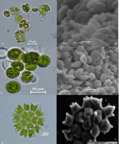
Samples were examined by light microscopy and later by SEM images. The samples were basically divided into two main groups. These, 3 species are Chlorophyta phylum and 4 species are Cyanobacteria. Two species from Chlorophyta are especially in the form of single-celled, 2-cell, 4-cell colonial cells. Cells shapes are elongate, fusiform or cylindrical. The dimensions of the cells are 4-8 x 8-11 μ and 5-45 x 10-50 μm. The cell walls are flat, thickening is visible. Chloroplasts are parietal and bowl-shaped. Based on these characteristics, it was thought that these species were in the order of Chlorococcales (Figure 1 A, B, C, D). The other sample
was observed to be morphologically quite different from the others and it is in colonial form. There are 8 to 32 cells in the colony. The colony is flat and there are pronounced horn shaped protrusions at the tip (Figure 1 E, F). Based on these features, it is thought to be in the order of Sphaeropleales as in the other two examples. The morphological characteristics of these species were given in Table 2.
Divisio : Chlorophyta Classis : Chlorophyceae Ordo : Chlorococcales
Family : Scenedesmaceae (Prescott, 1973) Divisio : Chlorophyta
Classis : Chlorophyceae Ordo : Sphaeropleales
Family : Hydrodictyaceae (Prescott, 1973)
Table 1. PCR primers and protocols used in this study for Chlorophyta and Cyanobacteria ITS1/ITS4 Hanen et al. (2002) 27F-1492R Hay et al. (2001) 18S rRNA 16S rRNA
Cycle Time Temperature Cycle Time Temperature
Initial denaturation 2 min 95 °C 1 min 94 °C
Denaturation 30 s 95 °C 45 s 94 °C
Annealing ´35 30 s 55 °C ´30 45 s 55 °C
Extension 1 min 72 °C 45 s 72 °C
Final extension 10 min 72 °C 10 min 72 °C
Figure 1. Light and electron microscope (SEM) images of green algae A, B Desmodesmus sp. C, D Chodatodesmus mucranulatus E, F Pediastrum duplex.
Table 2. Morphological characteristics of the green algae isolated from various habitats of lakes in Turkey Chodatodesmus
mucronulatus
Pediastrum duplex Desmodesmus sp.
Vegetative cells Single celled, cell wall smooth; cells elliptical
The cell bodies are polygonal, are granulated and have horn-like projections cells wall smooth, without granulations
cells are ovoid to ellipsoid, with rounded apices, and bearing long spines or teeth: the cell wall is granular, spiny or toothed, with wart-like projections and/or ribs present
zoospor nearly spherical biflagellate zoospore for each cell in the parent colony
subspherical Cell size
Vegetative cells 4-8 x 8-11 μ marginal cells 7-24-28x6-24-28 µm, inner cells 4-6-21-26x4-5-21-30 µm
5-45×10-50 µm
zoospor 5-10 µm 2.5-8 µm
Chloroplast shape parietal and striated parietal parietal
Pyrenoids single pyrenoid 2-4 pyrenoids single pyrenoid
Nucleus position central central central
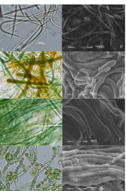
Figure 2. Light and electron microscope (SEM) images of cyanobacteria A, B Leptolyngbya sp. C, D Rivularia sp. E, F Phormidium autumnale G, H Fischerella ambigua.
4 species determined to be in the Cyanobacteria phylum, 2 species were considered to be very similar or even the same species according to their morphological characteristics. These two types have a common characteristic of filamentous, very thin sheath (Figure 2 A, B) or without sheath, no real branching, false branching, filaments with trichome organization, hormogonium present, which is characteristic features at the end part (Figure 2 E, F). The morphological characteristics of these species were given in Table 3.
Divisio : Cyanophyta Classis : Cyanophyceae Ordo : Oscillatoriales
Family : Oscillatoriaceae (Prescott, 1973)
The most important feature that distinguishes the other two species from the others is the presence of heterocyst. Heterocysts are only in one species at the end (Figure 2 C, D). In the other species, it was found to be in different parts of the filament (Figure 2 G, H). As a result of these investigations, these two species were thought to be in the order of Nostocales. The morphological characteristics of these species were given in Table 3.
Divisio : Cyanophyta Classis : Cyanophyceae Ordo : Nostocales
Family : Nostocaceae (Prescott, 1973)
Three different DNA isolated with using Chlorophyta phylum-specific primers (ITS1 / ITS4) and four DNA isolated with using universal primer pair bacterium and cyanobacterial primers (27F-1492R) and they were amplified by PCR.
Subsequently, the PCR products obtained were sent to REFGEN Gene Research and Biotechnology
Laboratory for sequence analysis. According to the sequence results, the phylogenetic map made been formed by NCBI-BLAST analysis and the species identification process was completed.
A phylogeny.fr platform was used to create the phylogenetic tree (Dereeper et al., 2008). For this program, the results from NCBI were recorded as FASTA. Then phylogenetic trees were created by loading on the online database. The phylogenetic trees were constructed by applying the "A la Carte" mode.
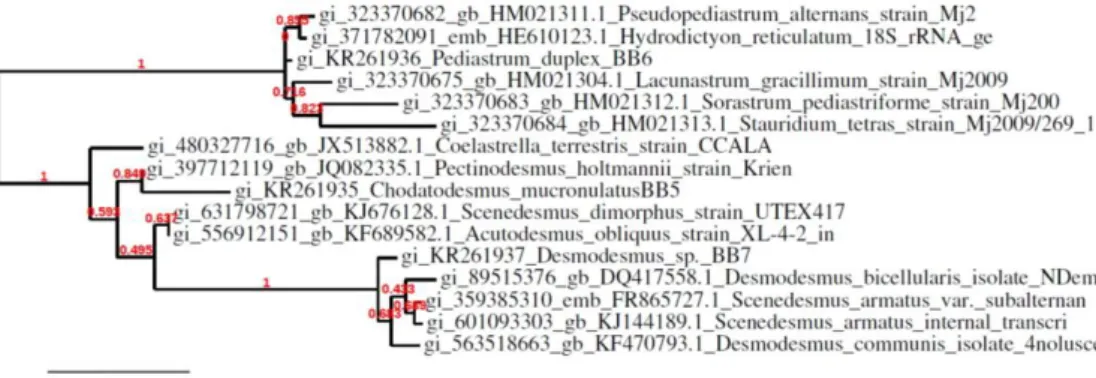
According to http://www.ncbi.nlm.nih.gov/ the results of the partial 18S rRNA sample of green algae isolated from the Apa Dam Lake are 1168/1328 base pairs in an adjacent sequence of the species determined under the order of Sphaeropleales that 97% Chodatodesmus mucranulatus, green algae isolated from the Beykavağı pond are 1062/1392 base pairs in an adjacent sequence of the species determined under the order of Chlorococcales that 98% Desmodesmus sp., green algae isolated from the Eber Lake are 1127/1363 base pairs in an adjacent sequence of the species determined under the order of Sphaeropleales that 97% Pediastrum duplex, were determined (Figure 3).
According to http://www.ncbi.nlm.nih.gov/ the results of the partial 16S rRNA sample of Cyanobacteria isolated from the Apa Dam Lake are 1546/1546 base pairs in an adjacent sequence of the species determined under the order of Nostocales that 99% Fischerella ambigua, 1415/1445 base pairs in an adjacent sequence of the species determined under the order of Oscillatoriales that 97% Leptolyngbya sp., Cyanobacteria isolated from the Beykavağı pond are 1509/1513 base pairs in an adjacent sequence of the species determined under the order of Oscillatoriales that 99% Phormidium
Table 3. Morphological characteristics of the cyanobacteria isolated from various habitats of lakes in Turkey Fischerella ambigua Phormidium
autumnale Leptolyngbya sp. Rivularia sp.
Vegetative cells subspherical conical and end cell
capitate coiled or spiral
hemispherical or
subspherical
heterocyst ovoid and mostly in the
main axis - - basal heterocyst
hormogonium - straight or slightly
formed as an ordered release of lengths of the trichome under conditions
-
Fibrous and
unbranched - unbranched unbranched unbranched
Fibrous and
pseudobranched Pseudo-branched - -
sometimes false
branching
sheath very thin sheath firm or
mucilaginous
sheath usually
colourless and thin
often frayed at the apical end
color distinct brown dark blue-green or
olive-green
yellow-brown or other
colour yellow-brown
Cell size
Vegetative cells 6-7.5 µm vegetative cells 4-7-9
µm wide, 2-5-7 µm long 4-6 µm
cells 5-7 µm wide, 2-5 µm long
heterocyst 4 µm - - 4-6 µm
autumnale and Cyanobacteria isolated from the Eber Lake are 1535/1535 base pairs in an adjacent sequence of the species determined under the order of Nostocales that 99% Rivularia sp., were determined (Figure 4).
Discussions
Among the first users of molecular techniques in Turkey, Tüney & Sukatar (2005) applied DNA isolation methods on Cystoseira mediterranea, a macroscopic brown marine algae. In microscopic freshwater algae, one of the first studies was made by Soylu and Gönülol in 2012 and observed the morphological diversity of green algae and determined the phylogenetic relationships of these strains according to the 18S rRNA gene sequences. According to DNA sequence analysis, two isolates were found to be similar to Scenedesmus subspicatus (98%), Desmodesmus sp. (98%) and Desmodesmus sp. (100%). Similar to this study, Baytut, Gürkanli, Özkoç & Gönülol (2013) showed that the diversity of single-celled Chlorophytes isolated from various freshwater habitats in the Central Black Sea Region is associated with three distinct lineages that related with the Chlamydomonadaceae (isolates 103I2 and C301), Chlorellaceae (isolate B4Riv) and Scenedesmaceae (isolates SIN-CON and N603) by
phylogenetic analyses. In this study, some of the isolates were similar to Scenedesmus and Desmodesmus genus. According to Komárek, Fott & Huber-Pestalozzi (1983), the Scenedesmus genus form colony between 2 and 32 cells, with a straight 1 or 2 rowed, cell shapes are always elongated, and unicellular and two or three colony forms of this genus shown in Desmodesmus sp. cultures. However, studies have shown that the Scenedesmus genus is separated from Desmodesmus genus (An, Friedl & Hegewald, 1999). While Scenedesmus genus has a cell wall that is very smooth, Desmodesmus genus cell wall has many characteristic structures. These are mainly teeth, rosettes, warts and spines (Hegewald, 2000). It may be difficult to identify these characters under light microscopy. Thus, researchers change the old, artificial classification system with a new, more natural, phylogenetic system. Especially this situation valid for coccoid green algae (Krienitz & Bock, 2012). These initial molecular phylogenetic studies have generally supported high-level classification of green algae and 18S phylogenies have made way for multi-gene and genome scale analyses (Leliaert et al., 2012; McCourt, 1995). In our study, morphologically identified species isolated were identified by molecular techniques and so isolated three species are green algae (KR261935
Figure 3 Tree formation of green algae according to 18 S rRNA sequences
Chodatodesmus mucranulatus, KR261936 Pediastrum duplex, KR261937 Desmodesmus sp.).
Because of isolation and replication of the blue green algae are more difficult than green algae, there are very few studies on this subject. As an example, blue-green algae (cyanobacteria) obtained from thermal facilities in İzmir province and surrounding area were isolated in laboratory environment and species determination was made with molecular methods by Yüksel et al. 2009. According to the sequence analysis, three of the species included in the genus Geitlerinema and the other one is the uncultered species (Yüksel, Demirel, Koçyiğit, & Sukatar, 2009).
In this study, filamentous, non-branched filamentous and false branching cyanobacteria species were found. Usually single-stranded, branched or non-branched filamentous forms are 1-4 μm wide, single-row, cylindrical cells, constructed in opposite walls, and form in contracting towards at the end. Cells are cylindrical, elongated, more or less isodiametric, later cells length more than cells width. The end cells are rounded or narrowed and contracted, sometimes hooked shaped, point shaped, narrow-round, sometimes ends with a round shaped calyptra. Cells may be matt blue-green, yellowish green, olive green and bright green colour. Heterocyst and hormogonium are present, no akinete. 16S rRNA sequence data have been used for many years to investigate the phylogenetic relationships of cyanobacteria in all taxonomic levels (Turner, 1997; Wilmotte & Herdman, 2001). The 16S rRNA gene is particularly valuable for phylogenetic studies because it has high conserved regions (for the evaluation of distant relationships) and some variable regions (for evaluation of closer relationships) (Řeháková, Johansen, Casamatta, Xuesong & Vincent, 2007). There is still considerable debate about what criteria should be used to identify species in cyanobacteria (Johansen & Casamatta, 2005). Sometimes, it is inadequate to safely identify morphological species. This is true for populations in the natural environment that share similar, but not identical, morphological characteristics found in similar biotopes. So should these similar populations be diagnosed morphologically or molecularly? We think that the answer to this question is to use it in both ways. In our study, morphologically identified species isolated were identified by molecular techniques (KR261931 Fischerella ambigua, KR261932 Phormidium autumnale, KR261933 Leptolyngbya sp., KR261934 Rivularia sp.). As a result of our study, 7 different algae from 3 different lakes were isolated and reproduced in BG-11 medium in the laboratory environment. In addition to the morphological characterization of green algae (Chodatodesmus mucranulatus, Pediastrum duplex, Desmodesmus sp.) and cyanobacteria species (Fischerella ambigua, Phormidium autumnale, Leptolyngbya sp., Rivularia sp.), it has been supported by molecular studies for accurate identification. Thus,
this investigation can provide a contribution to the field of molecular biological research in the area of phycology in Turkey.
References
An, S., Friedl, T., & Hegewald, E. (1999). Phylogenetic relationships of Scenedesmus and Scenedesmus‐like coccoid green algae as inferred from ITS‐2 rDNA sequence comparisons. Plant Biology, 1(4), 418-428. http://dx.doi.org/10.1111/j.1438-8677.1999.tb00724.x Baytut, Ö., Gürkanli, C. T., Özkoç, İ., & Gönülol, A. (2013).
Assessing 18S rDNA Diversity of the Chlorophytes among Various Freshwaters of the Central Black Sea Region. Turkish Journal of Fisheries and Aquatic Sciences, 13(5), 811-818. http://dx.doi.org/10.4194/1303-2712-v13_5_04
Castenholz, R. (1989). Oxygenic photosynthetic bacteria. Bergey's manual of systematic bacteriology, 3, 1710-1806.
Coat, G., Dion, P., Noailles, M.-C., De Reviers, B., Fontaine, J.-M., Berger-Perrot, Y., & Loiseaux-De Goër, S. (1998). Ulva armoricana (Ulvales, Chlorophyta) from the coasts of Brittany (France). II. Nuclear rDNA ITS sequence analysis. European journal of phycology, 33(1), 81-86.
Dereeper, A., Guignon, V., Blanc, G., Audic, S., Buffet, S., Chevenet, F., . . . Lescot, M. (2008). Phylogeny. fr: robust phylogenetic analysis for the non-specialist. Nucleic
acids research, 36(suppl_2), W465-W469.
http://dx.doi.org/10.1093/nar/gkn180
Fawley, M., Fawley, K., & Buchheim, M. (2004). Molecular diversity among communities of freshwater microchlorophytes. Microbial Ecology, 48(4), 489-499. http://dx.doi.org/10.1007/s00248-004-0214-4
Galhano, V., De Figueiredo, D. R., Alves, A., Correia, A., Pereira, M. J., Gomes-Laranjo, J., & Peixoto, F. (2011).
Morphological, biochemical and molecular
characterization of Anabaena, Aphanizomenon and Nostoc strains (Cyanobacteria, Nostocales) isolated from Portuguese freshwater habitats. Hydrobiologia, 663(1), 187-203. http://dx.doi.org/10.1007/s10750-010-0572-5 Gantt, E., & Conti, S. (1966). Granules associated with the chloroplast lamellae of Porphyridium cruentum. The Journal of cell biology, 29(3), 423-434.
Hay, A. G., Dees, P. M., & Sayler, G. S. (2001). Growth of a bacterial consortium on triclosan. FEMS Microbiology
Ecology, 36(2-3), 105-112.
http://dx.doi.org/10.1111/j.1574-6941.2001.tb00830.x Hegewald, E. (2000). New combinations in the genus
Desmodesmus (Chlorophyceae, Scenedesmaceae). Archiv fur Hydrobiologie-Supplementband Only, 131, 1-18.
Johansen, J. R., & Casamatta, D. A. (2005). Recognizing cyanobacterial diversity through adoption of a new species paradigm. Algological studies, 117(1), 71-93. https://doi.org/10.1127/1864-1318/2005/0117-0071 John, D. M., Whitton, B. A., & Brook, A. J. (2002). The
freshwater algal flora of the British Isles: an identification guide to freshwater and terrestrial algae (Vol. 1): Cambridge University Press.
Katırcıoğlu, H., Aslım, B., Türker, A. R., Atıcı, T., & Beyatlı, Y. (2008). Removal of cadmium (II) ion from aqueous system by dry biomass, immobilized live and heat-inactivated Oscillatoria sp. H1 isolated from freshwater
(Mogan Lake). Bioresource Technology, 99(10), 4185-4191.
Komárek, J., & Anagnostidis, K. (1989). Modern approach to the classification system of Cyanophytes 4-Nostocales.
Algological Studies/Archiv für Hydrobiologie,
Supplement Volumes, 247-345.
Komárek, J., Fott, B., & Huber-Pestalozzi, G. (1983). Das Phytoplankton des Süßwassers. Systematik und Biologie-Teil 7, 1. Hälfte.
Komárek, J., & Mareš, J. (2012). An update to modern taxonomy (2011) of freshwater planktic heterocytous cyanobacteria. Hydrobiologia, 698(1), 327-351. http://dx.doi.org/10.1007/s10750-012-1027-y
Krienitz, L., & Bock, C. (2012). Present state of the systematics of planktonic coccoid green algae of inland waters.
Hydrobiologia, 698(1), 295-326.
http://dx.doi.org/10.1007/s10750-012-1079-z
Larkum, A. W., & Vesk, M. (2003). Algal plastids: their fine structure and properties. In Photosynthesis in algae (pp. 11-28): Springer.
Leliaert, F., Smith, D. R., Moreau, H., Herron, M. D., Verbruggen, H., Delwiche, C. F., & De Clerck, O. (2012). Phylogeny and molecular evolution of the green algae. Critical Reviews in Plant Sciences, 31(1), 1-46. http://dx.doi.org/10.1080/07352689.2011.615705 McCourt, R. M. (1995). Green algal phylogeny. Trends in
ecology & evolution, 10(4), 159-163.
https://doi.org/10.1016/S0169-5347(00)89027-8 Oren, A. (2004). A proposal for further integration of the
cyanobacteria under the Bacteriological Code. International journal of systematic and evolutionary
microbiology, 54(5), 1895-1902.
http://dx.doi.org/10.1099/ijs.0.03008-0
Prescott, G. (1973). Algae of the Western Qreat Lakes area Otto Koeltz Science Publishers, 977 p. In: Germany. Rajaniemi, P., Hrouzek, P., Kaštovska, K., Willame, R., Rantala,
A., Hoffmann, L., . . . Sivonen, K. (2005). Phylogenetic and morphological evaluation of the genera Anabaena, Aphanizomenon, Trichormus and Nostoc (Nostocales, Cyanobacteria). International journal of systematic and
evolutionary microbiology, 55(1), 11-26.
http://dx.doi.org/10.1099/ijs.0.63276-0
Řeháková, K., Johansen, J. R., Casamatta, D. A., Xuesong, L., & Vincent, J. (2007). Morphological and molecular characterization of selected desert soil cyanobacteria: three species new to science including Mojavia pulchra gen. et sp. nov. Phycologia, 46(5), 481-502. https://doi.org/10.2216/06-92.1
Rippka, R. (1988). [1] Isolation and purification of cyanobacteria. In Methods in Enzymology (Vol. 167, pp. 3-27): Academic Press.
Sarsekeyeva, F. K., Usserbaeva, A. A., Zayadan, B. K., Mironov, K. S., Sidorov, R. A., Kozlova, A. Y., . . . Los, D. A. (2014). Isolation and characterization of a new cyanobacterial strain with a unique fatty acid composition. Advances in
Microbiology, 4(15), 1033.
http://dx.doi.org/10.4236/aim.2014.415114
Soylu, E. N., & Gönülol, A. (2012). Morphological and 18S rRNA analysis of coccoid green algae isolated from lakes of Kızılırmak Delta. Turkish Journal of Biology, 36(3), 247-254. http://dx.doi.org/10.3906/biy-1001-19
Turner, S. (1997). Molecular systematics of oxygenic photosynthetic bacteria. In Origins of Algae and their Plastids (pp. 13-52): Springer.
Tüney, I., & Sukatar, A. (2005). The DNA Extraction Protocol from one of the Brown algae Cystoseira mediteranea Sauvageau from Izmir Bay. Turkish Journal of Aquatic Life, 4, 100-103.
Van Hannen, E. J., Fink, P., & Lürling, M. (2002). A revised secondary structure model for the internal transcribed spacer 2 of the green algae Scenedesmus and Desmodesmus and its implication for the phylogeny of these algae. European journal of phycology, 37(2), 203-208. http://dx.doi.org/10.1017/S096702620200361X Wilmotte, A., & Herdman, M. (2001). Phylogenetic
relationships among the cyanobacteria based on 16S rRNA sequences. Bergey's Manual of Systematic Bacteriology. Volume One: The Archaea and the Deeply Branching and Phototrophic Bacteria, 487-493. Yüksel, K., Demirel, Z., Koçyiğit, A., & Sukatar, A. (2009). İzmir
İlinde Bulunan Termal Sularda Gelişen Bazı Termofilik Mavi-Yeşil Alglerin (Siyanobakterilerin) İzolasyonu ve Moleküler Tayini. E.U. Journal of Fisheries & Aquatic Sciences, 4, 267-270.