POLYTETRAFLUOROETYLENE TAPE AS TEMPORARY RESTORATIVE
MATERIAL: A FLUID FILTRATION STUDY ⃰
Geçici Restoratif Dolgu Materyali Olarak Politetrafloroetilen Bant (Ptfe):
Sıvı Filtrasyon Çalışması
Keziban Olcay1, liviu SteIer2, Hilal ErdoĞan3, Sema BElli3 received: 25/01/2015
accepted:30/04/2015
ABSTRACT
Purpose: the purpose of this study was to compare the sealing ability of temporary restorative materials at 24 hrs and 1 week. Materials and Methods: endodontic access cavities were prepared in 56 extracted lower incisor-teeth and divided into 5 groups (n=10). Standard 5 mm deep access preparations were completed and root canals were prepared to size ISO #30 file. the access cavities were restored as follows: Group 1: temporary restorative material (ceivitron); Group 2: glass ionomer cement (Fuji II); Group 3: zinc oxide-eugenol cement (IrM); Group 4: zinc phosphate cement (adhesor); Group 5: polytetrafluoroetylene tape (PtFe). the quality of the coronal sealing of each specimen was measured (24 hrs and 1 week) using fluid transport model. the data was analysed with repeated measurements of anoVa, Tukey, Paired samples T-Tests. Results: a significant difference was found among the groups at all time-periods (p<0.05). at 24 hrs, PtFe showed similar leakage with ceivitron, IrM, and Fuji II but it showed higher leakage than adhesor. at 1 week, ceivitron showed higher leakage than PtFe, meanwhile PtFe showed similar leakage with IrM, Fuji II, and adhesor (p>0.05). Sealing ability of IrM and PtFe groups significantly increased by time (p<0.05 and p<0.001 respectively). Conclusion: Within the limitations of this study, PtFe showed an acceptable short-term sealing capability when compared to the other commonly used temporary restorative materials at 1 week measurements.
Keywords: Dental leakage; polytetrafluoroethylene; temporary dental fillings
ÖZ
Amaç: Bu çalışmanın amacı, geçici restoratif materyallerin 24 saat ve 1 hafta süresinde sızdırmazlık kabiliyetlerinin karşılaştırılmasıdır.
Gereç ve Yöntem: 56 adet çekilmiş alt çene kesici dişin endodontik giriş kaviteleri açıldı. dişler 5 gruba ayrıldı (n=10). Giriş kaviteleri standart 5 mm derinlikte olacak şekilde açıldı ve kök kanalları ISo #30 numaraya kadar genişletildi. Giriş kaviteleri şu şekilde restore edildi: Grup 1: geçici restorasyon materyali (ceivitron); Grup 2: cam iyonomer siman (Fuji II); Grup 3: çinko oksit öjenol siman (IrM); Grup 4: çinko fosfat siman (adhesor); Grup 5: politetrafloroetilen bant (PtFe). Her örneğin koronal sızdırmazlık kalitesi sıvı filtrasyon yöntemi kullanılarak ölçüldü (24 saat ve 1 hafta). Veriler hesaplandı (lp), tekrarlayan ölçümlerle anoVa, Tukey ve bağımlı gruplarda P testi kullanılarak değerlendirildi. Bulgular: Tüm zamanlar için gruplar arasında anlamlı fark bulundu (p<0.05). 24 saatte, PtFe; ceivitron, IrM ve Fuji II ile benzer; fakat adhesor den daha fazla sızıntı değeri gösterdi. 1 haftada; Ceivitron, PTFE den daha fazla sızıntı değeri gösterdi, aynı zamanda PTFE; IrM, Fuji II ve adhesor ile benzer sızıntı değeri gösterdi (p>0.05). IrM ve PTFE gruplarının sızıntı değeri zamanla önemli derecede azaldı (p < 0.05 ve p < 0.001 sırasıyla).
Sonuç: Bu çalışmanın sınırları dâhilinde, PTFE, 1 haftalık ölçümlerde, diğer sıklıkla kullanılan geçici dolgu materyalleri ile kıyaslandığında, kabul edilebilir bir kısa-dönem sızdırmazlık kabiliyeti göstermiştir.
Anahtar kelimeler: dental sızıntı; politetrafloroetilen; geçici dolgu maddeleri
J Istanbul Univ Fac Dent 2015;49(3):17-22. orIGInal rESEarCH
1 Department of Endodontics Faculty of Dentistry Istanbul Medipol University
2 Dr.Med.Dent. Associate Clinical Professor, Specialist in Endodontics. Course director MsC in Endodontics; Warwick Dentistry, Institute of Clinical Education, Warwick Medical School, Medical School Building. The University of Warwick
3 Department of Endodontics Faculty of Dentistry Selçuk University
Introduction
The importance of the coronal leakage on the results of root canal treatment has been widely accepted (1-3). Microbial infection is one of the principle factors associated with endodontic failure (4). Therefore, the major aims of the root canal treatment are removing microorganisms from the root canal by chemomechanical debridement, and sealing of the root canal system against irritants such as; percolation of fluids, microorganisms, saliva and other debris from the oral cavity (5). These irritants may induce periapical pathosis (6, 7), thus all effort should be spent to prevent microbial contamination of
the pulp spacein every step of endodontic treatment.
Previous studies have demonstrated that the coronal seal as important as the quality of root canal filling for periapical health (2, 8). Microorganisms may able to pass root canal filling (9) and coronal leakage may occur within a few days (8). Consequently, temporary restorative material has to be applied. Temporary restorative materials are often used during endodontic treatment to seal the root canal between sessions or until a permanent restoration is placed. An ideal temporary restorative material should exhibit minimal or no leakage, good abrasion and compression resistance, lack of porosity, lack of dimensional changes, good aesthetic appearance and it must also be easily manipulated or removed while being effective in a moist environment (10). Cavit and IRM are the most commonly used temporary
restorations among specialists (11),followed by
glass-ionomer cement (GIC) (10)and zinc phosphate
cement (12). Conventional GICs were considered as suitable materials for temporary sealing (13) especially because of their adhesive properties (14). In recent publications, the use of polytetrafluoroethylene tape (PTFE) has been advocated in dentistry in several fields such as management of access channels in implant-supported prosthesis (15), for repairing abutment teeth (16), matrix to prevent etching and bonding of the adjacent teeth (17), spacer material (18), for repairing a damaged cast post and core restoration (19), for eliminating subgingival cement adhesion to implant abutments (20), guided bone regeneration barrier material (21) and dental floss (22). The purpose of this study was to compare short-term sealing ability of PTFE with four commonly used temporary restorative materials. The tested hypothesis was there is no significant difference among the sealing ability of the tested materials.
Materials and Methods
Single-rooted sound permanent human mandibular incisors with straight root canals stored in physiological saline solution were used in the study. Root canal morphologies were radiographically examined; tissue remnants and calculus were removed. Fifty six teeth with similar dimensions were selected (14mm±0.5mm long root; 7mm±0.5mm crown length). Five mm deep standard access cavities were made with #4 bur (Detsply Tulsa Dental, Tulsa OK). Patency of the root canals was verified with #10 K-file (Dentsply Maillefer, Tulsa, OK). The working lengths were determined by placing #10 K-file into the root canal until it was visible at the apical foramen and subtracting 1mm from the working length. Six mm coronal part of roots was flared using Gates Glidden drills (sizes 2-3, Maillefer, Ballaigues, Switzerland). The roots were instrumented with K-file (Dentsply Maillefer, Tulsa, OK) to size #30 using step-back technique. After completing the instrumentation, in order to standardize the leakage, each root was exited 1 mm out of the apical foramen with #20 K-file (Dentsply Maillefer, Tulsa, OK). Between each file use, the canals were irrigated with 1 ml, 5.25% NaOCl solution. The canals were dried with paper points and all specimens were randomly divided into five groups (n=10). The access cavities were then restored as follows:
Group 1: Ceivitron (Triune Med Tec, Cambridgeshire, UK);
Group 2: Fuji II (Fuji II LC, GC Corp, Tokyo, JAPAN); Group 3: IRM (IRM; Dentsply Caulk, Milford, DE); Group 4: Adhesor (Adhesor, Spofa Dental, Frankfurt, Germany).
Group 5: Polytetrafluoroethylene (PTFE) tape (Oatey Co, Cleveland, OH, ABD).
In group 1, the access cavity was filled with Ceivitron totally by using a hand plugger. In group 2, Fuji II was prepared with mechanical mixer
(ProMix,Dentsply International, York, PA, USA).
After inserting into the access cavity, Fuji II light cured for 20s using a curing unit (Bluephase 800
mW/cm2, Ivoclar, Vivadent AG, Liechtenstein,
Austria) under moisture free environment. In group 3 and 4, IRM and Adhesor cements were applied as recommended by the manufacturer. In the last group, 8mm long PTFE tape was used. The material was cut and the access cavity was filled with PTFE by compacting the material with hand plugger (Maillefer, Ballaigues, Switzerland) until the cavity was totally filled (Figure 1).
Figure 1. The access cavity was filled with PTFE by compacting the material with plugger until the cavity was totally filled.
Negative control (n=3) The cavities and apical
openings were filled with Clearfil AP-X (Kuraray, Tokyo, Japan) after cavity conditioning with Clearfil SE Bond (Kuraray, Tokyo, Japan) in negative control group and then covered with two layers of nail varnish.
Positive control (n=3) Three of roots were
instrumented to size #30 using step-back technique and each root was exited 1 mm out of the apical foramen with a #20 K-file and no coronal restoration was performed.
Evaluation of the leakage A fluid transport system
(23) was used. Coronal parts of the teeth were inserted 3mm into silicone tubing having an internal diameter of 3mm and attached to the outer surface of the tube with cyanoacrylate-adhesive. The tube was then connected to fluid transport model as described by
Derkson et al. (24)and as modified by Wu et al. (25).
A polyethylene tubing (Fisher Scientific, Pittsburg, PA) was used to connect the pressure reservoir to a 25 μl micropipette (Microcaps, Fisher Scientific, Philadelphia, PA). Additional tubing was used to connect the micropipette to a microsyringe (Gilmont Instruments Inc, Great Lakes, NY) and the silicone tube to the attached root. An air bubble was introduced into the system using the micro syringe and the bubble was moved inside the micropipette.
All tubing, pipette and syringe were filled with distilled water under a pressure of 220 kPa via use
of O2 gas. The sealing capability of the samples
was quantitated by following the progress of this tiny air bubble traveling within the micropipette. System leakage is determined as 5 minutes for each sample depending on negative control teeth which do not leak. The cavities and apical openings were filled with Clearfil AP-X (Kuraray, Tokyo, Japan) in combination with Clearfil SE Bond (Kuraray, Tokyo, Japan) in negative control group and then covered with two layers of nail varnish. The fluid flow rate through the three unsealed roots which were prepared for positive control was measured by weighing the amount of water that could flow through the needle in 1min. This value served both as a positive control and as 100% leakage case. The samples were kept in 100% humid conditions at 37°C throughout the experimental period. 0.02% sodium azide was added to the storing solution to prevent bacterial colonization. Measurements of fluid movement were recorded at 2min intervals for 8min and the results were averaged. The sealing quality of each specimen was measured at 24 hrs and 1 week.
Statistical analysis
The data was calculated as Lp. Repeated measurements of ANOVA and Tukey tests were performed to evaluate the difference among the leakage values of the groups at 24 hrs and 1 week. Paired Samples T-Test (SPSS 16.0) was also completed to evaluate the differences in each material’s leakage by time.
Results
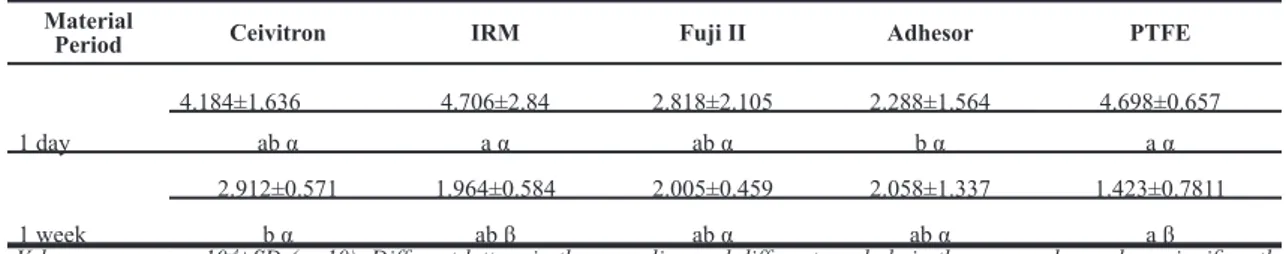
The positive controls had grossly leaked (40±0.5 µl); the varnish-coated negative controls had no measurable bubble movement at all time periods. The mean and standard deviations of the leakage values are shown in Table 1.
Table 1. Coronal leakage measurement values expressed in lp (μl / min-1 cm-2 cm H
2O-1) for all test groups at 1 day and 1 week.
Material
Period Ceivitron IRM Fuji II Adhesor PTFE
1 day 4.184±1.636 4.706±2.84 2.818±2.105 2.288±1.564 4.698±0.657 ab α a α ab α b α a α 1 week 2.912±0.571 1.964±0.584 2.005±0.459 2.058±1.337 1.423±0.7811 b α ab β ab α ab α a β
Values are means x10-4±SD (n=10). Different letters in the same line and different symbols in the same column show significantly
A significant difference was found among the groups at all time periods (p<0.05). PTFE showed similar leakage with Ceivitron, IRM, and Fuji II (p>0.05) but higher leakage than Adhesor at 24 hrs (p<0.05); and it showed less leakage than Ceivitron at 1 week (p<0.05). No significant difference was found among the leakage of PTFE, IRM and Fuji II at the end of 1 week (p>0.05). Sealing ability of IRM and PTFE groups significantly increased by the time (p<0.05 and p<0.001 respectively). Leakage of the groups sealed with Ceivitron, Fuji II and Adhesor did not change from 24 hrs to 1 week (p=0.113, p=0.306 and p=0.756 respectively).
Discussion
In this in vitro study, sealing ability of PTFE was compared with four commonly used temporary
filling materials using a fluid transport model.Coronal
leakage values for all test groups are shown in Table 1. A good marginal seal between tooth and temporary restorative material to prevent the entry of saliva and microorganisms is a very important factor to minimize contamination during the endodontic therapy (26, 27). The thickness of the temporary filling material is another important factor which contributes to its sealing ability. In a previous study, 3.5mm thickness of temporary material was used as the minimum thickness necessary to prevent total leakage of the dye
molecule (28).Temporary restorative materials need
adequate retention to prevent dislodgement between sessions therefore a thickness of 5 mm restorative
material was inserted in this study (29, 30).The
roots were kept empty to disregard the effect of root canal filling material. There have been many studies comparing the sealing ability of temporary filling material Cavit, IRM, Fuji II and Adhesor cement (10,
13, 30-33).Some authors indicated that there is no
difference in marginal leakage perspective between Cavit and IRM (30, 32), while others reported that IRM was able to seal against bacteria better than Cavit, possibly as a result of the antibacterial
properties of eugenol (31, 34).In the studies where
thermocycling was used (35, 36), Cavit showed better sealing than IRM. This is explained as a result of the hydrophilic property of the material that allowed compensation for microgaps that might open during
the temperature changes (37).Cavit has hydroscopic
properties causing it to expand and set when it comes
to contact with moisture (38).This property permits
the material to adapt better against dentinal walls (38).
Use of Cavit has been suggested because of its ease of use and the cost. In the present study, Ceivitron was used. Ceivitron is a calcium sulfate based filling material like Cavit and showed similar leakage at 24 hrs with PTFE (p>0.05). PTFE is also an inexpensive material and previously used in dentistry in different fields (15-22). It was reported that PTFE can be sterilized, it is radiopaque and easy to manipulate (15). Twenty four hours results indicated that this material can be an alternative to Ceivitron and 1 week results indicated that it can be an alternative to IRM, Fuji II and Adhesor as a temporary filling.
IRM is a reinforced zinc oxide eugenol material. It is known as hydrophobic as a result of its polymethylmethacrylate ingredient (37). Although some studies indicated that Cavit and Cavit like materials sometimes show better sealing than other temporary restorative materials (26, 27, 32, 39) Friedman et al. (40) found the opposite. In the present study, Ceivitron showed similar leakage when compared to IRM, Fuji II and Adhesor at 24 hours. However according to Friedman et al. (40), Ceivitron showed higher leakage than other tested materials after 1 week.
GICs are very effective biomaterials for adhesion to tooth tissue. Other important advantages of GICs include fluoride release and antimicrobial activity (13, 41). In this study Fuji II LC was used. Fuji II LC is resin modified glass ionomer cement formed by 2-hydroxymethyl methacrylate blended with a polyalkenoic acid liquid. The results indicated that the sealing ability of Fuji II LC did not change at the end of 1 week and showed a similar sealing performance with PTFE, IRM and Adhesor. Many factors in oral conditions may affect the performance of the materials such as thermal cycling and loading. This laboratory study did not mimic actual clinical conditions such as thermal changes or mechanical loading. Friedman
et al.(40) did not use thermocycling and reported less leakage of IRM compared to Cavit. In the present study, Ceivitron showed similar leakage with IRM at 1 week. Under different conditions, calcium sulfate based sealers can lose their sealing abilities due to deterioration (42). The sealing performance of the materials is expected to be different if all these factors were added to the testing protocol.
This study evaluated short term leakage of temporary restorative materials. The rationale for testing the materials at 24h and 1 week was that these are frequently used time intervals in dental practice either between sessions or while the permanent
restoration is placed after the root canal is obturated. Siquiera et al. (43) used 1 week period between the visits to test calcium hydroxide/camphorated paramonochlorophenol paste as an intracanal dressing. Furthermore, the average time for both contaminations of access cavities closed with IRM and Cavit-G was reported as 12.95 and 9.80 days, respectively (32). On the other hand, sometimes the circumstances may change and the period between the visits may extend. Therefore long term sealing ability of PTFE should be tested. This is one of the limitations of this study. The other limitation is that the cavity of the tooth may change due to previous history of the tooth. PTFE retained by friction and may not be applicable in some other conditions. These factors should be evaluated with further studies.
Conclusion
Within the limitations of this short term in vitro study, enclosed results were drawn:
PTFE showed an acceptable sealing performance when compared to the other commonly used temporary restorative materials;
PTFE showed similar leakage performance with IRM, Fuji II and Adhesor at the end of 1 week.
Source of funding
None declared
Conflict of interest
None declared
References
1. Hommez GM, Coppens CR, De Moor RJ. Periapical health related to the quality of coronal restorations and root fillings. Int Endod J 2002;35(8):680-689.
2. Ray HA, Trope M. Periapical status of endodontically treated teeth in relation to the technical quality of the root filling and the coronal restoration. Int Endod J 1995;28(1):12-18. 3. Siqueira JF, Jr., Rocas IN, Favieri A, Abad EC,
Castro AJ, Gahyva SM. Bacterial leakage in coronally unsealed root canals obturated with 3 different techniques. Oral Surg Oral Med Oral Pathol Oral Radiol Endod 2000;90(5):647-650. 4. Schwartz RS, Fransman R. Adhesive dentistry
and endodontics: Materials, clinical strategies and procedures for restoration of access cavities: A review. J Endod 2005;31(3):151-165. 5. Ricucci D, Siqueira JF, Jr. Recurrent apical
periodontitis and late endodontic treatment failure related to coronal leakage: A case report. J Endod 2011;37(8):1171-1175.
6. Cardoso AS, Silva NC, Silva JM, Herrera DR, Neves AA, Leal Silva EJ. Assessment of coronal leakage of a new temporary light-curing filling material in endodontically treated teeth. Indian J Dent Res 2014;25(3):321-324.
7. Siqueira JF, Jr., Rocas IN, Alves FR, Campos LC. Periradicular status related to the quality of coronal restorations and root canal fillings in a brazilian population. Oral Surg Oral Med Oral Pathol Oral Radiol Endod 2005;100(3):369-374. 8. Moreno JO, Alves FR, Goncalves LS, Martinez
AM, Rocas IN, Siqueira JF, Jr. Periradicular status and quality of root canal fillings and coronal restorations in an urban colombian population. J Endod 2013;39(5):600-604. 9. Torabinejad M, Ung B, Kettering JD. In vitro
bacterial penetration of coronally unsealed endodontically treated teeth. J Endod 1990;16(12):566-569.
10. Ciftci A, Vardarli DA, Sonmez IS. Coronal microleakage of four endodontic temporary restorative materials: An in vitro study. Oral Surg Oral Med Oral Pathol Oral Radiol Endod 2009;108(4):e67-70.
11. Vail MM, Steffel CL. Preference of temporary restorations and spacers: A survey of diplomates of the american board of endodontists. J Endod 2006;32(6):513-515.
12. Hosoya N, Cox CF, Arai T, Nakamura J. The walking bleach procedure: An in vitro study to measure microleakage of five temporary sealing agents. J Endod 2000;26(12):716-718.
13. BM. C. Glass-ionomer dental restoratives. . Prog Polym Sci 2001;26(4):577–604.
14. Naoum HJ, Chandler NP. Temporization for endodontics. Int Endod J 2002;35(12):964-978. 15. Moraguez OD, Belser UC. The use of
polytetrafluoroethylene tape for the management of screw access channels in implant-supported prostheses. J Prosthet Dent 2010;103(3):189-191. 16. Chan DC. Technique for repair of multiple
abutment teeth under preexisting crowns. J Prosthet Dent 2003;89(1):91-92.
17. Dunn WJ, Davis JT, Casey JA. Polytetrafluoroethylene (ptfe) tape as a matrix in operative dentistry. Oper Dent 2004;29(4):470-472.
18. Paranjpe A, Jain S, Alibhai KJ, Wadhwani CP, Darveau RP, Johnson JD. In vitro microbiologic evaluation of ptfe and cotton as spacer materials. Quintessence Int 2012;43(8):703-707.
19. Arabolu M, Nair KC, Raheel SA, Tarakji B, Azzeghaiby SN, Nassani MZ. Using an existing
crown to repair a damaged cast post and core restoration. J Int Oral Health 2014;6(5):111-113. 20. Hess TA. A technique to eliminate subgingival
cement adhesion to implant abutments by using polytetrafluoroethylene tape. J Prosthet Dent 2014;112(2):365-368.
21. Takata T, Wang HL, Miyauchi M. Migration of osteoblastic cells on various guided bone regeneration membranes. Clin Oral Implants Res 2001;12(4):332-338.
22. Tascón J.E GJC, Madrid A, Gallego M.D.P. . Price and the efficacy of the polytetrafluoroethylene (ptfe) tape for the removal of the proximal dental biofilm compared to the common nylon dental floss in adolescents and young adults. Columbia Medica 2006;37(4):287-292.
23. Pashley DH, Depew DD. Effects of the smear layer, copalite, and oxalate on microleakage. Oper Dent 1986;11(3):95-102.
24. Derkson GD, Pashley DH, Derkson ME. Microleakage measurement of selected restorative materials: A new in vitro method. J Prosthet Dent 1986;56(4):435-440.
25. Wu MK, De Gee AJ, Wesselink PR, Moorer WR. Fluid transport and bacterial penetration along root canal fillings. Int Endod J 1993;26(4):203-208.
26. Lai YY, Pai L, Chen CP. Marginal leakage of different temporary restorations in standardized complex endodontic access preparations. J Endod 2007;33(7):875-878.
27. Lee YC, Yang SF, Hwang YF, Chueh LH, Chung KH. Microleakage of endodontic temporary restorative materials. J Endod 1993;19(10):516-520.
28. Webber RT, del Rio CE, Brady JM, Segall RO. Sealing quality of a temporary filling material. Oral Surg Oral Med Oral Pathol 1978;46(1):123-130.
29. Balto H. An assessment of microbial coronal leakage of temporary filling materials in endodontically treated teeth. J Endod 2002;28(11):762-764.
30. Zmener O, Banegas G, Pameijer CH. Coronal microleakage of three temporary restorative materials: An in vitro study. J Endod 2004;30(8):582-584.
31. Blaney TD, Peters DD, Setterstrom J, Bernier WE. Marginal sealing quality of irm and cavit as assessed by microbiol penetration. J Endod 1981;7(10):453-457.
32. Imura N, Otani SM, Campos MJ, Jardim Junior EG, Zuolo ML. Bacterial penetration through temporary restorative materials in root-canal-treated teeth in vitro. Int Endod J 1997;30(6):381-385.
33. Saunders WP, Saunders EM. Coronal leakage as a cause of failure in root-canal therapy: A review. Endod Dent Traumatol 1994;10(3):105-108. 34. Hume WR. The pharmacologic and toxicological
properties of zinc oxide-eugenol. J Am Dent Assoc 1986;113(5):789-791.
35. Kazemi RB, Safavi KE, Spangberg LS. Assessment of marginal stability and permeability of an interim restorative endodontic material. Oral Surg Oral Med Oral Pathol 1994;78(6):788-796.
36. Tamse A, Ben-Amar A, Gover A. Sealing properties of temporary filling materials used in endodontics. J Endod 1982;8(7):322-325. 37. Koagel SO, Mines P, Apicella M, Sweet M. In
vitro study to compare the coronal microleakage of tempit ultraf, tempit, irm, and cavit by using the fluid transport model. J Endod 2008;34(4):442-444.
38. Barkhordar RA, Stark MM. Sealing ability of intermediate restorations and cavity design used in endodontics. Oral Surg Oral Med Oral Pathol 1990;69(1):99-101.
39. Naseri M, Ahangari Z, Shahbazi Moghadam M, Mohammadian M. Coronal sealing ability of three temporary filling materials. Iran Endod J 2012;7(1):20-24.
40. Friedman S, Shani J, Stabholz A, Kaplawi J. Comparative sealing ability of temporary filling materials evaluated by leakage of radiosodium. Int Endod J 1986;19(4):187-193.
41. Barkhordar RA, Pelzner RB, Stark MM. Use of glass ionomers as retrofilling materials. Oral Surg Oral Med Oral Pathol 1989;67(6):734-739. 42. Liberman R, Ben-Amar A, Frayberg E,
Abramovitz I, Metzger Z. Effect of repeated vertical loads on microleakage of irm and calcium sulfate-based temporary fillings. J Endod 2001;27(12):724-729.
43. Siqueira JF, Jr., Magalhaes KM, Rocas IN. Bacterial reduction in infected root canals treated with 2.5% naocl as an irrigant and calcium hydroxide/camphorated paramonochlorophenol paste as an intracanal dressing. J Endod 2007;33(6):667-672.
Corresponding Author: Keziban OLCAY
Department of Endodontics Faculty of Dentistry Istanbul Medipol University Unkapanı / Istanbul-TURKEY Phone: +90 212 453 48 00 (4958) e-mail: [email protected]
View publication stats View publication stats