Email: [email protected]
ORIGINAL ARTICLE
Corresponding author: Yasemin Basbinar, MD, PhD. Dokuz Eylul University, Institute of Oncology, Department of Translational Oncology 35350, Inciralti, Mithatpasa Str.56/13, Izmir, Turkey.
Tel: +90 232 412 58 90, Fax: +90 232 278 94 95, Email: [email protected] Received: 27/12/2018; Accepted: 02/02/2019
Curcumin effects on cell proliferation, angiogenesis and
metastasis in colorectal cancer
Gizem Calibasi-Kocal
1*, Ahu Pakdemirli
2*, Serdar Bayrak
3, Nazli Mert Ozupek
4, Tolga
Sever
4, Yasemin Basbinar
1, Hulya Ellidokuz
5, Turkan Yigitbasi
61Dokuz Eylul University, Institute of Oncology, Department of Basic Oncology, Izmir, Turkey; 2Dokuz Eylul University, Vocational School of Health Services, Department of First and Emergency Aid, Izmir, Turkey; 3Dokuz Eylul University, Faculty of Medicine, Department of Cardiovascular Surgery, Izmir, Turkey; 4Dokuz Eylul University, Institute of Health Sciences, Department of Oncology, Izmir, Turkey; 5Dokuz Eylul University, Institute of Oncology, Department of Preventive Oncology, Izmir, Turkey; 6Istanbul Medipol University, Faculty of Medicine, Department of Biochemistry, Istanbul, Turkey.
*These authors contributed equally to this study.
Summary
Purpose: Curcumin is a natural phytopolyphenol com-pound isolated from the root of turmeric (Curcuma longa) and possesses a wide range of biological properties. The purpose of this study was to evaluate the antiprolifera-tive, wound healing, anti-invasive and anti-migrative ef-fects of curcumin on HCT-116 and LoVo colorectal cancer cell lines.
Methods: The antiproliferative activity of 2.5-75 µM curcumin was tested on HCT-116 and LoVo colorectal cell lines and the viability of the cells was tested with WST-1 reagent by using ELISA plate reader at 450 nm. xCELLigence RTCA DP system was used for the
de-tection of anti-invasive and anti-migrative effects of curcumin.
Results: The IC50 of curcumin was 10±0.03 for HCT-116 and
20±0.05 µM for LoVo cell lines. The IC50 of curcumin (10µM
for HCT-116 and 20 µM for LoVo) showed anti-metastatic activity on these cell lines.
Conclusion: This study showed that curcumin could be evaluated as a promising anti-cancer agent for human colo-rectal cancer.
Key words: colorectal cancer, curcumin, invasion, migra-tion, proliferation
Introduction
Curcumin ((1E,6E)-1,7-bis(4-hydroxy-3-meth-oxyphenyl) hepta-1,6-diene-3,5-dione) is a hydro-phobic polyphenol-structured secondary metabo-lite of dried rhizome of turmeric (Curcuma longa and Curcuma spp.) [1,2] and its empirical formula is C21H20O6 [2]. It is a yellow-orange colored sub-stance. Moreover, curcumin is the most abundant component of turmeric and the other metabolites found in the plant are demethoxycurcumin and bis-demethoxycurcumin [3]. Since their natural path-ways are the same and they convert to each other,
their biological properties are quite similar [2]. Ac-cording to the experiments on both animals and humans, the daily oral intake of curcumin is up to 12 g [4]. Also, curcumin has biological activities such as angiogenic, inflammatory, anti-oxidant and anticancer [2]. Colorectal cancer is the third most common cancer type around the world which leads to cancer-related death [5]. However, if diagnosed in the early stages, the survival of colorectal cancer is increased [6]. There are several factors (age, familial cancer syndromes,
inflamma-tory bowel disease, dietary factors, and hereditary polyposis conditions) effect that can contribute to the development of colorectal cancer [7]. The main components of colorectal cancer therapy are chemo-therapy (5-fluorouracil and oxaliplatin), radiothera-py and surgery [8,9]. As natural therapeutic agents, irinotecan (a derivative of camptothecin) and poly-saccharide K are used for colorectal cancer [10].
The purpose of this study was to investigate the anticancer activities of curcumin, i.e. antiprolifera-tive, anti-invasive, anti-migrative and wound heal-ing properties on colorectal cancer cell lines in vitro.
Methods
Cell culture
Human colorectal carcinoma HCT116 (ATCC CCL-247) cell line was obtained from American Type Culture Collection (ATCC) (Rockville, CT, USA). This cell line was cultured in McCoy’s-5a modified medium (Biochrom GbmH, Berlin, Germany) supplemented with 15% fetal bovine serum (FBS, Cegrogen Biotech GmbH, Stadtal-lendorf, Germany) and 1% penicillin/streptomycin (Biochrom GbmH, Berlin, Germany). Human metastatic colorectal adenocarcinoma, LoVo (ATCC CCL-229) cell line was obtained from ATCC (Rockville, CT, USA). This cell line was cultured in RPMI-1640 (Biochrom GbmH, Berlin, Germany) containing 10% FBS (Cegrogen Bio-tech GmbH, Stadtallendorf, Germany), 1% L-glutamine (Biochrom GbmH, Berlin, Germany) and 1% penicillin/ streptomycin (Biochrom GbmH, Berlin, Germany). Cells were kept at 37 °C in humidified 5% CO2 incubator.
Cell viability
The antiproliferation activity of curcumin (C7727, Sigma-Aldrich, St Louis, MO, USA) was tested on HCT-116 and LoVo cell lines. These cell lines were seeded in 96-well plates (1×10⁴ cells/well). After 48 h of incubation
with 2.5-75 µM curcumin, cell viability was evaluated with water soluble tetrazolium salt-1 (WST-1) assay (Version 16 Cell Proliferation Reagent WST-1, Roche Di-agnostics, Indianapolis, USA). The assay was performed according to the manufacturer’s protocol.
Wound healing assay
Cell migration capability of HCT-116 and LoVo cells were also detected by using wound healing assay. These cell lines (30×104 cells/well) were seeded in 6-well
plates. After 90% confluence of cells on the plates, the wells were scratched with a pipette tip across the center of the well and the IC50 and IC10 doses of curcumin were
added. McCoy’s-5A modified medium (Biochrom GBMH, Berlin, Germany) was used for HCT-116 cell line and RPMI-1640 (Biochrom GBMH, Berlin, Germany) was used for LoVo cell line as control. JuLI Br live cell movie analyzer was used for monitoring the wounds. ImageJ software 1.49 was used for measuring the wound gaps. Cell invasion and migration assay
Cell invasion and migration activity were measured by using xCELLigence RTCA Dual Plate (RTCA-DP) in-strument according to the manufacturer’s recommen-dations with CIM-plate 16 (Roche Diagnostics GmbH, Mannheim, Germany). For detection of cell migration, electrical impedance changes were measured at a gold microelectrode plated on the bottom of a membrane
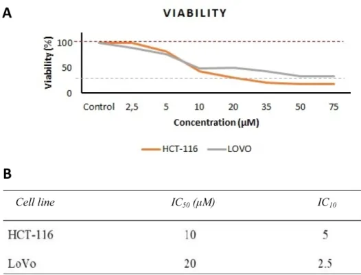
Figure 1. A: Dose-dependent cytotoxicity of curcumin on HCT-116 (orange) and LoVo (gray) cells incubated with increas-ing concentrations of curcumin (p<0.05). B: IC50 and IC10 of curcumin on HCT-116 and LoVo cell lines.
Figure 2. Invasion effect of curcumin on A: HCT-116 and B: LoVo cell lines after 24-h incubation (p<0.05).
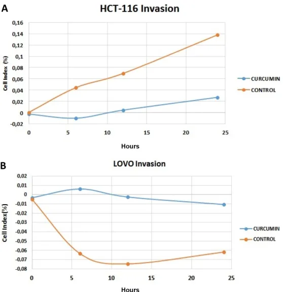
Figure 3. Cell invasion results of curcumin on A: HCT-116 and B: LoVo cell lines at 24 h (orange control, blue curcumin). The results were obtained from xCelligence RTCA DP.
separating the upper and lower chambers. The lower compartment was supplemented with 10% FBS-contain-ing medium for migration assay. 4×10⁴ cells suspended
in serum-free medium were supplemented to the up-per compartment of the plate. The upup-per compartment was coated with 1:40 diluted Matrigel (Basement Mem-brane Matrix, growth factor reduced, BD Biosciences, Erembodegem, Belgium) and the lower compartment was supplemented with 10% FBS-containing medium for invasion analysis. IC50 of curcumin found by WST-1
assay analysis was added to the upper compartments for both invasion and migration assays for each cell line. The impedance was recorded per 15 min and the total analysis time was set at 48 h. xCELLigence RTCA soft-ware vs.1.2.1 was used for data analysis. At 6th, 12th and 48th h migration and invasion of cells were calculated as the percentage of CI compared to control group. Statistics
Mann-Whitney U test was used to determine the significance between curcumin and its activity. SPSS (Version 15.0; SPSS, Inc., Chicago, IL, USA) was used for all analyses. The level of statistical significance was set at p<0.05. The data in the figures represent the
mean±standard error of the mean (SEM). In this study, all tests were performed in triplicate.
Results
The effect of curcumin on cell proliferation of colorectal cancer cells
The in vitro proliferation activity doses of cur-cumin on HCT-116 and LoVo cells were measured by using WST-1 reagent. After 48-h incubation with curcumin, IC50 values were 10 µM for
HCT-116 and 20 µM for LoVo cell lines. Also, IC10 values
were 5 µM for HCT-116 and 2.5 µM for LoVo cell lines (Figure 1A AND 1B).
Wound healing activity of curcumin on colorectal can-cer cell lines
After 24-h incubation, the control group exhib-ited 16.2% closure for HCT-116 cell line. IC10 dose
of curcumin (5 µM) showed 25.6% closure. On the other hand, the wound opened up 14.2% at IC50 dose
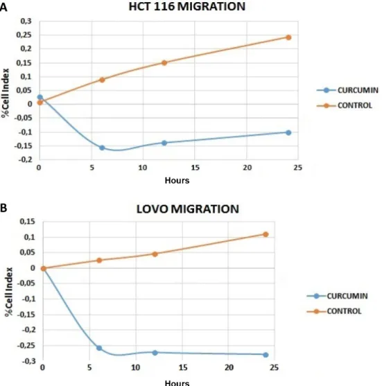
Figure 4. Cell migration results of curcumin on A: HCT-116 and B: LoVo cell lines at 24 h (Orange control, blue cur-cumin). The results were obtained from xCelligence DP (p<0.05).
of curcumin (10 µM). For the control group of LoVo cell line, IC10 dose of curcumin (2.5 µM) and IC50
dose of curcumin (20 µM) showed 10.1, 18.4 and 7.3% closure, respectively. The results are shown in Figure 2A and B.
Cell invasion and migration analysis
The invasion and migration activities of cur-cumin on HCT-116 and LoVo cell lines were inves-tigated at the 48th hour. According to these results, curcumin showed anti-migrative effect on HCT-116 and LoVo cell lines. (Figures 3 and 4).
Statistical analysis
P<0.05 was used to denote significant differ-ence between curcumin and control groups for each cell line. As shown in Figure 3, curcumin inhibited the invasion of HCT-116 (Figure 3A) and LoVo (Fig-ure 3B) cell lines at 24th h. For invasion, curcumin
had the best invasion inhibition activity on HCT-116 cell line (Figure 3A). In Figure 4, the migra-tion activity of curcumin on HCT-116 and LoVo cell lines is shown. Similarly, curcumin inhibited the migration of HCT-116 (Figure 4A) and LoVo (Figure 4B) cell lines at 24th h. For migration,
cur-cumin had a significant activity on each cell line.
Discussion
In this study, the antiproliferative, anti-meta-static and wound healing activity of curcumin was studied. Previous studies have revealed that NF-KB activation is critical for the proliferation, survival and metastasis of colon cancer cells; therefore, in-hibition of NF-KB activation is a potential antitu-mor strategy [11].
Curcumin has been suggested to inhibit the STAT-3 and NF-ΚB signal pathways (key compo-nents on cancer progression and development) [12]. Guo et. al. have shown that curcumin induces apoptosis by affecting the ubiquitin-proteasome pathway. The released LDH from LoVo cells was increased along with the increase of curcumin. Curcumin could activate caspase-3 and caspase-9 in LoVo cells. Therefore, curcumin-induced apop-tosis of LoVo cells was accompanied by increased activities of caspase-3 and caspase-9, which then stimulated the downstream apoptotic molecular cascade [13]. Also, this molecule plays an impor-tant role in colorectal cancer by reducing CD24 ex-pression [2]. Curcumin up-regulates the E-cadherin expression and so, it can be characterized as an inhibitor of epithelial-mesenchymal transition. In colorectal cancer, curcumin down-regulates Sp-1 and FAK. Not only curcumin but also curcumin-related compounds decrease phospho-extracellular
signal-regulated kinase (Erk)1/2 and phospho-Akt in different cell lines [14].
The main way of the cytotoxic activity of a compound is inhibiting or blocking mitosis or growth of cancer cells. In this study, the cytotoxic activity of curcumin in colorectal cancer cell lines was also studied. According to the results, IC50 of
curcumin in HCT-116 and LoVo cell lines were 10 µM and 20 µM, respectively. In the study of Ono et al., after 72-h incubation, IC50 of curcumin was
25 µM on HAG-1 gallbladder adenocarcinoma [15]. Khatwani et al. carried out a study on the antiprolif-erative activity of curcumin, tetrahydrocurcumin, bisdemethoxycurcumin and dimethoxycurcumin on DLD-1 colon cancer cell line [16]. Their results revealed that IC50 values of
bisdemethoxycurcum-in, tetrahydrocurcumin and dimethoxycurcumin were 10 µM, 50 µM and 2 µM, respectively [17]. Also, the results of the study of Kaverimanian et al. showed that IC50 of curcumin was 12.5 µM in
DLD-1 colon cell line [17]. Chen et al. carried out a study on seven different colorectal cancer cell lines. Their results revealed that the IC50 of curcumin on
HCT-116 cell line was almost 50 µM after 24-h in-cubation [18]. As it is shown, the results presented in this study are well in-line with the literature.
One of the most dangerous parts of cancer biol-ogy is metastasis. Anti-metastatic agents’ investi-gation from natural sources is fundamental. In this study, the activity of curcumin on invasion and mi-gration was also studied. According to the results, curcumin had the highest invasion and anti-migration activity in HCT-116 cell line. Chen et al. studied the anti-migration activity of curcumin in colorectal cancer cell lines and observed that the highest anti-migration levels were shown in HCT-116 cell line. [18]. Their results are quite similar to ours.
In this study, wound healing assay was per-formed to observe the effect of curcumin on wound healing and migration. Curcumin exerted its func-tion against wound healing (a kind of migrafunc-tion assay) on two different colorectal cancer cell lines. After 24-h incubation, the results showed that IC50
of curcumin closes the wounds. A similar study by Chen et al. showed that curcumin inhibits the migration on HCT-116 cell line [18].
Conclusion
A synergistic combination of curcumin and resveratrol has been also described by Majumdar et al. Both agents, acting together, inhibited the con-stitutive activation of EGFRs and IGF-1R in HCT-116 colorectal cancer cells. A test with a xenograft mouse model of colorectal cancer showed that the
combination of resveratrol and curcumin (at doses of 50 and 500 mg/kg, respectively, administered by gavage for 3 weeks) was highly effective in inhib-iting tumor growth and stimulating apoptosis of colorectal cancer cells in vivo, through attenuation of NF-κB activity [19].
Curcumin seems to be an ideal agent because significant evidence has indicated its potential against several chronic diseases. Also, curcumin targets several of molecular pathways without any associated toxicity or resistance. Despite its effica-cy and safety, curcumin has not yet been approved as a therapeutic agent, in part perhaps because of lack of intellectual rights to it.
In this study, the anticancer activity of cur-cumin on colorectal cancer cell lines was studied. According to the results, curcumin showed
antipro-liferation activity in HCT-116 and metastatic colo-rectal cancer cell lines. Also, curcumin inhibited migration and invasion on HCT-116 and LoVo cell lines. The wound healing assay revealed that cur-cumin plays an important role in wound healing on HCT-116 and LoVo cell lines. In conclusion, curcum-in is a promiscurcum-ing agent curcum-in metastasis and cancer.
Acknowledgement
This work was supported by Dokuz Eylul Uni-versity Scientific Research Coordination Unit (Pro-ject Number: 2018.KB.SAG.029).
Conflict of interests
The authors declare no conflict of interests.
References
1. Kolev TM, Velcheva EA, Stamboliyska BA, Spiteller M. DFT and experimental studies of the structure and vibrational spectra of curcumin. Inter J Quant Chem V 2005;102:1069.
2. Nong H, Hung LX, Thang PN et al. Fabrication and vibration characterization of curcumin extracted from turmeric (Curcuma longa) rhizomes of the northern Vietnam. SpringerPlus 2016;5:1147.
3. Vallianou NG, Evangelopoulos A, Schizas N, Kazazis C . Potential anticancer properties and mechanisms of action of curcumin. Anticancer Res 2015;35:645-51. 4. Lao CD, Ruffin MT 4th, Normolle D et al. Dose
escala-tion of a curcuminoid formulaescala-tion. BMC Complement Altern Med 2006;6:10.
5. World Health Organization, Cancer [homepage on the Internet]. 2018, Geneva, [updated 2018 Sep 12; cited 2002 Aug 12]. Available from: http://www.who.int/ news-room/fact-sheets/detail/cancer
6. Haggar F, Boushey RP. Colorectal Cancer Epidemiology: Incidence, Mortality, Survival, and Risk Factors. Clin Colon Rectal Surg 2009;22:191-7.
7. Thélin C, Sikka S. Epidemiology of Colorectal Cancer - Incidence, Lifetime Risk Factors Statistics and Tem-poral Trends. In: Ettarh R (Ed): Screening for Colorectal Cancer with Colonoscopy. IntechOpen, 2015, pp 61-77. 8. Haraldsdottir S, Einarsdottir HM, Smaradottir A,
Gunn-laugsson A, Halfdanarson TR. Colorectal Cancer (Re-view). Laeknabladid 2014;100:75-82.
9. Lee JG, McKinney KQ, Pavlopoulos AJ, Park JH, Hwang S. Identification of anti-metastatic drug and natural compound targets in isogenic colorectal cancer cells. J Proteomics 2015; 113:326-36.
10. Miura K, Satoh M, Kinouchi M et al. The use of natural products in colorectal cancer drug discovery. Expert Opin Drug Discov 2015;10:411-26.
11. Weihua Tong, Quan Wang, Donghui Sun, Jian Suo. Curcumin suppresses colon cancer cell invasion via AMPK-induced inhibition of NF-κB, uPA activator and MMP9. Oncol Lett 2016;12:4139-46.
12. Kasdagly M, Radhakrishnan S, Reddivari L, Veera-macheni DN, Vanamala J. Colon carcinogenesis: Influ-ence of Western diet-induced obesity and targeting stem cells using dietary bioactive compounds. Nutri-tion 2014;30:1242-56.
13. Guo LD, Chen XJ, Hu YH, Yu ZJ, Wang D, Liu JZ. Cur-cumin Inhibits Proliferation and Induces Apoptosis of Human Colorectal Cancer Cells by Activating the Mitochondrial Apoptotic Pathway. Phytother Res 2013;27:422-30.
14. Zhou DY, Zhang K, Conney AH. Synthesis and Evalua-tion of Curcumin-Related Compounds Containing Ben-zyl Piperidone for Their Effects on Human Cancer Cells. Chem Pharm Bull 2013;61:1149-55.
15. Ono M, Higuchi T, Takeshima M, Chen C, Nakano S. Antiproliferative and apoptosis-inducing activity of curcumin against human gallbladder adenocarcinoma cells. Anticancer Res 2013;33:1861-6.
16. Khatwani N, Adeyeni T, Ezekiel U. The anti-prolifera-tive effects of curcumin derivaanti-prolifera-tives, dimethoxycurcum-in, bisdemethoxycurcumin and tetrahydrocurcumdimethoxycurcum-in, on DLD-1 colon cancer cells. FASEB J 2016;30:1 (Suppl). 17. Kaverimanian A, Khatwani N, Ezekiel U.
Antiprolifera-tive effect of curcumin and kaempferol on colon cancer cells. FASEB J 2016;30:1 (Suppl).
18. Chen CC, Sureshbabul M, Chen HW et al. Curcumin suppresses metastasis via Sp-1, FAK inhibition, and E-cadherin upregulation in colorectal cancer. Evid Based Complement Alternat Med 2013;2013:541695. 19. Majumdar AP, Banerjee S, Nautiyal J et al. Curcumin
synergizes with resveratrol to inhibit colon cancer. Nutr Cancer 2009;61:544-53.