ARAŞTIRMA
NEUROPATHIC PAIN AND DISABILITY IN
PATIENTS WITH LUMBAR SPINAL STENOSIS
LOMBER SPİNAL STENOZLU HASTALARDA
NÖROPATİK AĞRI VE YETİ KAYBI
Introduction: In this study, we aimed to determine the characteristics of neuropathic pain
and clarify the relationships between the clinical signs, symptom characteristics and disability in patients with lumbar spinal stenosis.
Materials and Method: Seventy-nine patients diagnosed with clinical and radiological
lumbar spinal stenosis were included in the study. Pain severity was assessed using a self-administered visual analog scale (0–10 cm). Subjective disability was measured using the Oswestry Disability Index and Istanbul Low Back Pain Disability Index. To identify neuropathic pain, the physician-administered Douleur Neuropathique 4 questionnaire was utilized.
Results: A total of 79 patients (35 males, 44 females) with a mean age of 56.88±12.13 (range,
35–89) years were included in the study. The average symptom duration was 24.5 months. The Douleur Neuropathique 4 score was ≥4 in 41 patients (51.8%), indicating that they had neuropathic pain. A significant correlation was observed between the Douleur Neuropathique 4 score and age (R=0.350, p=0.002), body mass index (R=0.239, p=0.034), and visual analog scale (R=0.453, p< 0.001).
Conclusion: Approximately half of all patients with lumbar spinal stenosis in our cohort
had neuropathic pain. Higher age, body mass index and increased pain scores demonstrate important relationships with neuropathic pain.
Keywords: Neuralgia; Spinal Stenosis; Pain; Aged
Giriş: Bu çalışmada, lomber spinal stenoz hastalarında nöropatik ağrı karakterini belirlemeyi
ve klinik semptom, bulgu ve yeti kaybı ile olan ilişkisini araştırmayı amaçladık.
Gereç ve Yöntem: Çalışmaya klinik ve radyolojik olarak lomber spinal stenoz tanısı alan
yetmiş dokuz hasta alındı. Ağrı düzeyi için visüel analog skala (0-10 cm), fonksiyonel durum için Oswestry Dizabilite İndeksi ve İstanbul Bel Ağrısı Dizabilite İndeksi ölçüldü. Nöropatik ağrıyı belirlemek için Douleur Neuropathique 4 anketi dolduruldu.
Bulgular: Ortalama yaşı 56.88±12.13 (35-89 yaş aralığı) olan 79 hasta (35 erkek, 44 kadın)
çalışmaya dahil edildi. Ortalama semptom süresi 24,5 ay idi. Hastaların 41’inde (%51.8) Douleur Neuropathique 4 skoru ≥4 (nöropatik ağrı) idi. Douleur Neuropathique 4 sonucu ile yaş (R=0.350, p=0.002), vücut kitle indeksi (R=0.239, p=0.034) ve visüel analog skala (R=0.453, p< 0.001) arasında anlamlı ilişki görüldü.
Sonuç: Çalışmamızda, hastaların yaklaşık yarısında nöropatik ağrı gözlendi. İleri yaş, yüksek
vücut kitle indeksi ve artmış ağrı skorları ile nöropatik ağrı arttığı sonucuna ulaşıldı.
Anahtar sözcükler: Nöralji; Spinal stenoz; Ağrı; Yaşlı
A
BSTRACTÖ
ZTurkish Journal of Geriatrics DOI: 10.31086/tjgeri.2018137967 2018;21 (1):56-61
Mehmet AĞIRMAN1 Ayşe Serap AKGÜN2
CORRESPONDANCE Mehmet AĞIRMAN İstanbul Medipol University,
Faculty of Medicine, Department of Physical Medicine and Rehabilitation
İstanbul, Turkey Phone: 4447070
e-mail: [email protected] Received: 14/02/2018
Accepted: 05/03/2018 1 İstanbul Medipol University,
Faculty of Medicine, Department of Physical Medicine and Rehabilitation
İstanbul, Turkey
2 İstanbul Medipol University Department of Radiology İstanbul, Turkey
INTRODUCTION
Lumbar spinal stenosis (LSS) is one of the most common conditions, which causes pain and disabilities in older people (1). This condition is commonly associated with lumbar degenerative disc disease and spondylosis so-called acquired lumbar stenosis (2). It results in the narrowing of the spinal canal, lateral nerve roots, and intervertebral neural foramina at one or more levels. Clinical signs occur because of compression of the spinal cord, nerve roots, and vascular elements of the lumbar spine (3). These symptoms include pain in the back, buttocks, and lower leg; numbness in the leg or foot, weakness in the lower extremities, and neurogenic claudication (4).
There are several classifications and types of pain, including nociceptive, chronic, radicular, and neuropathic. Understanding these can aid in diagnosis and planning appropriate treatment (5). Neuropathic pain (NeP) has been defined as pain caused by a lesion or disease of the somatosensory system. Patients usually complain of burning and electrical sensations, with pain often resulting from typically non-painful stimulation (e.g., light touch) (6).
LSS can be diagnosed based on clinical, neurological, and radiological findings. However, radiological LSS can be detected using magnetic resonance imaging (MRI) or computed tomography in asymptomatic patients. A clinical diagnosis of LSS requires both the characteristic clinical presentation and radiographic evidence of LSS (7). Neurogenic claudication and radicular pain are best described in the literature for patients with LSS (7,8). However, there are few studies on NeP in patients with spinal stenosis (9,10).
In this study, we aimed to determine the characteristics of NeP and clarify the relationships between the clinical signs and symptom characteristics in patients with LSS.
MATERIALS AND METHOD Participants
One-hundred and sixteen patients with LSS symptoms were included in the study between
February 2016 and November 2017. All patients signed written informed consent forms to participate in the study, which was approved by the local ethics committee. This study was conducted in accordance with the principles of the Declaration of Helsinki.
All patients were recruited from the outpatient clinic of the Department of Physical Medicine and Rehabilitation. Clinical LSS was diagnosed based on symptoms of pain in the buttocks or thighs, pain in the lower leg induced by lumbar spine extension, numbness or tingling in the leg or foot,
and neurogenic claudication. Patients diagnosed
with clinical LSS were included if lumbar spine MRI showed signs of LSS. Radiological LSS was accepted as absolute if the sagittal spinal canal diameters
measured ≤10 cm in at least one intervertebral disc
(11). Initially, seventy nine patients with both the
characteristic clinical presentation and radiographic
evidence of LSS were collected. Patients were
excluded if they had previously undergone spine
(n=3), hip, and/or lower limb-related surgery (n=3);
diabetes (n=7); peripheral vascular disease (n=1);
and/or inflammatory disorders (n=2). Furthermore,
we also excluded those with a symptom duration of<1
month (n=7) and those taking drugs for NeP, such as
pregabalin, gabapentin, tricyclic antidepressants, or selective serotonin–norepinephrine reuptake inhibitors (n=14).
Assessment of symptoms and functional disability
All questionnaires and clinical evaluations were recorded by a blinded physician to the clinical and radiological findings. A detailed medical history was taken and physical examination was performed, and age, gender, height, weight, body mass index (BMI), and symptom duration were recorded for each patient.
Low back pain severity was assessed using a self-administered visual analog scale (VAS; range, 0–10 cm). Subjective disability was measured using the Oswestry Disability Index (ODI; range 0-100) and Istanbul Low Back Pain Disability Index (ILBPDI; range 0-90). ODI is a 10-item, self-administered questionnaire that is commonly used to assess
limitations in activities of daily living because of low back pain. Disability increases as the total ODI score increases. ILBPDI focuses on activities required to function in daily life, such as transport, sitting, standing, dressing, and hygiene, and where they are done (e.g., room, kitchen, car, or street).
To identify NeP, the physician-administered Douleur Neuropathique 4 (DN4) questionnaire was utilized. This questionnaire contains seven items related to symptoms and three related to clinical examination. Each item is scored as 1 (positive) or 0 (negative), and a total score of ≥4 out of 10 suggests NeP. The questionnaires have Turkish validity and reliability (12,13).
Statistical analysis
All qualitative variables are reported as frequencies and percentages. Continuous variables are expressed as means±standard deviations. The correlations between DN4, ODI, ILBPDI, VAS, and other clinical signs were calculated using the Spearman’s correla tion coefficient. IBM SPSS for Windows, Version 21 (IBM Corp., Armonk, NY, USA) was used for all statistical analyses. P-values<0.05 were considered to be statistically significant.
RESULTS
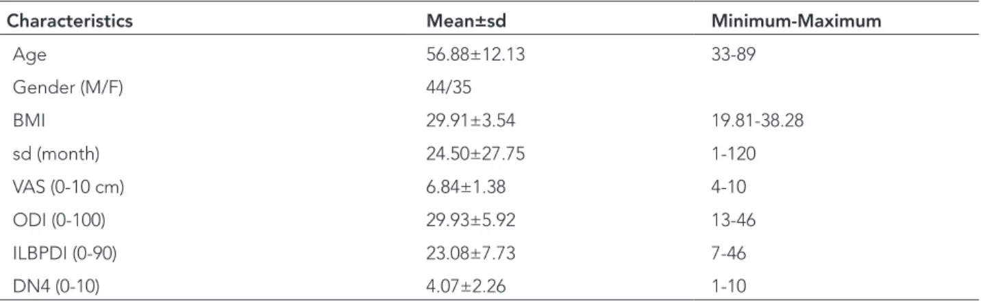
The clinical and demographic characteristics of the participants are summarized in Table 1. A
total of 79 patients (35 males, 44 females) with a mean age of 56.88±12.13 (range, 35–89) years were included in the study. All patients were clinically and radiologically diagnosed with LSS. The average symptom duration was 24.5 months (Table 1).
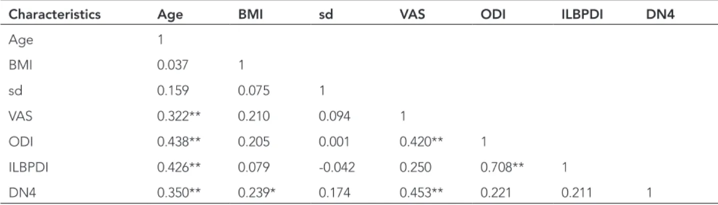
In terms of the questionnaires, the average VAS, ODI, ILBPDI, and DN4 scores were 6.84±1.38, 29.93±5.92, 23.08±7.73, and 4.07±2.26, respectively. The DN4 score was ≥4 in 41 patients (51.8%), indicating that they had NeP. A significant correlation was observed between the DN4 score and age (R=0.350, p=0.002), BMI (R=0.239,
p=0.034), and VAS (R=0.453, p< 0.001). There was
also a significant correlation between the ODI score and age (R=0.438, p< 0.001), VAS score (R=0.420,
p< 0.001), and ILBPDI score (R=0.708, p<0.001).
Finally, there was a significant correlation between the ILBPDI score and both age (R=0.426, p<0.001) and VAS score (R=0.250, p=0.026). However, the symptom duration was not significantly correlated with any of the questionnaires (Table 2).
Claudication was present in 41.8% of patients. Using DN4, most patients complained of tingling (67.1%) and numbness (51.9%), with the least number of patients complaining of itching (13.9%; Table 3). In the physical examination, hypoesthesia to touch, hypoesthesia to pinprick, and increased pain by brushing were present in 61.9%, 65.8%, and 21.5% of the patients, respectively.
Table 1. The clinical and demographic characteristics of patients.
Characteristics Mean±sd Minimum-Maximum
Age 56.88±12.13 33-89 Gender (M/F) 44/35 BMI 29.91±3.54 19.81-38.28 sd (month) 24.50±27.75 1-120 VAS (0-10 cm) 6.84±1.38 4-10 ODI (0-100) 29.93±5.92 13-46 ILBPDI (0-90) 23.08±7.73 7-46 DN4 (0-10) 4.07±2.26 1-10
BMI: Body mass index; SD: Symptom duration; VAS: Visual analog scale; ODI: Oswestry Disability Index; ILBPDI: Istanbul Low Back Pain Disability Index; DN4: Douleur Neuropathique 4
Table 2. Correlations of demographical findings, pain, subjective disability and neuropathic pain (r-values).
Characteristics Age BMI sd VAS ODI ILBPDI DN4
Age 1 BMI 0.037 1 sd 0.159 0.075 1 VAS 0.322** 0.210 0.094 1 ODI 0.438** 0.205 0.001 0.420** 1 ILBPDI 0.426** 0.079 -0.042 0.250 0.708** 1 DN4 0.350** 0.239* 0.174 0.453** 0.221 0.211 1
BMI: Body mass index; SD: Symptom duration; VAS: Visual analog scale; ODI: Oswestry Disability Index; ILBPDI: Istanbul Low Back Pain Disability Index; DN4: Douleur Neuropathique 4
*. Correlation is significant at the 0.05 level **. Correlation is significant at the 0.01 level
Table 3. The percentage of neurogenic claudication and positive signs and symptoms assessed by DN4 questionnaire in LSS patients.
Characteristics Yes (%) No (%) Claudication 41.8 58.2 DN4 questions Burning 43.0 57.0 Painful cold 25.3 74.7 Electric shocks 39.1 60.9 Tingling 67.1 32.9
Pins and needles 45.6 54.4
Numbness 51.9 48.1 Itching 13.9 86.1 Hypoesthesia to touch 61.9 38.1 Hypoesthesia to prick 65.8 34.2 Brushing 21.5 78.5 DISCUSSION
In this study, we showed that most patients with LSS had NeP associated with age; BMI; and ODI, VAS, and ILBPDI scores. The prevalence of degenerative disc disease and neuropathic pain syndromes
are known to increase with both aging and spinal stenosis (14,15). In our study, a correlation was observed between increased age and DN4, ODI, and ILBPDI scores. These results might have resulted from increased degeneration and stenosis typically associated with increased aged.
Lumbar spinal stenosis has heterogeneous and nonspecific signs and symptoms and may even be asymptomatic, with symptoms assumed to result from nerve compression, local vascular insufficiency, or both (1). Indeed, radiological findings do not prove that symptoms arise from nerve root compression (16). Neurogenic claudication, back and leg pain, comorbidities, and other nociceptive pain indexes are well studied in patients with LSS (16-19). It has also been reported that patients with LSS have less intense leg pain than those with lumbar disc herniation and that their symptoms are more often increased by lumbar extension and relieved by bending forward or sitting (2,17,20).
The pain in LSS can be divided into two main categories: nociceptive and neuropathic. NeP is defined as the pain caused by a lesion or disease of the somatosensory system, and it is estimated to affect 15%–25% of patients with chronic pain (8,21). Although NeP is a clinical diagnosis, there are several diagnostic criteria. In Turkey, DN4 has been the best performing screening tool with good validity and reliability (13). This tool assesses symptoms of burning, tingling, sensitivity to touch, pain caused by mild pressure, electric shock-like pain, pain to cold or heat, and numbness, which are characteristic NeP symptoms.
In our study, 41 patients (51.8%) had NeP according to the DN4 questionnaire, and this was shown to be correlated with increased BMI. In another study of 102 patients, the painDETECT questionnaire was used to show that nociceptive, neuropathic, and unclear pain were present in 57.9%, 17.6%, and 24.5% of patients, respectively (9). Even if the unclear pain is combined with the NeP group, the NeP rate still only reached 42.1%. Our high values of NeP, may have resulted from using the DN4 questionnaire. In the above study, they showed that the group with NeP had a significantly poorer
quality of life than the other groups. However, ODI, ILBPDI and symptom duration did not significantly correlate with DN4 questionnaire (Table 2).
In patients with LSS, pain mostly spreads to the buttock or thighs and to both lower legs, with patients also complaining of numbness and tingling (19). For patients with chronic low back pain and LSS, other researchers have also reported that buttock and leg pain were significantly associated with NeP (10,22). Our results are consistent with these results, showing that most subjects complained of leg symptoms (e.g., tingling in 67.1% and numbness in 51.9%), including claudication (41.8%), and that very few complained of itching (Table 3).
There are conflicting results about the association between radiological and clinical findings. In a multicenter study, no relationship was shown between the radiological severity and the presence of back or leg pain in patients with LSS (23). In another research, the opposite was shown to some extent (24). Although we believe that the radiological distinction could be ignored because of the selection of only symptomatic patients in our study, further research is needed in relation to this topic.
Except the radiological distinction there are other limitations in the present study. Although the frequency of neurogenic claudication determined in the study, the walking distance/duration was not measured. The other limitation is that the quality of life was not evaluated.
In conclusion, approximately half of all patients with LSS in our cohort had NeP. Higher age, BMIs and increased VAS scores demonstrate important relationships with NeP.
Conflict of interest
The authors have no conflict of interest to declare.
REFERENCES
1. Backstrom KM, Whitman JM, Flynn TW. Lumbar spinal stenosis-diagnosis and management of the aging spine. Man Ther 2011;16(4):308-17. (PMID:21367646).
2. Gilbert JW, Martin JC, Wheeler GR, et al. Lumbar stenosis rates in symptomatic patients using weight-bearing and recumbent magnetic resonance imaging. J Manipulative Physiol Ther 2011;34(8):557-61. (PMID:21907413).
3. Ishimoto Y, Yoshimura N, Muraki S, et al. Associations between radiographic lumbar spinal stenosis and clinical symptoms in the general population: the Wakayama Spine Study. Osteoarthritis Cartilage 2013;21(6):783-8. (PMID:23473979).
4. Battié MC, Jones CA, Schopflocher DP, Hu RW. Health-related quality of life and comorbidities associated with lumbar spinal stenosis. Spine J 2012;12(3):189-95. (PMID:22193054).
5. Nalamachu S. An overview of pain management: the clinical efficacy and value of treatment. Am J Manag Care 2013;19(14):261-6. (PMID:24494608).
6. Cohen SP, Mao J. Neuropathic pain: mechanisms and their clinical implications. BMJ 2014;348:f7656. (PMID:24500412).
7. Suri P, Rainville J, Kalichman L, Katz JN. Does this older adult with lower extremity pain have the clinical syndrome of lumbar spinal stenosis? JAMA 2010;304(23):2628-36. (PMID:21156951).
8. Djurasovic M, Glassman SD, Carreon LY, Dimar JR. Contemporary management of symptomatic lumbar spinal stenosis. Orthop Clin North Am 2010;41(2):183-91. (PMID:20399357).
9. Takahashi N, Shirado O, Kobayashi K, Mashiko R, Konno S. Classifying patients with lumbar spinal stenosis using pain DETECT: a cross-sectional study. BMC Fam Pract 2016;17:90. (PMID:27443164). 10. Orita S, Yamashita T, Ohtori S, et al. Prevalence
and Location of Neuropathic Pain in Lumbar Spinal Disorders: Analysis of 1804 Consecutive Patients With Primary Lower Back Pain. Spine (Phila Pa 1976) 2016;41(15):1224-31. (PMID:26967122).
11. Kalichman L, Cole R, Kim DH, et al. Spinal stenosis prevalence and association with symptoms: the Framingham Study. Spine J 2009;9(7):545-50. (PMID:19398386).
12. Duruöz MT, Özcan E, Ketenci A, Karan A. Development and validation of a functional disability index for chronic low back pain. J Back Musculoskelet Rehabil 2013;26(1):45-54. (PMID:23411648).
13. Unal-Cevik I, Sarioglu-Ay S, Evcik D. A comparison of the DN4 and LANSS questionnaires in the assessment
of neuropathic pain: validity and reliability of the Turkish version of DN4. J Pain 2010;11(11):1129-35. (PMID:20418179).
14. Zeifang F, Schiltenwolf M, Abel R, Moradi B. Gait analysis does not correlate with clinical and MR imaging parameters in patients with symptomatic lumbar spinal stenosis. BMC Musculoskelet Disord 2008;9:89. (PMID:18570636).
15. Kutsal YG, Eyigör S, Dogan A, et al. Neuropathic pain in elderly: a multicenter study. Turkish Journal of Geriatrics 2016;19(1):9-18.
16. Konno S, Kikuchi S, Tanaka Y, et al. A diagnostic support tool for lumbar spinal stenosis: a self-administered, self-reported history questionnaire. BMC Musculoskelet Disord 2007;8:102. (PMID:17967201).
17. Rainville J, Lopez E. Comparison of radicular symptoms caused by lumbar disc herniation and lumbar spinal stenosis in the elderly. Spine (Phila Pa 1976) 2013;38(15):1282-7. (PMID:23462576).
18. Kim YU, Kong YG, Lee J, et al. Clinical symptoms of lumbar spinal stenosis associated with morphological parameters on magnetic resonance images. Eur Spine J 2015;24(10):2236-43. (PMID:26292958). 19. Yamashita K, Aono H, Yamasaki R. Clinical
classification of patients with lumbar spinal stenosis based on their leg pain syndrome: its correlation with 2-year surgical outcome. Spine (Phila Pa 1976) 2007;32(9):980-5. (PMID:17450073).
20. Ozcan-Eksi EE, Yagci I, Erkal H, Demir-Deviren S. Paraspinal muscle denervation and balance impairment in lumbar spinal stenosis. Muscle Nerve 2016;53(3):422-30. (PMID:26138076).
21. Bouhassira D, Lantéri-Minet M, Attal N, Laurent B, Touboul C. Prevalence of chronic pain with neuropathic characteristics in the general population. Pain 2008;136:380-7. (PMID:17888574). 22. Park SY, An HS, Moon SH, et al. Neuropathic Pain
Components in Patients with Lumbar Spinal Stenosis. Yonsei Med J 2015;56(4):1044-50. (PMID:26069129). 23. Weber C, Giannadakis C, Rao V, et al. Is There
an Association Between Radiological Severity of Lumbar Spinal Stenosis and Disability, Pain, or Surgical Outcome?: A Multicenter Observational Study. Spine (Phila Pa 1976) 2016;41(2):E78-83. (PMID:26352747).
24. Sigmundsson FG, Kang XP, Jönsson B, Strömqvist B. Correlation between disability and MRI findings in lumbar spinal stenosis: a prospective study of 109 patients operated on by decompression. Acta Orthop 2011;82(2):204-10. (PMID:21434811).