Rezumat
Impactul disulfurii de alil asupra distrugerii oxidative æi regenerãrii hepatice pe un model experimental de hepatectomie Date generale: Am investigat efectele disulfurii de alil (un
extract de usturoi) asupra distrucåiei tisulare, regenerãrii, proliferãrii æi distrugerii oxidative pe un model experimental de rezecåie hepaticã.
Materiale æi metode: În acest studiu 24 de femele de æobolani
albi Wistar cu greutãåi de aproximativ 200-250 g au fost utilizaåi. Grupul 1: toåi æobolanii din acest grup experimental au fost supuæi unei hepatectomii de 70% din dimensiunea ficatului æi au fost hrãniåi cu disulfurã de alil (30 μg/kg/zi, disulfurã de alil, Sigma-Aldrich, formula: C6H10S2, Numãrul-CAS: 2179-57-9, greutate molecularã: 146.27 g/mol) suplimentar faåã de dieta normalã timp de câte o sãptãmânã pre- æi postoperator. Grupul 2: Æobolanii din grupul control au fost de asemenea supuæi unei hepatectomii în proporåie de 70% æi au primit un regim nutriåional æi de hidratare normale atât pre-, cât æi postoperator. Grupul 3: În grupul de control non-placebo toåi æobolanii au fost sacrificaåi la 7 zile dupã intervenåia chirurgicala. În vederea evaluãrii biochimice, AST, ALT, bilirubina, CRP æi malondialdehida sericã au fost studiate. În cadrul analizei histopatologice au fost examinate acumularea de åesut grãsos la nivel hepatic, existenåa fibrozei (macro-micro veziculare), pleomorfismul nucleilor hepa-tocitelor, inflamaåia portalã, existenåa de celule inflamatorii
intralobulare, dilatarea la nivelul sinusoidelor, congestia, con-gestia venei centrale, regenerarea, existenåa de celule Kupffer în lumenul sinusoidal æi indicele de proliferare ki-67 la nivelul hepatocitelor.
Rezultate: S-a observat o diferenåã semnificativã între grupurile
1 æi 2 în ceea ce priveæte prezenåa regenerãrii, (p:0.06), pleomorfismelor nucleare (p:0,001) æi statusului de activitate fibroblasticã (p:0.001). Diferenåe semnificative au fost gãsite între grupurile experimentale vizând creæterea numericã æi dilatarea celulelor Kupffer æi statusul hiperemic la nivelul lumenelor sinusoidelor (p:0.013 æi p:0.001 respectiv). În grupul cu administrare de disulfurã de alil indicele de proliferare a fost semnificativ mai ridicat decât în celelalte grupuri (p:0,001), în timp ce valoarea medie a malondialdehidei serice a fost mai scãzutã comparativ cu celelalte 2 grupuri (p: 0,042). Nu s-au înregistrat diferenåe semnificative între grupuri referitor la valorile de malondialdehidã tisularã (p:0,720). Nu s-au gãsit diferenåe importante între nivelurile de ALT æi AST din grupul 1 în comparaåie cu celelalte grupuri (p:0.247 æi p:0.539 respectiv). Valorile medii de bilirubinã totalã au fost de 0,12, 0,08 æi 0,04 în grupurile 1, 2 æi 3. Aceastã diferenåã dintre grupuri prezintã semnificaåie statisticã (p:0.001). Valorile medii ale bilirubinei directe au fost 0,06, 0,02 æi 0,02 în grupul experimental, grupul control æi grupul control non-placebo. Æi aceastã variaåie între grupuri este semnificativã din punct de vedere statistic (0.001).
Concluzii: Am observat cã suplimentarea de disulfurã de alil
dupã o hepatectomie majorã are un impact pozitiv asupra regenerãrii, proliferãrii æi distrugerii oxidative la nivel hepatic. Cuvinte cheie: Allium sativum, hepatectomie majorã, distrugere oxidativã, disulfurã de alil
Copyright© Celsius
Corresponding author: Oguzhan Karatepe, MD
Medipol University Kosuyolu Hospital, Department of Surgery, Turkey E-mail: drkaratepe@yahoo.com
Impact of Allyl Disulfide on Oxidative Damage and Liver Regeneration
in an Experimental Hepatectomy Model
M. Battal1
, A. Kartal1
, B. Çitgez1
, B. Yılmaz2
, A. Akcakaya, O. Karatepe3 1General Surgery Department, Sisli Etfal Training Hospital, Turkey
2
Pathology Department, Sisli Etfal Etfal Training Hospital, Turkey
Abstract
Backgraund: We investigated the effects of allyl disulfide (a
garlic extract) on tissue damage, regeneration, proliferation and oxidative damage in an experimental liver resection model.
Materials and Methods: In the study, 24 female Wistar albino
rats weighing approximately 200-250 g were used. Group 1: The rats in the experimental group all received a 70% hepatectomy and were fed an Allyl disulfide (30 μg/kg/day, Allyl disulfide, Sigma-Aldrich, formula: C6H10S2, CAS Number: 2179-57-9, formula weight: 146.27 g/mol) in supplement to a regular diet for 1 week both preoperatively and postoperatively. Group 2: The rats in the control group also underwent a 70% hepatectomy and were given regular food and water for 1 week both preop and postop. Group 3: In the sham group, all rats were sacrificed 7 days after surgery. For biochemical evaluation, SGOT, SGPT, bilirubin, CRP and MDA were studied. In a histopathological examination, the fattening of the liver tissue, existence of (macro-micro vesicu-lar), fibrosis, pleomorphism at hepatocyte nuclei, portal inflammation, existence of intralobular inflammatory cells, dilation at sinusoids, congestion, congestion at the central vein, regeneration, existence of Kupffer cells in the sinusoidal lumen and ki-67 proliferation index at hepatocytes were examined.
Results: A significant difference between group 1 and group
2 was observed regarding the existence of regeneration, (p:0.06), the occurrence of nuclear pleomorphisms (p:0,001) and the fibroblast activity status (p:0.001). Significant differences were found between the experimental groups in regard to Kupffer cell increase and dilation and the hyper-emia status in the sinusoid lumens (p:0.013 and p:0.001, respectively). In the Allyl disulfide group, the proliferation index was significantly higher than that of the other groups (p:0,001), while the average plasma MDA value was lower than that of the other groups (p: 0,042). No significant differences were found among the groups with respect to tissue MDA values (p:0,720). No significant difference was found for SGPT (ALT) and SGOT (AST) levels between Group 1 and the other groups (p:0.247 and p:0.539, respec-tively). The average total bilirubin (T. Bili) values were 0,12, 0,08 and 0,04 in the allyl disulfide group, control group and Sham group, respectively. This difference among the groups is statistically significant (p:0.001). The average direct bilirubin (D. Bili) values were 0,06, 0,02 and 0,02 in the allyl disulfide group, control group and Sham group, respec-tively. This variation among the groups is also statistically significant (0.001).
Conclusion: We observed that the use of Allyl disulfide
supplementation after major hepatectomy has a positive impact on liver regeneration, proliferation and oxidative damage.
Abbreviations: Postop: post-operative, Preop: pre-operative,
SGOT(AST): serum glutamic oxaloacetic transaminase, SGPT(ALT): serum glutamate-pyruvate transaminase, CRP: C- Reactive protein, MDA: Malondialdehyde, DAS: Garlic extract diallyl sulfide, AGE: aged garlic extract
Key words: Allium sativum, major hepatectomy, oxidative damage, Allyl disulfide
Introduction Introduction
Today, chronic liver disease, hepatosteatosis, and toxic effects of chemotherapy on the liver in oncological patients are com-mon occurrences. In concordance with a better understanding of liver anatomy, technical developments and improvements in the treatment of intensive care conditions, liver surgeries have become increasingly frequent procedures. There have been many studies that investigated potential methods to increase the capacity of liver regeneration or the impact of chemicals on the liver. The purpose of such studies is to reduce morbidity and mortality, which is a probable outcome during the treatment of such patients (1-6).
Garlic and garlic extract has been consumed by humans for thousands of years for medicinal purposes. The use of garlic for medical purposes extends through history to before the birth of Christ. It is known that during the 1500’s B.C., it was used in Egypt as a remedy for headaches, cardiac diseases and cancer (7). Many recent studies have experimentally demonstrated the disease preventing effects of garlic and garlic extracts. (8).
However, the effects of garlic and garlic extract Allyl disulfide consumption on liver regeneration remain unknown. The present study investigated the role of garlic extract in the diet after major liver resection on oxidative damage, prolifera-tion and regeneraprolifera-tion as well as on other impacts on treat-ment. From the findings obtained from this study, we acquired information about the practical use of Allyl disulfide after liver disease, liver transplantation and liver resection.
Materials and Methods Materials and Methods
The study was conducted with the approval of Istanbul University Animal Experiments Local Ethics Review Board no. 2012/155 dated 01.11.2012.
The study was conducted at Istanbul University Experimental Medicine Research Institute. Twenty-four albino, female Wistar rats weighing approximately 200-250 g and fed with standard laboratory feed were used. The rats were 8-10 weeks of age and were housed four per cage at room tempera-ture. The rats were fed a proper diet of regular pellet feed and water, were provided with 12 hour light-dark cycles and appropriate ambient temperature (22±1°C) and humidity (65-70%). Three groups of 8 rats each were created from the total number of rats included in the study.
Group 1: This group is the experimental group. The rats in this group each received a 70% hepatectomy and were fed Allyl disulfide (30 μg/kg/day, Allyl disulfide, Sigma-Aldrich Munich, Germany, formula: C6H10S2, CAS Number: 2179-57-9, formula weight: 146.27 g/mol) as a supplement to their regular diet for one week both preoperatively and post-operatively.
Group 2: A 70% hepatectomy was performed on each rat in this group, which was the control group. The rats were given regular food and water for one week both preop and postop.
Group 3: This group underwent surgery, which was terminated without resection after the abdomen was opened and the liver anatomy was revealed (sham group).
Rats were administered 40 mg/kg Ketamine HCl (Ketalar) and 10/mg/kg Ksilazin HCI anesthesia. After shaving the abdomens of the rats, a laparotomy was performed with a midline incision of approximately 3 cm in length. Sterile conditions were ensured throughout the procedure. After freeing the pedicles of the liver left lateral and median lobes, the coronary, left lateral and gastrohepatic ligaments were tied with 3/0 silk and a 70% hepatectomy was performed as described by Higgins and Anderson (9). Rats were sacrificed on day 7 after liver resection.
Assessment of biochemical results
Shortly before death, intra-cardiac blood samples were taken from subjects for biochemical analysis. The blood was kept in the collection tubes at room temperature for half an hour, and after coagulation, it was centrifuged and the serum was separated. Afterwards, serum was stored at -80 °C until analysis. On the day of biochemical analysis, the serums were brought to room temperature and AST, ALT, bilirubin and CRP levels were studied using a Roche Cobas c 701 device. AST and ALT were measured using a Tris buffer method, bilirubins were studied with a Dichlorophenyl diazonium method and CRP with an immunoturbidimetric method. The results were calculated in U/L for AST and ALT, mg/dL for bilirubin and mg/L for CRP.
Assessment of pathology results
Shortly before death, liver samples were taken from the rats for pathological analysis. After the liver tissues from rats in all three groups were fixed for 24 hours in a 10% formalin solution, the samples were passed through a series of 60%, 70%, 80%, 96% and 100% alcohol, followed by a dehydration process. Then, the tissues were passed through a xylol series to remove alcohol and make the samples pellucid. The tissues were kept in 3 separate molten paraffin containers for the removal of xylol from the tissue before the paraffin embedding process. Paraffin blocks were prepared, and 4 micron cross sections were obtained with a microtome. Cross sections were stained with hematoxylin and eosin and were then evaluated using an optical microscope (Olympus BX50) by an experi-enced pathologist specifically familiar with liver pathology. Additional immunohistochemical evaluation was performed on the same tissue sections using Masson's Trichrome and reticulin (Bio-Optica histochemical ready stain kit) stains.
The existence of fattening (macro-micro vesicular) in the liver parenchyma cell (hepatocyte) cytoplasms, fibrosis, pleo-morphism of hepatocyte nuclei, portal inflammation, intra-lobule inflammatory cells, dilation at sinusoids, general congestion, congestion at central veins, regeneration, Kupffer cells in sinusoid lumens and the ki-67 proliferation index at
hepatocytes were all evaluated.
A streptavidin-biotin staining technique was used for the immunostaining (IHC) method. For the purpose of determining Ki-67 expression, 4-micrometer thick cross sections cut from paraffin blocks were taken into ‘Poly-L-Lysine’ laminas. Immunohistochemical staining was performed with a Ki-67 (SP6) (Biocare U.S.) ready to use rabbit monoclonal anti-body.
From the laminas used in the assessment, 500 cells were counted at 400 large growth areas. All preparations were assessed by the same pathologist. The ratio of the number of cells with positive Ki-67 staining in the liver tissue to the total number of cells was calculated (10,11).
MDA measurement
A MDA analysis was conducted as an indicator of oxidative damage in liver tissue samples. For the analysis, liver tissue homogenates were prepared in cold 0,15 M KCl (10%, w/v) using glass homogenizers. The tissue MDA concentration as a lipid peroxidation agent was analyzed according to the method described by Beuge and Aust. The method is based on the principle of colorimetric measurement of an MDA and thio-barbituric acid complex. According to this method, mixtures prepared after the reaction of one volume of tissue homogenate sample with two volumes of stock reagent (containing 14% trichloroacetic acid and 0,375% thiobarbituric acid within 0,25 N HCI) were incubated in a boiling water bath for 15 minutes and were then cooled and centrifuged at 1000 g for 10 minutes. The absorbance of supernatants was spectrophoto-metrically measured at 535 nm against a blank. Calculated results were expressed in nmol MDA/gram tissue.
MDA measurement at plasma
Blood samples were taken for the MDA analysis as an indicator of oxidative damage. Reaction mixtures were prepared using one volume of sample with two volumes of stock reagent (containing 14% trichloroacetic acid and 0,375% thiobarbituric acid within 0,25 N HCI) and were incubated in a boiling water bath for 15 minutes, then cooled and centrifuged at 1000 g for 10 minutes. Absorbance of supernatants was spectrophotometrically measured at 535 nm against a blank. Calculated results were expressed in μmol MDA/L (12).
Statistical analysis
SPSS 17.0 software was used for the statistical analysis of results. A Chi-Square test was used for investigating the existence of significant differences between the groups as well as the categorical variables, namely, fattening, fibrosis, nuclear pleomorphism, portal inflammatory cell infiltration, dilation and hyperemia at sinusoids, increased Kupffer cells at sinusoid lumens, congestion at central veins and regeneration. The Kruskal-Wallis test, a non-parametric test group, was used to determine is the presence of any statistical differentiation between subject groups and averages of digital variables.
Results Results
The results of the present study determined statistically significant differences in the regeneration status between subject groups. Although liver tissue regeneration was observed in the control group, all of the rats with advanced regeneration at liver tissue were from Allyl disulfide group (p: 006).
Statistically significant differences in the presence of nuclear pleomorphism was determined between subject groups. All rats with advanced nuclear pleomorphism were from the Allyl disulfide group, while all rats without nuclear pleo-morphism were from the sham group (p:0,001). Even in cases with nuclear pleomorphism from sham group, the ratio was not at a high level. Nuclear pleomorphism was high particularly in the Allyl disulfide group, which, we interpreted this observa-tion interpreted in favor of the fact that mitotic activity, and therefore liver regeneration, after a liver hepatectomy is high.
The present study determined that there were statistically significant differences in fibroblast activity between subject groups. We observed that all rats with intense fibroblast activity were from the Allyl disulfide group (p: 0.001). Statistically significant differences were also found in Kupffer cell increases and in dilation as well as hyperemia across sinusoid lumens and between the experimental groups. All of the rats with an advanced level of Kupffer cell increase and dilation at sinusoid lumens as well as hyperemia were from the Allyl disulfide group. There was a reduction in this level in the sham group. Inflammatory cell infiltration was examined, and it was observed that inflammatory cell infiltration was less in the Allyl disulfide group, but the difference was not statisti-cally significant. These results were obtained at postop day 7, indicating that Allyl disulfide consumption increases fibroblast activity and induces collagen synthesis. Therefore, the use of Allyl disulfide has a positive impact on wound healing.
In rats fed with Allyl disulfide, the proliferation ratio was found to be significantly higher than in the other groups. This differentiation among groups was also statistically significant (p: 0,001). These findings demonstrate that the use of Allyl disulfide significantly accelerate hepatocyte proliferation after liver resection.
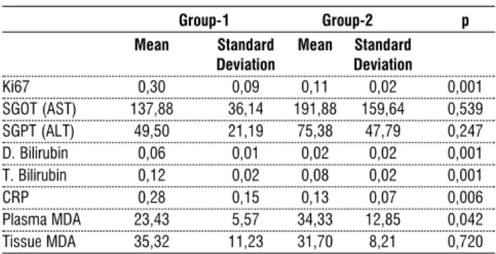
The average plasma MDA value was determined as 23,43 in the Allyl disulfide group, 34,33 in the control group and 31,19 in the sham group. This differentiation among groups was also statistically significant (p: 0,042). The average tissue MDA level was determined to be 35,32 in the Allyl disulfide group, 31,70 in the control group and 30,93 in the sham group. This differentiation among groups was not statistically significant (p: 0,720).
Although there were significant differences in the CRP level among groups in this study, the levels remained within regular laboratory values.
Although SGPT (ALT) and SGOT (AST) vales were determined to be lower in average at Allyl disulfide group (Group 1), the difference between other groups (groups 2-3) was not statistically significant (p:0.247, p:0.539).
The average T. Bili values were 0,12 in the Allyl disulfide
group, 0,08 in the control group and 0,04 in sham group. This differentiation among groups was statistically significant (p: 0.001). The average D. Bili value was determined to be 0,06 in the Allyl disulfide group, 0,02 in the control group and 0,02 in the sham group. This differentiation among groups was also statistically significant (0.001).
Table 1. Summary of the pathology findings
Group-1 Group-2 p
Fibroblast Activity N/A 2 8 ,001
11,1% 44,4%
Present 6 0
100,0% 0,0%
Nuclear Pleomorphism N/A 0 0 ,001
0,0% 0,0%
Present 1 8
7,1% 57,1%
advanced 7 0
100,0% 0,0%
Portal inflammatory N/A 6 8 ,319
cell infiltration
28,6% 38,1%
Present 2 0
66,7% 0,0%
Dilation, hyperemia N/A 3 8 ,013
at sinusoids 15,8% 42,1% Present 4 0 100,0% 0,0% Very advanced 1 0 100,0% 0,0%
Kupffer cell increase N/A 0 3 ,001
at sinusoid lumens 0,0% 30,0% Present 4 5 40,0% 50,0% Advanced 4 0 100,0% 0,0% 17,6% 35,3% Regeneration N/A 0 0 ,006 0,0% 0,0% Present 3 8 16,7% 44,4% advanced 5 0 100,0% 0,0%
Table 2. Summary of the Biochemical Parameters
Group-1 Group-2 p
Mean Standard Mean Standard
Deviation Deviation Ki67 0,30 0,09 0,11 0,02 0,001 SGOT (AST) 137,88 36,14 191,88 159,64 0,539 SGPT (ALT) 49,50 21,19 75,38 47,79 0,247 D. Bilirubin 0,06 0,01 0,02 0,02 0,001 T. Bilirubin 0,12 0,02 0,08 0,02 0,001 CRP 0,28 0,15 0,13 0,07 0,006 Plasma MDA 23,43 5,57 34,33 12,85 0,042 Tissue MDA 35,32 11,23 31,70 8,21 0,720
Discussion Discussion
The garlic plant is a member of the family Liliaceae and is known under the species name Allium sativum. The most important chemical compounds present in garlic are sulfur compounds, including aliin, allicin, thiosulfinates, gamma-glutamylcysteine peptides and various other sulfur compounds. Phyto-chemicals in garlic have potential pharmacological effects against many physiological events. Because of this component, garlic has been suggested to inhibit cancer and cardiovascular diseases and thus its therapeutic use has been studied (13). Many experimental cancer models show that Allil sulfide compounds inhibit both the formation and metastasis of tumors (14). Furthermore, the antioxidant, bacterial, antiviral, antifungal, immune stimulator, anti-agregan, cholesterol regulating, re-epitelizan, wound healing and detoxifying features of garlic are well recognized. Guyonnet et al. researched the impact of DAS, compound of garlic, on Aflatoxin B(1) genotoxicity in a study conducted on rats, which revealed that these compounds decrease the risk of liver carcinoma caused by aflatoxin B(1) (15). Additionally, epidemiological studies have shown an inverse relationship between consumption of garlic and gastric and colon cancer. Iimura et al. conducted an experimental study in which Helicobacter pylori along with 4% AGE (aged garlic extract) was administered to rats, and gastritis and gastric cancer formation was researched and compared with a control group. It was observed that each rat from the control group had gastritis and gastric cancer, while gastritis symptoms were mild and gastric cancer did not form in rats administered AGE. The authors deduced that garlic extracts may be used for the prevention of gastritis and gastric cancer that may be caused by H. pylori (16). Many other studies have revealed that garlic extract suppresses cancer development; however, a single study regarding the impact of garlic on metastasis has been conducted (Hu X et al.). That study studied the impact of AGE on rat sarcoma tumor migration and development. Depending on the dose of AGE, the authors observed an inhibition in the development of rat sarcoma cells and sarcoma cell metastasis (17). Another well-known effect of garlic is its broad antibacterial feature on both gram negative and gram positive bacteria (18). Furthermore, the antioxidant, antibacterial, antiviral, antifungal, immune stimulator, anti-agregan, cholesterol regulating, re-epitelizan, wound healing and detoxifying features of garlic are well recognized (19-22).
Despite its vast metabolic load, the liver is the organ with the broadest cell proliferation capacity. Only 0.0012% - 0.01% of hepatocytes undergo mitosis at any period of life (23-24). This low turnover rate in healthy livers changes in the case of toxic liver damage or surgical resection. Active cell replication starts within 24 hours after partial hepatectomy. Within the initial 10 days, regeneration occurs to a significant level, and this event is completed within 4-5 weeks. Regeneration mostly occurs in the form of the formation of new lobules and the expansion of residual lobules. Stimulants needed for hepatic regeneration are humoral factors that arise from the pancreas, other extrahepatic organs and the regenerated liver itself (25).
Many growth factors and cytokines are involved in liver regeneration. Hepatocyte growth factor (HGF): This is a growth factor involved in protein structure formation present in many tissue and plasma, primarily Ito and Kupffer cells. In rats, the plasma HGF concentration increases 20-fold within an hour after hepatectomy (26). In humans, the plasma HGF level reaches a maximum within days 1 to 3 following liver resection (27). It has been shown that liver DNA synthesis fails in sheep with TNF-α receptor deficiency and IL-6 gene deletion, which was determined using an anti-TNF-α antibody (28). Epidermal growth factor (EGF): This is the first factor to stimulate DNA synthesis in hepatocytes. The mitogenic effect of EGF has been demonstrated in hepatocyte cultures (29). Transforming Growth Factor Alpha (TGF-α): This factor is thought to become active after the first stage of liver regeneration. It affects the same receptor as EGF. It increases DNA synthesis in hepatocyte cultures (30). Norepinephrine: This hormone increases liver regeneration directly by way of α1-adrenergic receptors and indirectly by increasing EGF. Sympathetic denervation and α1 receptor blockage reduces DNA synthesis (29). It has been reported that factors such as fibroblast growth factor (FGF), insulin, vascular endothelium growth factor (VEGF), triiodothyronine (T3), retinoic acid, some medications (barbiturates, diazepam, hypolipidemic agents, antiepileptic agents), growth hormone, PGE2, siklo-sporin, FK506, vasopressin, estrogen and progesterone positively contribute to liver regeneration (31,32).
Under experimental conditions, we found that liver regeneration capacity was significantly higher in rats fed with Allyl disulfide after major hepatectomy in comparison with rats in the control and sham groups.
Nuclear pleomorphisms and the presence of double nuclei in hepatocytes were higher in the rats that were fed Allyl disulfide, which was interpreted in concordance with the theory that mitotic activity, and therefore liver regeneration, after a major hepatectomy is high.
The Kİ 67 proliferation ratio Allyl disulfide group was found to be significantly higher than in the other groups. These findings demonstrate that the use of Allyl disulfide increases hepatocyte proliferation after liver resection.
The relatively lower MDA levels that were observed in the rats that were fed Allyl disulfide led us to believe that Allyl disulfide consumption reduces the formation of free oxygen radicals. Such free radicals typically form and accumulate as a consequence of liver resection and lead to tissue damage.
Plasma AST and ALT levels were close to normal values in the rats that were given a Allyl disulfide, indicating a positive impact of Allyl disulfide on hepatocyte functions.
We observed that liver regeneration and proliferation was positively affected after a major hepatectomy in rats fed with an Allyl disulfide. Additionally, the Allyl disulfide had a protective effect against oxidative stress, which occurs after hepatectomy. Based on these results, we suggest that the addition of Allyl disulfide to the diet of liver resection patients during both pre-surgery and post-surgery periods would be beneficial. This beneficial supplementation is applicable to hepatectomy patients with various etiologies, including HCC, liver
metasta-sis, cholangioma and cancer, among other etiologies. The dose of garlic extract that confers an optimal impact and how it affects liver patients that do not require surgery (toxic hepatitis, viral hepatitis, fatty liver, etc.) requires further clarification. References
References
1. González MA, Contini Mdel C, Millen N, Mahieu ST. Role of melatonin in the oxidative damage prevention at different times of hepatic regeneration. Cell Biochem Funct. 2012 Dec; 30(8):701-8
2. Hung KC, Hsieh PM, Yang KL, Lin KJ, Chen YS, Hung CH. Effect of thalidomide on the expression of vascular endothe-lial growth factor in a rat model of liver regeneration. Oncol Lett. 2013 Mar; 5(3):852-856
3. El-Lakkany NM, Hammam OA, El-Maadawy WH, Badawy AA, Ain-Shoka AA, Ebeid FA Anti-inflammatory/anti-fibrotic effects of the hepatoprotective silymarin and the schistosomicide praziquantel against Schistosoma mansoni-induced liver fibrosis. Parasit Vectors. 2012 Jan 11; 5:9
4. Kim J, Ha HL, Moon HB, Lee YW, Cho CK, Yoo HS, Yu DY. Chemopreventive effect of Curcuma longa Linn on liver pathology in HBx transgenic mice. Integr Cancer Ther. 2011 Jun; 10(2): 168-77
5. Latha RC, Daisy P. Insulin-secretagogue, antihyperlipidemic and other protective effects of gallic acid isolated from Terminalia bellerica Roxb. in streptozotocin-induced diabetic rats. Chem Biol Interact. 2011 Jan 15;189(1-2):112-8
6. Karaman A, Kirimlioglu H, Tas E, Karadag N, Gülsul C, Fadillioglu E, Demircan M. Effect of leflunomide on liver regeneration after partial hepatectomy in rats. Pediatr Surg Int. 2010 Feb; 26(2):219-226
7. Borek C. Antioxidant health effects of aged Garlic extract. J. Nutr. 2001; 131: 1010-1015
8. Donald LM, Dale RR. Enhanced immunocompetence by Garlic: Role in bladder cancer and other malignancies. J. Nutr. 2001; 131: 1067–1070
9. Higgins GM and Anderson RM. Experimental pathology of the liver - restoration of the liver of the white rat following partial surgical removal. Arch Pathol 1931; 7: 187-202 10. Zeybek A, Ercan F, Cetinel Ş, Cikler E, Saglam B, Sener G.
Protective Effects of Aqueous Garlic Extract in Reducing Water Avoidance Stress-Induced Degeneration of the Stomach, Ileum, and Liver: Morphological and Biochemical Study, Dig Dis Sci (2007) 52: 2984–2992
11. Ana Cristina Aoun Tannuri, Uenis Tannuri, Maria Cecília Coelho, Neide Aparecida dos Santos e Evandro Sobroza de Mello Experimental models of hepatectomy and liver regeneration using newborn and weaning rats. Clinics 2007; 62(6):757-62
12. Eda S, Kaufmann J, Roos W et al. Development of a new microparticle-enhanced turbidimetric assay for C-reactive protein with superior features in analytical sensitivity and dynamicrange. J Clin Lab Anal 1998; 12: 137-144
13. Buege JA. Aust SD. Microsomal Lipid Peroxidation. Methods Enzymol. 1978; 52: 303-310
14. Taiichiro S, Takashi H, Tomomi HF, Kahoru I, Rie T, Anticancer effects of diallyl trisulfide derived from Garlic.
Asia Pac J Clin Nutr 2008; 17 (S1): 249-252
15. Guyonnet D, Belloir C, Suschetet M, Siess MH, Le Bon AM. Mechanisms of protection against aflatoxin B(1) genotoxicity in rats treated by organosulfur compounds from Garlic. 2002; Carcinogenesis. 23(8): 1335-41
16. Iimuro M, Shibata H, Kawamori T, Matsumoto T, Arakawa T, Sugimura T, Wakabayashi K. Suppressive effects of Garlic extract on Helicobacter pylori-induced gastritis in Mongolian gerbils. Cancer Lett. Dec 2001; 187(1-2): 61-8
17. Hu X, Cao BN, Hu G, He J, Yang DQ, Wan YS. Attenuation of cell migration and induction of cell death by aged garlic extract in rat sarcoma cells. Int J Mol Med.2002; 9(6):641-643 18. Tatara MR, Sliwa E, Dudek K, Gawron A, Piersiak T, Dobrowolski P, Mosiewicz J, Siwicki AK, Studzinski T. Aged garlic extract and allicin improve performance and gastro-intestinal tract development of piglets reared in artificial sow. 2008; Ann Agric Environ Med 15: 63–69
19. Borek C.Garlic reduces dementia and heart disease risk. J. Nutr. 2006; 136: 810–812
20. Zhang XH, Lowe D, Giles P, Fell S, Connock MJ, Maslin DJ. Gender may affect the action of garlic oil on plasma cholesterol and glucose levels of normal subjects. J. Nutr. 2001; 131: 1471–1478
21. Seki T, Hosono T, Fukao TH, Inada K, Tanaka R, Ogihara J, Ariga T. Anticancer effects of dailly trisulfide derived from garlic. J Clin Nutr 2008; 17 (S1): 249-252
22. Xiang L, Tan JW, Huang LJ, Jia L, Liu YQ, Zhao YQ, Wang K,Dong JH. Inhalation of hydrogen gas reduces liver injury during major hepatotectomy in swine. World J Gastroenterol 2002; 18(37): 5197-5204
23. Fausto N, Webber EM. Liver regeneration. In: Arias I, Boyer J, Fausto N, et al. eds. The liver:biology and pathobiology. New York: Raven Pres; 1994:1059- 1084
24. Holt DR, Thiel DV, Edelstein S, Brems JJ. Hepatic resections. Arch Surg 2000; 135:1353-1358
25. Perek S, Kapan S, Ed: Değerli Ü, Bozfakıoğlu Y. Cerrahi Gastroenteroloji. s. 194-208. 5. Basım, Nobel Tıp Kitabevleri, İstanbul, 2000
26. Nishizaki T, Takenaka K, Yoshizumi T et al. Alteration in levels of human hepatocyte growth factor following hepatectomy. J Am Coll Surg 1995;181: 6-10
27. Kinoshita T, Tashiro K, Nakamura T. Marked increase of HGF mRNA in nonparanchymal liver cells of rats treated with hepatotoxins. Biochem Biophys Res Commun 1989;165: 1229-34
28. Michalopoulos GK. Liver regeneration: molecular mechanism of growth control. FASEB J 1990; 4:176-87
29. Michalopoulos GK, De Frances MC. Liver regeneration. Science 1997; 276: 60- 66.
30. Hashimoto M, Kothary BC, Raper S. The effects of trans-forming growth factor alpha and somatostatin on regenerating hepatocytes in the rat. Regulatory peptides 1993; 44: 49-59 31. Francavilla A, Polimeno L, Barone M et al. Hepatic
regenera-tion and growth factors. J Surg Oncol 1993,13: 1-7
32. Ekberg S, Luther M, Nakamura T et al. Growth hormone promotes early initiation of hepatocyte growth factor gene expression in the liver of hypophysectomised rats after partial hepatectomy. J Endocrinol 1992; 135: 59-67
View publication stats View publication stats