E-Mail [email protected]
At the Cutting Edge
Neuroendocrinology 2014;100:95–102 DOI: 10.1159/000369072Agouti-Related Protein Neuron Circuits
That Regulate Appetite
Scott M. Sternson
a
Deniz Atasoy
b, c
a Janelia Research Campus, HHMI, Ashburn, Va. , USA; b Department of Physiology, School of Medicine, and c Research
Center for Regenerative and Restorative Medicine (REMER), Istanbul Medipol University, Istanbul , Turkey
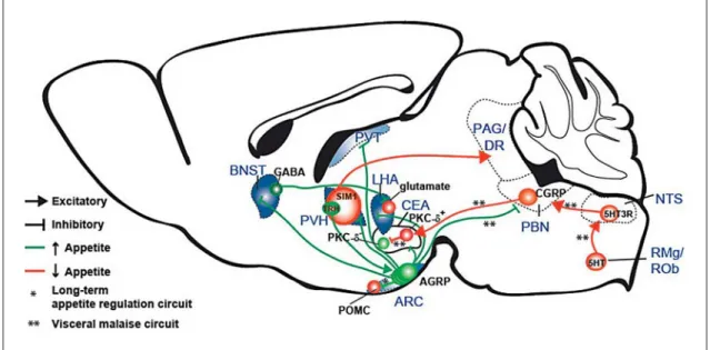
(e.g., hormones and metabolites) and either coordinate or suppress behavioral responses to seek and consume food. Much of the recent work has focused on the circuits in-volving Agouti-related protein (AGRP)-expressing neu-rons, a small molecularly defined population in the hypo-thalamic arcuate nucleus (ARC) that senses circulating signals of the energetic status. AGRP neurons are acti-vated by hormonal signals of energy deficit (e.g., ghrelin) [6, 7] and are inhibited by signals of energy surfeit (e.g., leptin) [7, 8] . Optogenetic and chemogenetic activation of AGRP neuron firing leads to voracious food seeking and consumption behaviors within minutes [9–11] . Con-versely, AGRP neuron ablation or chemogenetic inhibi-tion results in appetite suppression [10, 12] . Bidirecinhibi-tional control of feeding behavior by AGRP neurons makes them an attractive entry point to investigate the neural circuits that regulate feeding behavior, and, using new tools, multiple pathways have been identified ( fig. 1 ; ta-ble 1 ). Here we review emerging insights into the neural control of appetite.
Molecular Mediators of Appetite
AGRP neurons release a number of molecules that rapidly elicit intense food consumption when injected into the brain: AGRP [13] , neuropeptide Y (NPY) [14] ,
Key Words
Agouti-related protein · Appetite · Ghrelin
Abstract
New tools for mapping and manipulating molecularly de-fined neural circuits have improved the understanding of how the central nervous system regulates appetite. Studies that focused on Agouti-related protein neurons, a starva-tion-sensitive hypothalamic population, have identified multiple circuit elements that can elicit or suppress feeding behavior. Distinct axon projections of this neuron popula-tion point to different circuits that regulate long-term appe-tite, short-term feeding, or visceral malaise-mediated an-orexia. Here, we review recent studies examining these neu-ral circuits that control food intake. © 2014 S. Karger AG, Basel
Introduction
A longstanding goal of neuroscience is to understand the relationship between brain function and behavioral states such as hunger [1–3] . Appetite is controlled by multiple circuits that are comprised of distinct molecu-larly defined neuron populations [4, 5] . These networks integrate peripheral signals of metabolic and visceral state
Received: July 31, 2014
Accepted after revision: October 14, 2014 Published online: November 6, 2014
Scott M. Sternson
Janelia Research Campus, HHMI 19700 Helix Dr.
Ashburn, VA 20147 (USA) E-Mail sternsons @ janelia.hhmi.org
© 2014 S. Karger AG, Basel 0028–3835/14/1003–0095$39.50/0 www.karger.com/nen
and γ-aminobutyric acid (GABA) [15] . The relative role of these molecules for AGRP neuron-evoked feeding has recently been examined. The results of several experi-ments indicate that NPY and GABA mediate initiation of eating within minutes of elevated AGRP neuron activity, whereas the neuropeptide AGRP regulates feeding over longer timescales. Increased food intake resulting from optogenetic or chemogenetic activation ( fig. 2 ) of AGRP neurons was suppressed by either an NPY 1R antagonist or a GABA A receptor antagonist microinjected in the paraventricular hypothalamic nucleus (PVH) [11] . This showed that AGRP neurons require both NPY and GABA signaling to elicit acute feeding responses. A related result using mutant mice confirmed this finding, with some noteworthy differences. Chemogenetic activation of AGRP neurons ( fig. 2 c) in either Npy –/– mice or in mice
lacking GABA in AGRP neurons each rapidly evoked feeding behavior [16] . One difference with the pharma-cological analysis described above was that either NPY or GABA alone appeared sufficient for acute feeding. An ex-planation for this apparent discrepancy is indicated by a closer evaluation of Npy –/– mice in which
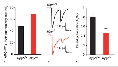
electrophysio-logical analysis showed dramatic compensatory upregu-lation of AGRP neuron synaptic output: connectivity and GABA release probability ( fig. 3 ) [11] . Therefore, it ap-pears that GABA and NPY are both required for acute
feeding, as revealed by pharmacological inactivation ex-periments, but they likely compensate for one another in mutant mouse models.
What is the role for the AGRP neuropeptide released by AGRP neurons? AGRP inhibits melanocortin recep-tors, which leads to increased food intake and body weight. However, AGRP neuron photostimulation still rapidly elicits feeding behavior in A y mutant mice [9] , in which
melanocortin receptors are constitutively blocked. This indicated that the AGRP neuropeptide was not required for acute AGRP neuron-evoked feeding behavior. Inter-estingly though, activation of AGRP neurons lacking both GABA and NPY increased feeding with a delay of several hours, and this was not observed if melanocortin receptor 4 was absent [16] . Together, these results from AGRP neu-ron activation studies revealed that unlike NPY and GABA, which mediate acute feeding, the neuropeptide AGRP and its interaction with melanocortin receptors are involved in long-term regulation of feeding behavior.
Circuit for Long-Term Regulation of Food Intake
Appetite is regulated on timescales of minutes for the execution of feeding behavior as well as over longer tim-escales associated with maintaining energy homeostasis.
Fig. 1. Summary diagram illustrating cell types and circuits that influence appetite. Blue shading represents sites associated with AGRP neuron-evoked food intake. Based on anatomical studies, AGRP neurons are shown as separate subpopulations defined by their axon projection sites. RMg = Raphe magnus nucleus; ROb = raphe ob-scurus nucleus; SIM1 = single-minded homolog 1; DR = dorsal raphe.
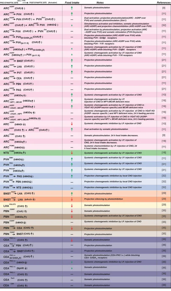
AGRP Neuron Circuits That Regulate Appetite Neuroendocrinology 2014;100:95–102 DOI: 10.1159/000369072 97 Food intake ARC AGRP ARC PVH Notes ARC AGRP AGRP (ChR2 ) ARCAGRP BNST (ChR2 )
ARCAGRP LHA (ChR2 )
ARCAGRP PVT (ChR2 )
ARCAGRP CEA (ChR2 )
ARCAGRP PBN (ChR2 )
ARCAGRP PAG (ChR2 )
ARC AGRP
ARCAGRP PVH (ChR2 ) + PVH Sim1 (ChR2 )
ARCAGRPPVH (ChR2 ) + PVH Oxt (ChR2 )
ARCAGRP PVH(bl-GABA R) (ChR2 )
A ARCAGRPPVH(bl-Y1 R) (ChR2 )
ARCAGRP (hM3Dq ) + PVH(bl-GABA R)A ARCAGRP (hM3Dq ) + PVH(bl-Y1 R)
(hM3Dq ) 'NPY, 'MC4R ARC AGRP (hM3Dq ) 'VGAT, 'MC4R ARC AGRP (hM3Dq ) 'VGAT, 'NPY
ARC AGRP'VGAT, 'NPY,'MC4R(hM3Dq )
ARC POMC
ARC POMC ARC POMC
NTS POMC PVH SIM1
ARCAGRP (ChR2 )+ ARCPOMC (ChR2 )
BNSTVGAT LHA (ChR2 )
BNSTVGAT LHA (eArch )
LHAVGLUT2 (ChR2 )
PVH SIM1 PAG
+ ARCAGRPPVH (hM4Di )
ARC AGRP (hM4Di ) PVH SIM1 PBN(hM4Di ) PVH SIM1 NTS(hM4Di ) PVH TRH (hM4Di ) (hM3Dq ) PVH PACAP(hM3Dq ) PBN (ChR2 ) CGRP PBN (hM3Dq ) CGRP PBN CGRP(hM4Di ) PBNCGRP CEA (ChR2 ) PBNCGRP BNST (ChR2 ) (ChR2 ) (ChR2 ) (hM3Dq ) (ChR2 ) (hM4Di ) (hM3Dq ) (hM3Dq ) A A ARC (hM4Di ) AGRP References [9] [11] [32] [21] [10] [16] [18] [31] [29] [35] [11] [11] [11] [11] [11] [11] [21] [21] [21] [11] [21] [16] [16] [16] [10] [11] [9] [11] [18] [11] [31] [32] [32] [32] [29] [29] [35] [35] [35] [35] PRE-SYNAPTIC SITE CELL TYPE
POST-SYNAPTIC SITE (Actuator)
Somatic photostimulation
Systemic chemogenetic activation by I.P. injection of CNO Projection photostimulation Projection photostimulation Projection photostimulation Projection photostimulation Projection photostimulation Projection photostimulation Projection photostimulation
Systemic chemogenetic activation by I.P. injection of CNO in NPY+MC4R deficient mice Systemic chemogenetic activation by I.P. injection of CNO in VGAT KO (AGRP-neuron specific) and MC4R deficient mice Systemic chemogenetic activation by I.P. injection of CNO in VGAT KO (AGRP- neuron specific ) and NPY deficient mice, 24 h feeding persists Systemic activation by I.P. injection of CNO in VGAT KO (AGRP- neuron specific) and NPY + MC4R deficient mice, 24 h feeding persists
Somatic photostimulation. 24 h food intake decreases.
Systemic chemoinhibition by I.P. injection of CNO, 24 h food intake increases.
Systemic chemogenetic activation by I.P. injection of CNO Systemic chemogenetic activation by I.P. injection of CNO
Projection chemogenetic inhibition by local CNO injection Projection chemogenetic inhibition by local CNO injection Projection chemogenetic inhibition by local CNO injection Systemic chemogenetic activation by I.P. injection of CNO Systemic chemogenetic activation by I.P. injection of CNO
Systemic chemogenetic inhibition by I.P. injection of CNO Dual activation; projection photostimulation(ARC - AGRP over PVH) and somatic photostimulation (Sim1)
Simultaneous activation and inhibition, somatic photostimulation (ARC-AGRP) and projection chemoinhibition (ARC-AGRP over PVH) Dual activation by photostimulation; projection activation (ARC - AGRP over PVH) and somatic stimulation (PVH-Oxytocin) Projection photostimulation (ARC-AGRP over PVH) while blocking PVH - GABA receptors
Projection photostimulation (ARC-AGRP over PVH) while blocking PVH - Y1R receptors
Systemic chemogenetic activation by I.P. injection of CNO (ARC-AGRP) while blocking PVH - GABA receptors Systemic chemogenetic activation by I.P. injection of CNO (ARC-AGRP) while blocking PVH - Y1R receptors
Dual activation by somatic photostimulation
Systemic chemogenetic activation by I.P. injection of CNO, 24 h food intake decreases.
Projection photostimulation Projection silencing by photoinhibition
Somatic photostimulation Somatic photostimulation
Systemic chemogenetic activation by I.P. injection of CNO
Projection photostimulation Projection photostimulation
Systemic chemogenetic inhibition by I.P. injection of CNO
CEAPKC-G+ (ChR2 ) (ChR2 ) [36] [36] [36] [36] [36] [36] Somatic photoinibition Somatic photostimulation Somatic photostimulation CEAPKC-(bl-GABA R)G+ A
Somatic photostimulation (CEA-PKC G+ ) while blocking
CEA- GABA receptorsA
CEAPKC-G+(hM4Di ) Systemic chemogenetic inhibition by I.P. injection of CNO
CEAPKC-G+(NpHR ) CEAPKC-G- (ChR2 ) [36] Somatic photostimulation Somatic photostimulation CEATac2 (ChR2 ) CEACRF (ChR2 ) (ChR2 ) [36] [36] CEAPKC-G+ CEAPKC-G+ BNST (ChR2 ) PBN Projection photostimulation Projection photostimulation
Table 1. Cell type-specific neu-ronal circuit manipulations that regulate food intake
Distinct regulation of acute and long-term control over appetite has been found with manipulation of molecu-larly defined cell types and circuits. Within the ARC, AGRP neurons are intermingled with POMC neurons that release the neuropeptide α-MSH, which has been shown to strongly suppress food intake. Cell type-specif-ic POMC neuron activation over 24 h showed reduced body weight and food intake [9, 17] , and POMC neuron inhibition correspondingly increased food intake over 24 h [11] . AGRP neurons form local inhibitory synaptic con-nections onto POMC neurons [18] , indicating a possible circuit through which AGRP neurons can influence ap-petite. In addition, functional studies have demonstrated that this interaction powerfully suppresses POMC neu-ron firing when AGRP neuneu-rons are activated [11] . How-ever, neither POMC neuron gain-of-function nor loss-of-function manipulations significantly affected food intake over the 1-hour timescale associated with voracious AGRP neuron-evoked food intake [9, 17, 19] . The mis-match in the timescales for AGRP and POMC neuron control over feeding indicated a role for POMC neurons in long-term regulation of appetite and also that AGRP neurons likely do not control acute induction of feeding behavior through suppression of POMC neuron activity. This was confirmed with neural circuit epistasis anal-ysis in which AGRP neurons were photostimulated si-multaneously with POMC neurons [11] . In this experi-ment, AGRP inhibition of POMC neurons was over-come by coincident depolarization of POMC neurons with channelrhodops2 to test the necessity of this in-hibitory interaction for acute control over food con-sumption. Mice consumed as much food with AGRP neuron activation alone as they did with coactivation of AGRP and POMC neurons [11] , consistent with the con-clusion that ARC AGRP → ARC POMC does not regulate short-term food intake. Together with the long timescale for POMC neuron activation and silencing to affect food intake, this indicates that AGRP neuron inhibition of POMC neurons may be important for long-term regula-tion of appetite but not necessarily for the short-term feeding behavior that is elicited upon activation of AGRP neurons.
Neural Circuits That Rapidly Elicit Food Seeking and Consumption Behavior
The second-order circuit nodes through which AGRP neurons influence behavior and physiology can be visual-ized by their axon projections using immunochemistry a
b
c
Fig. 2. Optogenetic [37] and chemogenetic [38] tools for neuronal activity manipulation. a Rapid reversible manipulation of neuronal membrane potential can be achieved using optogenetic tools. Channelrhodopsin-2 (ChR2) is a cation channel activated by blue light, which depolarizes neurons in response to blue light to trigger action potentials with millisecond precision. Halorhodopsin (NpHR) and Archaerhodopsin (Arch) are activated by yellow wavelengths and pump chloride ions and protons, respectively. These channels hyperpolarize neurons and inhibit activity with millisecond precision. b , c Variations on chemogenetic silencing tools; direct manipulation of ionic conductances ( b ) or G-protein-coupled intracellular pathways ( c ). Chimeric PSAM (pharmaco-logically selective actuator module)-based ligand gated ion chan-nels: PSAM-5HT3 or PSAM-GlyR acutely modulate cation or chlo-ride membrane conductances when activated by their cognate synthetic ligands (PSEMs = pharmacologically selective effector molecules), enabling rapid reversible activation or inhibition of neuronal activity, respectively. Similarly, engineered variants of human muscarinic acetylcholine receptors, hM3Dq and hM4Di, have reduced acetylcholine sensitivity but are responsive to an oth-erwise inert synthetic ligand, Clozapine-N-oxide (CNO). Delivery of CNO activates hM3Dq and hM4Di signaling through their downstream Gα q protein or Gα i protein intracellular pathways, cul-minating in neuron activation or neuron inhibition (in part through increased inward rectifier K + channel conductance), respectively.
AGRP Neuron Circuits That Regulate Appetite
Neuroendocrinology 2014;100:95–102
DOI: 10.1159/000369072 99
for the neuropeptide AGRP [20] or transgenic expression of a fluorescent protein [19, 21] . The long-range neural circuit connections of AGRP neurons that mediate acute elevation of appetite have been investigated by optoge-netic activation of axon projections [11, 21] ( fig. 1 ). These studies identified the anterior portion of the bed nucleus of the stria terminalis (aBNST), the lateral hypothalamus suprafornical division (LHAs), and the PVH as sufficient to increase food intake to a level similar to that achieved by activation of AGRP neuron somata. Activation of AGRP neuron projections to the paraventricular thala-mus (PVT) modestly increased food intake. Axon projec-tions to the central nucleus of the amygdala (CEA), peri-aqueductal gray (PAG), and parabrachial nucleus (PBN) did not elicit increased food intake. Therefore, a subset of brain areas contacted by AGRP neuron projections is suf-ficient to induce food consumption behavior. Two of these brain areas, the PVH [22] and the LHA [2] , had al-ready been extensively investigated for the feeding behav-iors evoked; however, the importance of the LHAs subdi-vision had only been proposed based on anatomical con-nectivity [23] . In addition, injection of a GABA A receptor agonist into the PVT was found to acutely increase feed-ing [24] . The BNST had been implicated in obesity based on a lesion study [25] , but the role of the anterior subdivi-sions of the BNST to acutely regulate feeding behavior had not been shown. Therefore, analysis of the axon pro-jections of AGRP neurons enabled identification of a sub-set of brain areas that were sufficient to elicit food intake. In addition to functional studies, anatomical analysis of AGRP neuron axon projections has also provided im-portant insights into the organization of feeding circuits.
Individual axon projection fields appear to arise from dis-tinct subpopulations of AGRP neurons, and axon collat-eralization was not detected [21] . Therefore, molecularly defined AGRP neurons can be subdivided into separate populations defined by their axon projection targets ( fig. 1 ). It is a configuration that is seen in some neuro-modulatory systems, such as midbrain dopamine neu-rons [26] , but not in other populations, such as noradren-ergic cells in the locus coeruleus [27] . This is suggestive of specialized roles for these distinct AGRP neuron cir-cuits. One example of this comes from the analysis of leptin receptor (Lepr) expression as detected by a
Lepr-IRES-Cre mouse line, which indicated that AGRP
neu-rons that project outside of the hypothalamus express leptin receptor, while those that project within the hypo-thalamus do not [21] . In contrast, responsiveness to ghre-lin or to food deprivation was observed in both intrahy-pothalamic and extrahyintrahy-pothalamic AGRP neuron pro-jection subpopulations.
Because AGRP neuron axon projections arise from separate neuronal subpopulations, their activation indi-vidually identifies target areas that are independently suf-ficient to initiate feeding (aBNST, LHAs, PVH, and PVT). Nevertheless, anatomical analyses have shown substan-tial interconnectivity between these brain areas, indicat-ing a network across which feedindicat-ing may be coordinated. For example, optogenetic activation of GABA-releasing BNST neurons (BNST GABA ) elicits voracious eating [28] . Using optogenetic circuit mapping in conjunction with single-cell RT-PCR, this was found to involve an inhibi-tory synaptic projection to the LHA that selectively tar-gets glutamate neurons ( fig. 1 ). Consistent with this,
a b c
Fig. 3. Compensatory adaptations in ARC AGRP → PVH circuit in Npy –/– mice.
a Observed connectivity rate of the ARC AGRP → PVH projection in Npy +/+ and
Npy –/– mice. b , c Representative whole-cell voltage clamp recordings showing paired pulse depression of ARC AGRP → PVH syn-apses in Npy +/+ and Npy –/– mice ( b ) and the average paired pulse response ( c ). In-creased paired pulse depression in Npy –/– mice indicates increased GABA release probability. A 1 = Amplitude of first pulse; A 2 = amplitude of second pulse. Data is represented as mean ± SEM and is from Atasoy et al. [11] .
LHA glutamate neuron inhibition evokes feeding [28] . How-ever, the precise relationship to AGRP neuron projec-tions is not yet clear. Although the BNST and LHA neu-ronal manipulation covered broad areas of each region, the targeted areas were both posterior relative to the respective AGRP axon projection fields targeted for photostimulation ( fig. 1 ). Further work is required to es-tablish the relationship between ARC AGRP → aBNST, ARC AGRP → LHAs, and BNST GABA → LHA glutamate circuit elements.
The PVH is another brain area that has a well-estab-lished role in regulating feeding behavior [29] , and activa-tion of GABA-releasing AGRP neuron axons in the PVH robustly evoked feeding [11] . Cell type-specific chemoge-netic neuronal silencing of SIM1 neurons in the PVH (en-compassing most PVH neurons) also rapidly increased eating as well as instrumental food seeking behavior [11] . Interestingly, a recent study has shown that a subset of PVH neurons that express thyrotropin-releasing hormone (TRH) and release glutamate from synaptic contacts onto AGRP neurons can activate food intake [30] . Because si-lencing the PVH elicits eating (and this likely includes the TRH subpopulations that project to AGRP neurons), it in-dicates that the PVH circuit responsible for orchestrating feeding behavior also involves PVH axon projections else-where in the brain. To identify this projection, a cell type-specific chemogenetic synaptic silencing tool, based on an axon-selective variant of the Gα i protein-coupled hM4D receptor ( fig. 2 c), has been used to examine downstream PVH axon projections for the regulation of appetite. This study found that PVH inhibition acts to increase food in-take through projections to the region around the caudal ventrolateral PAG and dorsal raphe [31] ( fig. 1 ). Further experiments are required to determine the underlying cell types in these areas that control appetite.
Neural Circuits for Anorexia
One of the striking characteristics of AGRP neurons is that their rapid ablation in adult mice leads to anorexia, ultimately resulting in death by starvation [12] . Although this was first attributed to the important role of AGRP neurons to positively regulate appetite, further investiga-tion has revealed that suppression of eating may be due to a function of AGRP neurons that is distinct from the process by which these neurons acutely elicit voracious food intake. Using localized intracranial injections of benzodiazepines (GABA A receptor potentiators), it was found that infusion into the PBN restored feeding in mice
after AGRP neuron ablation, and this was not seen in oth-er areas, such as the PVH [32] . Thoth-erefore, AGRP neuron projections to the PBN appear to play an essential role for maintaining appetite through their release of GABA. In light of this, it was surprising when activation of AGRP axon projections to the PBN did not elicit feeding in the same manner as the ARC AGRP → PVH projection did [11] . However, the distinction between these two pathways has become increasingly clear with further analysis of the PBN appetite regulatory circuit, which appears to be a visceral malaise anorexia circuit and not an acute activa-tor of voracious eating.
Using rescue of AGRP neuron ablation-induced an-orexia as a readout, a series of hindbrain manipulations was identified that restored eating in mice lacking AGRP neurons. These experiments revealed a circuit from hind-brain serotonin neurons to serotonin receptor 3 (5HT3 receptor)-expressing neurons in the nucleus of the soli-tary tract (NTS) that, in turn, activates glutamatergic NTS neurons that project to the PBN [33] ( fig. 1 ). The involve-ment of 5HT3 receptor signaling in the NTS and a finding that the antiemetic ondansetron (a 5HT3 receptor antag-onist) restored eating after AGRP neuron ablation indi-cated that this circuit may also be activated by signals that lead to nausea. Indeed, further investigation has shown that a subset of PBN neurons that express the peptide cal-citonin gene-related peptide (CGRP) mediate the re-sponse to many signals associated with nausea or illness and are known to suppress appetite [34] . Activation of PBN CGRP axon projections to the CEA but not to the BNST strongly reduces appetite [34] . Moreover, silencing PBN CGRP neurons leads to modest reduction in anorexia due to nausea agents such as LiCl; however, basal food intake is not elevated [34] . Furthermore, a recent report found that neurons that express protein kinase C-δ (PKC-δ + ) in the lateral division of CEA are likely respon-sible for the anorexigenic effect of PBN CGRP activation [35] . PKC-δ + neurons receive direct synaptic input from PBN CGRP neurons. Moreover, increasing PKC-δ + neuron activity robustly reduced food intake, whereas inhibition of these neurons modestly increased food intake [35] . A local inhibitory connection made by PKC-δ + neurons onto other CEA neurons (PKC-δ – ) likely mediates this effect, although the precise molecular identity of these PKC-δ – neurons remains to be determined [35] .
What then is the role of the ARC AGRP → PBN pro-jection? It appears that a visceral malaise anorexia circuit involving RMg 5HT /ROb 5HT → NTS 5HT3R → PBN glutamate(CGRP) → CEA GABA(PKC-δ+) → CEA PKC–δ– sup-presses appetite and is modulated by AGRP neurons
AGRP Neuron Circuits That Regulate Appetite
Neuroendocrinology 2014;100:95–102
DOI: 10.1159/000369072 101
( fig. 1 ). The loss of the ARC AGRP → PBN inhibitory input appears to upset the excitatory/inhibitory balance in PBN neurons that mediates anorexia related to circulating and neural signals associated with visceral malaise. Therefore, ARC AGRP → PBN axon projections play an essential role for gating visceral inputs and suppressing a visceral mal-aise state that causes anorexia. It will also be important to investigate the possibility of a similar role for the ARC AGRP → CEA circuit, which was not found to evoke food intake [21] , but it may be positioned to gate the an-orexigenic output of the CEA.
Conclusions
Overall, the emerging picture is that AGRP neurons influence appetite through multiple processes. Based on investigations with cell type-specific and
projection-specific manipulations, these include (1) long-term reg-ulation of feeding behavior through POMC neurons, (2) acute control of appetite through an interconnected ‘core forebrain feeding circuit’ [21] involving aBNST, PVH, LHAs, and the PVT, and (3) gating of visceral sig-nals that include those associated with illness and nau-sea. It is noteworthy that nearly all of the brain regions associated with long-term feeding, acute appetite, or visceral malaise are also interconnected. Identification of additional cell types, cell type-specific perturbations, and circuit mapping techniques will be needed to eluci-date the role of these seemingly distinct but shared net-works in regulating different aspects of appetite. Fur-thermore, access to these different neural circuits will facilitate the understanding of the underlying behavior-al mechanisms by which AGRP neurons elicit gobehavior-al-di- goal-di-rected motivated behaviors associated with food seek-ing [36] .
References
1 Hess WR: The Functional Organization of the Diencephalon. New York, Grune & Stratton, 1957.
2 Delgado JM, Anand BK: Increase of food in-take induced by electrical stimulation of the lateral hypothalamus. Am J Physiol 1953; 172: 162–168.
3 Hetherington AW, Ranson SW: Hypotha-lamic lesions and adiposity in the rat. Anat Rec 1940; 78: 149–172.
4 Saper CB, Chou TC, Elmquist JK: The need to feed: homeostatic and hedonic control of eat-ing. Neuron 2002; 36: 199–211.
5 Gao Q, Horvath TL: Neurobiology of feeding and energy expenditure. Annu Rev Neurosci 2007; 30: 367–398.
6 Cowley MA: The distribution and mecha-nism of action of ghrelin in the CNS demon-strates a novel hypothalamic circuit regulat-ing energy homeostasis. Neuron 2003; 37: 649–661.
7 van den Top M, Lee K, Whyment AD, Blanks AM, Spanswick D: Orexigen-sensitive NPY/ AgRP pacemaker neurons in the hypothalam-ic arcuate nucleus. Nat Neurosci 2004; 7: 493– 494.
8 Fioramonti X, Contie S, Song Z, Routh VH, Lorsignol A, Penicaud L: Characterization of glucosensing neuron subpopulations in the arcuate nucleus: integration in neuropeptide Y and pro-opio melanocortin networks? Dia-betes 2007; 56: 1219–1227.
9 Aponte Y, Atasoy D, Sternson SM: AGRP neurons are sufficient to orchestrate feeding behavior rapidly and without training. Nat Neurosci 2011; 14: 351–355.
10 Krashes MJ, Koda S, Ye C, Rogan SC, Adams AC, Cusher DS, Maratos-Flier E, Roth BL, Lowell BB: Rapid, reversible activation of AgRP neurons drives feeding behavior in mice. J Clin Invest 2011; 121: 1424–1428. 11 Atasoy D, Betley JN, Su HH, Sternson SM:
Deconstruction of a neural circuit for hunger. Nature 2012; 488: 172–177.
12 Luquet S, Perez FA, Hnasko TS, Palmiter RD: NPY/AgRP neurons are essential for feeding in adult mice but can be ablated in neonates. Science 2005; 310: 683–685.
13 Ollmann MM, Wilson BD, Yang YK, Kerns JA, Chen Y, Gantz I, Barsh GS: Antagonism of central melanocortin receptors in vitro and in vivo by agouti-related protein. Science 1997; 278: 135–138.
14 Clark JT, Kalra PS, Crowley WR, Kalra SP: Neuropeptide Y and human pancreatic poly-peptide stimulate feeding behavior in rats. Endocrinology 1984; 115: 427–429.
15 Kelly J, Grossman SP: GABA and hypotha-lamic feeding systems. II. A comparison of GABA, glycine and acetylcholine agonists and their antagonists. Pharmacol Biochem Behav 1979; 11: 647–652.
16 Krashes MJ, Shah BP, Koda S, Lowell BB: Rap-id versus delayed stimulation of feeding by the endogenously released AgRP neuron media-tors GABA, NPY, and AgRP. Cell Metab 2013; 18: 588–595.
17 Zhan C, Zhou J, Feng Q, Zhang JE, Lin S, Bao J, Wu P, Luo M: Acute and long-term sup-pression of feeding behavior by POMC neu-rons in the brainstem and hypothalamus, re-spectively. J Neurosci 2013; 33: 3624–3632.
18 Cowley MA, Smart JL, Rubinstein M, Cerdan MG, Diano S, Horvath TL, Cone RD, Low MJ: Leptin activates anorexigenic POMC neurons through a neural network in the arcuate nu-cleus. Nature 2001; 411: 480–484.
19 Atasoy D, Aponte Y, Su HH, Sternson SM: A FLEX switch targets Channelrhodopsin-2 to multiple cell types for imaging and long-range circuit mapping. J Neurosci 2008; 28: 7025–7030.
20 Haskell-Luevano C, Chen P, Li C, Chang K, Smith MS, Cameron JL, Cone RD: Character-ization of the neuroanatomical distribution of agouti-related protein immunoreactivity in the rhesus monkey and the rat. Endocrinolo-gy 1999; 140: 1408–1415.
21 Betley JN, Cao Zhen Fang H, Ritola Kimberly D, Sternson Scott M: Parallel, redundant cir-cuit organization for homeostatic control of feeding behavior. Cell 2013; 155: 1337–1350. 22 Leibowitz SF: Paraventricular nucleus: a
pri-mary site mediating adrenergic stimulation of feeding and drinking. Pharmacol Biochem Behav 1978; 8: 163–175.
23 Hahn JD, Swanson LW: Distinct patterns of neuronal inputs and outputs of the juxtapara-ventricular and suprafornical regions of the lateral hypothalamic area in the male rat. Brain Res Rev 2010; 64: 14–103.
24 Stratford TR, Wirtshafter D: Injections of muscimol into the paraventricular thalamic nucleus, but not mediodorsal thalamic nuclei, induce feeding in rats. Brain Res 2013; 1490: 128–133.
25 King BM, Rollins BL, Grundmann SJ, Olivier LG: Excessive weight gains in female rats with transections of the stria terminalis. Physiol Behav 2003; 78: 563–568.
26 Prensa L, Parent A: The nigrostriatal pathway in the rat: a single-axon study of the relation-ship between dorsal and ventral tier nigral neurons and the striosome/matrix striatal compartments. J Neurosci 2001; 21: 7247– 7260.
27 Simpson KL, Altman DW, Wang L, Kirifides ML, Lin RC, Waterhouse BD: Lateralization and functional organization of the locus coe-ruleus projection to the trigeminal somato-sensory pathway in rat. J Comp Neurol 1997; 385: 135–147.
28 Jennings JH, Rizzi G, Stamatakis AM, Ung RL, Stuber GD: The inhibitory circuit archi-tecture of the lateral hypothalamus orches-trates feeding. Science 2013; 341: 1517–1521.
29 Kelly J, Rothstein J, Grossman SP: GABA and hypothalamic feeding systems. I. Topograph-ic analysis of the effects of mTopograph-icroinjections of muscimol. Physiol Behav 1979; 23: 1123–1134. 30 Krashes MJ, Shah BP, Madara JC, Olson DP,
Strochlic DE, Garfield AS, Vong L, Pei H, Wa-tabe-Uchida M, Uchida N, Liberles SD, Low-ell BB: An excitatory paraventricular nucleus to AgRP neuron circuit that drives hunger. Nature 2014; 507: 238–242.
31 Stachniak TJ, Ghosh A, Sternson SM: Chemo-genetic synaptic silencing of neural circuits localizes a hypothalamus → midbrain pathway for feeding behavior. Neuron 2014; 82: 797– 808.
32 Wu Q, Boyle M, Palmiter R: Loss of GABAer-gic signaling by AgRP neurons to the parabra-chial nucleus leads to starvation. Cell 2009; 137: 1225–1234.
33 Wu Q, Clark MS, Palmiter RD: Deciphering a neuronal circuit that mediates appetite. Na-ture 2012; 483: 594–597.
34 Carter ME, Soden ME, Zweifel LS, Palmiter RD: Genetic identification of a neural circuit that suppresses appetite. Nature 2013; 503: 111–114.
35 Cai H, Haubensak W, Anthony TE, Anderson DJ: Central amygdala PKC-delta(+) neurons mediate the influence of multiple anorexigen-ic signals. Nat Neurosci 2014; 17: 1240–1248. 36 Sternson SM, Nicholas Betley J, Cao ZF:
Neu-ral circuits and motivational processes for hunger. Curr Opin Neurobiol 2013; 23: 353– 360.
37 Fenno L, Yizhar O, Deisseroth K: The devel-opment and application of optogenetics. Annu Rev Neurosci 2011; 34: 389–412. 38 Sternson SM, Roth BL: Chemogenetic tools to
interrogate brain functions. Annu Rev Neu-rosci 2014; 37: 387–407.
![Fig. 2. Optogenetic [37] and chemogenetic [38] tools for neuronal activity manipulation](https://thumb-eu.123doks.com/thumbv2/9libnet/5418815.103002/4.892.63.438.96.624/fig-optogenetic-chemogenetic-tools-neuronal-activity-manipulation.webp)