OPTIMIZATION OF SAMPLE PREPARATION METHODS FOR THE MECHANICAL CHARACTERIZATION OF TISSUE ARCHITECTURES
A DISSERTATION SUBMITTED TO
THE GRADUATE SCHOOL OF ENGINEERING AND SCIENCE OF BILKENT UNIVERSITY
IN PARTIAL FULFILLMENT OF THE REQUIREMENTS FOR THE DEGREE OF
DOCTOR OF PHILOSOPHY IN
MATERIALS SCIENCE AND NANOTECHNOLOGY
By
ALPER DEVRİM ÖZKAN August 2017
OPTIMIZATION OF SAMPLE PREPARATION METHODS FOR THE MECHANICAL CHARACTERIZATION OF TISSUE ARCHITECTURES
By Alper Devrim Özkan August 2017
We certify that we have read this thesis and that in our opinion it is fully adequate, in scope and in quality, as a thesis for the degree of Doctor of Philosophy.
………. Ayşe Begüm Tekinay (Principal Advisor) ………. Aykutlu Dâna (Co-advisor) ………. Memed Duman ………. Tamer Uyar ………. Michelle Adams ………. Lokman Uzun
Approved for the Graduate School of Engineering and Science:
………. Ezhan Karaşan Director of the Graduate School
i
ABSTRACT
OPTIMIZATION OF SAMPLE PREPARATION METHODS FOR THE MECHANICAL CHARACTERIZATION OF TISSUE ARCHITECTURES
Alper Devrim Özkan
PhD in Materials Science and Nanotechnology Advisor: Ayşe Begüm Tekinay
Co-advisor: Aykutlu Dâna August 2017
There is now mounting evidence that mechanical signals are as crucial as genetic and biochemical feedback mechanisms for directing and organizing complex cellular behaviors, and material characterization tools are routinely being employed in biomedical research to investigate the physical aspects of cell-to-cell communication. Atomic force microscopy (AFM) is a surface characterization tool that is compatible with aqueous environments and has recently emerged as an important technique for the mechanical analysis of biomaterials such as proteins, nucleic acids, cells and tissues. However, owing to the natural heterogeneity of biological materials and the diversity of sample preparation methods, AFM results in the literature are characterized by large discrepancies between individual studies, which prevents the drawing of general conclusions from the existing research. While the effects of factors such as AFM probe morphology and fixation length on measurement quality have been described individually in the literature, previous studies typically focus on the analysis of bacterial and eukaryotic cells rather than tissues. Consequently, a detailed comparison of tissue preparation methods for AFM analysis is lacking.
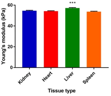
The present thesis describes the mechanical characteristics of four murine tissues (heart, liver, spleen and kidney) following sample preparation using three commonly employed methods (paraffin-embedding, cryosectioning and agarose-embedding). Fixatives used in the paraffin-embedding process are observed to greatly increase the elastic modulus of tissues due to the irreversible cross-linking of the tissue extracellular matrix. Cryosectioning and agarose-embedding, in contrast, provide elasticity values that are more consistent with the live condition of the tissue, but suffer from high tip-sample adhesion that must be compensated through the use of high (~10 μm) measurement distances and/or non-adhesive AFM probes. In addition, agarose-embedded sections are subject to stringent measurement conditions due to the lack of long-term storage options. While cryosectioning is found to be an ideal compromise between data quality and ease of measurement, perfusion with 4% PFA is observed to increase tissue elasticity even under cryosectioning, showing that perfusion is not a recommended step in tissue preparation for AFM.
ii
ÖZET
DOKU YAPILARININ MEKANİK İNCELENMESİ İÇİN ÖRNEK HAZIRLAMA YÖNTEMLERİNİN OPTİMİZASYONU
Alper Devrim Özkan
Malzeme Bilimi ve Nanoteknoloji, Doktora Tez danışmanı: Assist. Prof. Dr. Ayşe Begüm Tekinay
Eş danışman: Assist. Prof. Dr. Aykutlu Dâna Ağustos 2017
Günümüzde hücrelerin davranışlarının genetik ve biyokimyasal mekanizmalar kadar mekanik sinyallerle de kontrol edildiği hakkında detaylı bilimsel kanıt bulunmaktadır ve malzeme analiz yöntemleri biyomedikal alanda hücre-hücre iletişiminin fiziksel yönlerini araştırmak için sıklıkla kullanılmaktadır. Atomik kuvvet mikroskopu (atomic force microscope, AFM) sıvıda ölçüm alabilen bir yüzey karakterizasyon cihazıdır ve son yıllarda proteinler, nükleik asitler, hücreler ve dokular gibi biyomalzemelerin mekanik analizinde önemli bir yöntem olarak ortaya çıkmıştır. Ancak biyolojik malzemelerin doğal olarak heterojen olmaları ve örnek hazırlama yöntemlerinin çeşitliliği nedeniyle, literatürdeki çalışmalar ve araştırma grupları arasında AFM sonuçları büyük farklar göstermektedir. Bu durum ise yapılan çalışmalardan genel sonuçlar çıkarılmasını engellemektedir. Her ne kadar literatürde ölçüm frekansı ve fiksatif kullanım süresi gibi faktörlerin ölçüm kalitesine etkisi detaylandırılmış olsa da, önceki bu çalışmalar genellikle bakteri ve ökaryot hücreleri hakkında yapılmış olup, dokuların ölçümü konusu ise incelenmemiş bulunmaktadır. Dolayısıyla, AFM analizi için kullanılan doku hazırlama yöntemlerinin karşılaştırması, literatürde eksik olan bir konudur.
Bu çalışma dört fare dokusunun (kalp, karaciğer, dalak ve böbrek) üç sık kullanılan örnek hazırlama yönteminden (parafin blok, dondurarak kesme (cryosectioning) ve agaroz blok) sonraki mekanik özelliklerini araştırmaktadır. Parafin blok yönteminde kullanılan fiksatif kimyasalların, dokudaki hücrelerarası matris yapısını cross-link ederek elastik modül değerlerini kayda değer bir miktarda arttırdığı gözlemlenmiştir. Parafin blok yönteminin aksine, dondurarak kesme ve agaroz blok yöntemleri canlı dokuya daha yakın elastikiyet sonuçları vermekte, ancak AFM ucunun örneğe yapışması sebebiyle yüksek ölçüm uzaklıkları (~10 μm) veya yapışmayan AFM uçlarının kullanılmasını gerektirmektedir. Ayrıca, agaroz blok örneklerinde uzun süre saklanama imkanı bulunmadığı için hızlı ölçüm yapılmalıdır. Dondurarak kesme data kalitesi ve ölçüm kolaylığı açısından ideal bir yöntem olarak görülmektedir, ancak %4 PFA ile perfüzyon bu yöntemde bile doku elastikiyetini artırdığı için AFM için perfüze doku kullanımını tavsiye edilmemektedir.
iii
ACKNOWLEDGEMENTS
During these past six years I have toiled hard and often, as many will attest, though not always to further my thesis, as many will attest also. It has been an exceedingly long journey, and I could not have made it without the contributions of friends, family and collagues who have helped me through every step of the way. It is here that I address those who have supported me through thick and thin.
My advisor, Dr. Ayşe Begüm Tekinay, has my endless gratitude for guiding me and my fellow graduate students with competence through these difficult years, and my co-advisor, Dr. Aykutlu Dâna, will always be appreciated for his keen mind and timely advice. I also owe much to Dr. Turgay Tekinay, who first welcomed me to UNAM and fostered many a long-lasting collaboration, Dr. Mustafa Özgür Güler, whose measured feedback has delivered us from countless critical mistakes, and Dr. Emine Deniz Tekin, from whom I have learned much in the way of molecular simulation.
I had the good fortune to share my research environment with many brilliant men and women of science. I would be a much more competent scientist, I think, if I had some of Hakan’s capacity for independent thinking, Rashad’s talent to get his ideas across, Büşra’s careful and methodological way of working, Gözde’s sharp eye and stable hand in surgery, Berna’s ability as a judge of character, Yasin’s coolness in precarious situations, Öncay and Ceren’s willingness to tackle difficult research with little time and resources, Murat’s devotion to his life choices, Mustafa’s sense of justice, Gülistan’s research ethic, Melis and Göksu’s broad-minded understanding of people and the literature alike, Arif and Ruslan’s peerless expertise in all things chemical, Melike’s aptitude to do cutting-edge research while running an entire laboratory on
iv
her own, Elif’s skill at turning wide-ranging data into coherent manuscripts, Fatih and Nurcan’s ability to perform a doctorate’s worth of work in the duration of an MS, Nuray’s commitment to do good science regardless of the required effort, Özge’s flair for handling umpteen different research topics with gusto, Oğuz’s cheerfulness in the face of lengthy and daunting work, Canelif’s frankness, İdil’s perseverance and good humor, and Ahmet Emin’s way of living in the moment. I hope that they, too, will remember this humble biologist and his knack for words – my e-mail address is always open to those who would like a bit of proofreading or some insect trivia, though my mobile phone might not be so!
As for my circle of close friends; Tolga Tarkan Ölmez, Ahmet Emin Topal, Ebuzer Kalyoncu and Ömer Faruk Sarıoğlu, thanks for your companionship for all these years. But don’t think you’ll get kind words like the folks above! I will never forget Tolga’s conviction in upholding what he thinks is right (but is egregiously wrong), Ebuzer’s crafty get-rich-quick schemes, Ahmet Emin’s strange non-sequiturs and Ömer Faruk’s indispensable role as a voice of reason in our unruly little group.
But it is my family that deserves the biggest thanks of all, and I would not have made it anywhere without the endless support of my mother Ayşe and my sister Jülide. You both know that I am no good at expressing sentimentality, but I dearly hope that you understand how much your encouragement has meant to me in difficult times.
Lastly, I would like to thank TÜBITAK for providing me with financial support through the TÜBİTAK BİDEB 2211-C programme, which has made my doctoral studies possible.
v
TABLE OF CONTENTS
TABLE OF CONTENTS ... v
LIST OF FIGURES ... vii
LIST OF TABLES ... ix
CHAPTER 1 ... 1
Introduction ... 1
1.1. The atomic force microscope ... 2
1.1.1. Principle and relevance to biological sciences ... 2
1.1.2. Analysis of tip-sample interactions ... 4
1.2. Use of AFM in biomechanical characterization ... 8
1.2.1. Use of AFM in the characterization of prokaryotic cells and exudates ... 8
1.2.1.1. Bacterial cell surfaces ... 12
1.2.1.2. Bacterial secretions, exudates and biofilms ... 15
1.2.2. Use of AFM in the characterization of eukaryotic cells and tissues ... 17
1.2.2.1. Cancer diagnosis and characterization ... 21
1.2.2.2. Diagnosis of other diseases... 24
1.2.2.3. Stem cell differentiation ... 28
1.2.2.4. Extracellular secretions and tissue microenvironments ... 30
1.3. Importance of mechanical signaling in the context of biological research ... 33
1.3.1. Mechanotransduction pathways ... 33
1.3.1.1. Role of membrane-associated elements ... 36
1.3.1.2. Role of cytoplasmic, nuclear and extracellular matrix elements ... 38
1.3.2. Mechanical organization of cells and tissues ... 40
1.4. The importance of comparative studies in the mechanical characterization of biomaterials ... 42
CHAPTER 2 ... 43
Optimization of sample preparation methods for the AFM analysis of tissues ... 43
2.1. INTRODUCTION ... 43
2.2. MATERIALS & METHODS... 44
vi
2.2.2. Paraffin-embedding and deparaffinization ... 44
2.2.3. Cryosectioning ... 45
2.2.4. Agarose-embedding and vibrotomy ... 45
2.2.5. Atomic force microscopy analyses... 46
2.2.6. Curve-fitting analyses ... 47
2.2.7. Statistical analyses ... 47
2.3. RESULTS AND DISCUSSION ... 48
2.3.1. AFM analysis of deparaffinized mouse tissues ... 48
2.3.2. AFM analysis of cryosectioned mouse tissues ... 53
2.3.3. AFM analysis of agarose-embedded mouse tissues ... 60
2.4. CONCLUSIONS ... 63
CHAPTER 3 ... 68
All-Atom Molecular Dynamics Simulation of the Dependence of Peptide Assembly and Thermal Stability on Counterion Identity ... 68
3.1. INTRODUCTION ... 69
3.2. MATERIALS & METHODS... 73
3.2.1. Simulation details ... 73
3.3. RESULTS ... 76
3.4. DISCUSSION ... 90
3.5. CONCLUSION ... 93
CHAPTER 4 ... 95
Conclusion and future perspectives ... 95
BIBLIOGRAPHY ... 98
APPENDIX A ... 118
vii
LIST OF FIGURES
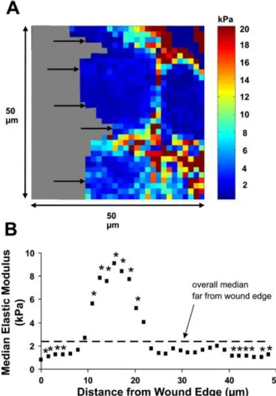
Figure 1.1. Comparison of micro- and nanoindentation for the identification of changes associated with aging in the cartilage of C57BL/6 mice. ... 3 Figure 1.2. Approach and retraction curves showing the binding interaction between HL-60 cells and HUVECs. ... 6 Figure 1.3. AFM topographs and associated force-distance curves of Klebsiella terrigena ATCC33527 cells following three fixation methods. ... 9 Figure 1. 4. The effect of an antimicrobial peptide (CM15) on E. coli cell walls, as observed by high-speed AFM. ... 14 Figure 1. 5. Changes associated with mammary tumor formation, as quantified by AFM. .. 22 Figure 1. 6. Young’s modulus measurements of erythrocytes from young and healthy; old and healthy and old and type-II diabetic individuals. ... 25 Figure 1. 7. Elastic moduli of 16HBE human bronchial epithelial cells, as expressed in relation to the distance to a scratch wound in cell culture, show a transitory increase in stiffness at the wound edge. ... 31 Figure 1. 8. A general overview of mechanotransduction pathways and the associated signaling cascades. ... 34 Figure 2. 1. Comparison of Young’s moduli associated with deparaffinized tissues. ... 48 Figure 2. 2. Representative height, adhesion, elastic modulus maps, appearance under light microscopy, and a sample force-distance curve for deparaffinized heart tissue. ... 49 Figure 2. 3. Young’s modulus distribution associated with deparaffinized heart tissue. ... 49 Figure 2. 4. Representative height, adhesion, elastic modulus maps, general appearance under light microscopy, and a sample force-distance curve for deparaffinized kidney tissue. ... 50 Figure 2. 5. Young’s modulus distribution of deparaffinized kidney tissue. ... 50 Figure 2. 6. Representative height, adhesion, elastic modulus maps, general appearance under light microscopy, and a sample force-distance curve for deparaffinized liver tissue. 51 Figure 2. 7. Young’s modulus distribution associated with deparaffinized liver tissue... 51 Figure 2. 8. Representative height, adhesion, elastic modulus maps, general appearance under light microscopy, and a sample force-distance curve for deparaffinized spleen tissue. ... 52 Figure 2. 9. Young’s modulus distribution associated with deparaffinized spleen tissue. .... 52 Figure 2. 10. Comparison of Young’s moduli associated with cryosectioned tissues. ... 53 Figure 2. 11. Comparison of Young’s moduli associated with cryosectioned tissues under different deflection thresholds. ... 54 Figure 2. 12. Comparison of Young’s moduli associated with cryosectioned tissues of adult and newborn (day 11) mice. ... 55 Figure 2. 13. Comparison of Young’s moduli associated with cryosectioned tissues with and without perfusion. ... 56
viii
Figure 2. 14. Representative height, adhesion, elastic modulus maps, general appearance under light microscopy, and a sample force-distance curve for cryosectioned kidney tissue.
... 57
Figure 2. 15. Young’s modulus distribution associated with cryosectioned kidney tissue. .. 57
Figure 2. 16. Representative height, adhesion, elastic modulus maps, general appearance under light microscopy, and a sample force-distance curve for cryosectioned heart tissue. 58 Figure 2. 17. Young’s modulus distribution associated with cryosectioned heart tissue. .... 58
Figure 2. 18. Representative height, adhesion, elastic modulus maps, general appearance under light microscopy, and a sample force-distance curve for cryosectioned liver tissue. 59 Figure 2. 19. Young’s modulus distribution associated with cryosectioned liver tissue. ... 59
Figure 2. 20. Representative height, adhesion, elastic modulus maps, general appearance under light microscopy, and a force-distance curve for cryosectioned spleen tissue. ... 60
Figure 2. 21. Young’s modulus distribution associated with cryosectioned spleen tissue.... 60
Figure 2. 22. Comparison of Young’s moduli associated with agarose-embedded tissues. . 61 Figure 2. 23. Representative height, adhesion, elastic modulus maps, general appearance under light microscopy, and a sample force-distance curve for agarose-embedded kidney tissue. ... 62
Figure 2. 24. Young’s modulus distribution of agarose-embedded kidney tissue... 62
Figure 2. 25. Representative height, adhesion, elastic modulus maps, general appearance under light microscopy, and a sample force-distance curve for agarose-embedded liver tissue. ... 63
Figure 2. 26. Young’s modulus distribution associated with agarose-embedded liver tissue. ... 63
Figure 2. 27. Comparison of Young’s moduli associated with all tissues under all sample preparation methods tested. ... 65
Figure 3. 1. Chemical structures of lauryl-VVAGERGD at pH 7 and pH 2. ... 75
Figure 3. 2. RMSD graphs of PA1, PA2, and PA3 nanofibers. ... 77
Figure 3. 3. Snapshots of PA1-based nanofibers at 338 K. ... 78
Figure 3. 4. Snapshots of the PA1-based nanofiber at 358 K. ... 79
Figure 3. 5. RMSD graph of simulation I for the PA1 nanofiber. The initial structure was taken from 338 K and heated to 358 K. ... 81
Figure 3. 6. Snapshots of simulation I for the PA1 nanofiber. ... 81
Figure 3. 7. RMSD graph of PA1 nanofiber gradually heated from 300 K to 358 K. ... 82
Figure 3. 8. Snapshots of simulation II for the PA1 nanofiber. ... 83
Figure 3. 9. Extended simulation for the RMSD values and number of H-bonds associated with the gradual heating of the PA1 nanofiber. ... 84
Figure 3. 10. Snapshots of the PA2 nanofiber at 338 K. ... 84
Figure 3. 11. Snapshots of the PA2 nanofiber at 358 K. ... 85
Figure 3. 12. Snapshots of the PA3 nanofiber at 338 K. ... 86
Figure 3. 13. Snapshots of the PA3 nanofiber at 338 K. ... 87
Figure 3. 14. Number of H-bonds formed between PA molecules as a function of simulation time. ... 89
ix
LIST OF TABLES
Table 1. 1. Mechanical characterization of single-celled organisms by AFM ... 10 Table 1. 2. Mechanical characterization of mammalian cells and tissues by AFM... 18 Table 3. 1. Percentage of β-sheet and random coil conformations associated with PA systems under each temperature. ... 88
1
CHAPTER 1
Introduction
This chapter has been modified from the following publications:
Ozkan AD, Topal AE, Dana A, Guler MO, Tekinay AB (2016) Atomic force microscopy for the investigation of molecular and cellular behavior. Micron 89:60-76.
Ozkan AD, Topal AE, Dikecoglu FB, Guler MO, Dana A, Tekinay AB (2017) Atomic force microscopy probe modification methods and their applications in the imaging of biological materials. Seminars in Cell and Developmental Biology (accepted manuscript).
2
1.1. The atomic force microscope
1.1.1. Principle and relevance to biological sciences
At its simplest, atomic force microscopy (AFM) is a characterization method that measures the topology and material properties of surfaces by recording the deflection of a metallic probe (or “tip”) as it moves over the target surface. AFM can be operated under three principal modes: In contact mode, the tip is dragged directly over the surface and deflects away due to repulsive Coulombic interactions, while in non-contact mode it is held at a short (typically <100 nm) distance over the sample and oscillates at a frequency that depends on the attractive van der Waals forces acting upon it. In tapping or intermittent contact mode, the tip is kept oscillating above the sample, and the oscillation frequency changes as the tip approaches the surface at regular intervals1. Contact and intermittent modes are particularly suitable for the
probing of biological samples due to their applicability in liquid media2: Despite considerable losses in resolution, a liquid environment allows cellular imaging in a native environment and, more importantly, permits the direct investigation of mechanical changes on a live cell surface in response to an introduced stimulus3. Time-lapse elastographs taken in this fashion have been utilized for a diverse array of applications, including to visualize the formation of amyloid4 or collagen5 fibers under different environmental conditions, determine how membrane integrity is altered in the presence of antibiotics6, or record the production and dissolution of cytoskeletal elements during cell movement7. In addition, it is possible to utilize the AFM tip as a stimulus to elicit a response from the target cell, and the probe itself can be
3
functionalized with ligand molecules to determine the affinity of the cell membrane to a particular biological moiety.
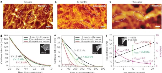
Figure 1.1. Comparison of micro- and nanoindentation for the identification of
changes associated with aging in the cartilage of C57BL/6 mice. Microindentation results could detect no difference between 1, 10 and 19-months old individuals, while nanoindentation was able to determine that the cartilage elasticity of non-arthritic mice changes with age, demonstrating the importance of tip morphology for the acquisition of accurate AFM results. Replicated with permission from Stolz et al.8
Despite this flexibility, cells and tissues are exceptionally susceptible to measurement errors in AFM, as biological materials are far from ideal substrates for this technique. AFM was originally developed for the imaging of nano-to-microscale samples with low surface roughness, while eukaryotic cells on flat surfaces may measure in excess of 10 μm in height and 100 μm in diameter, which is beyond the vertical and horizontal ranges of many commercial systems. In addition, mammalian cells do not survive in the absence of certain environmental conditions, and while the necessary temperature
4
and CO2 ranges can be maintained by specialized attachments for biological imaging,
measurements must still be performed in a growth or differentiation medium that contains a wide range of proteins and growth factors. These biomolecules readily attach to available surfaces and create a corona that may interfere with measurement, while physical contact with the sample may also foul or abrade the probe during contact-mode imaging or force-displacement measurements, especially for complex tissue samples9. Furthermore, probe morphology and measurement conditions have been reported to significantly alter AFM results: Chiou et al., for example, found that effective Young’s moduli of NIH3T3 and 7-4 cells were two-folds higher when measured by sharp tips compared to flat or bead-attached cantilevers10, while Carl and Schillers similarly demonstrated that spherical probes produce significantly lower Young’s modulus values compared to conventional, sharp-tipped AFM probes for the elasticity analysis of Chinese hamster ovary cells, despite exhibiting consistent results over a wide range of probe radii (0.5-26 μm)11. Consequently, despite the ability of AFM to characterize material surfaces with excellent resolution and high repeatability, results associated with cells and tissues should always be considered in the context of their measurement conditions.
1.1.2. Analysis of tip-sample interactions
AFM is not limited to the surface characterization and can be used to determine the elastic modulus of a broad range of biological and non-biological samples. In these measurements, the AFM tip indents the sample until the repulsive forces acting against it exceed a pre-determined threshold, and the strength of these forces, when expressed in relation to the distance by which the tip is forced into the sample, allows the
5
calculation of the sample elastic modulus. In addition to indentation, forces associated with tip retraction are also measured by AFM, and adhesive interactions between the tip and the sample can be quantified by evaluating the difference between tip approach and retraction curves (which corresponds to the work performed by adhesive forces between the two materials). A variety of contact models have been used to describe the relationship between tip forces and the depth of indentation, taking into account factors such as surface roughness, tip morphology and force-induced deformation of the tip and the sample. In the context of AFM imaging, principal among these are the Hertz model, which assumes that both indenting and indented objects are uniform, smooth, non-deforming and linearly elastic12; the Sneddon model, which is a modification of the Hertz model for conical indenters; and the DMT and JKR models, which include additional force terms to account for tip-sample adhesion13. As the JKR model is more suitable for highly deformable surfaces, it is generally preferred for the AFM analysis of biological materials, although other models will also yield comparable elastic moduli under most conditions. It should also be noted that, in addition to model choice, parameters associated with each model (such as spring constants and tip radii) should be known for accurate fitting.
Other types of information can be derived from force-distance curves; for example, the sudden detachment of the sample (or a part thereof) manifests as a sudden “bump” in the retraction curve and can be used for the domain analysis of proteins. Accordingly, the unfolding of each domain of a complex protein is reflected as a single detachment event, and the comparative strength of each domain can be evaluated through the order in which they unfold14. Similarly, the adhesive forces between two
6
biologically affine molecules, such as a receptor and its ligand or an antigen and its corresponding antibody, can be quantified through force-distance curve analysis. Even entire cells can be used as probes to determine the affinity between two distinct cell types, and AFM is frequently employed in this capacity for the analysis of immune cell interactions – Zhang, Wojcikiewicz and Moy, for example, reported that Jurkat cells (T-lymphocytes) formed stronger adhesions to HUVECs with longer interaction times, and used antibody blocking to demonstrate that, following 0.25 seconds of binding, the adhesion molecules E-cadherin, ICAM-1 and VCAM-1 were responsible for 18%, 39% and 41% of the interaction between the two cell types15.
Figure 1.2. Approach and retraction curves showing the binding interaction between
HL-60 cells and HUVECs. Top, middle and bottom curves show the strong interaction between the two cell types, loss of this interaction in the presence of an anti-VCAM-1 antibody, and relatively weak binding of HL-60 to BSA. Replicated with permission from Zhang et al.16
7
In another study, this group also demonstrated that ICAM-1 and VCAM-1 partially mediated the interactions between monocytic human promyelocytic leukemia cells (HL-60 cells) and HUVECs, while β1-integrins played a comparatively stronger role
in attachment and αVβ3-integrins had no significant role in this process16. In addition,
the leukocyte-endothelial cell association was inhibited through the disruption of VLA-4/VCAM-1 binding by the cRGD sequence, which is a well-known motif for cell attachment and presumably competes with adhesive cell membrane proteins for binding sites17. Cellular detachment was associated in both cases with multiple rupture events that correspond to the stretching and/or breaking of receptor-ligand bonds.
Adhesive interactions are also of fundamental importance for tumorigenesis and cancer metastasis, and Yu et al. have found that surface-coated ephrin-A1 (but not soluble ephrin-A1) stimulates collagen I binding in PC3 prostate cancer cells by enhancing the adhesive capacity of β1-integrins, which may be relevant to the metastasis of these cells into collagen I-rich bone tissue18. In addition to mammalian cell lines, the adhesion of bacterial and single-celled eukaryotic cells can also be quantified through cell-functionalized probes; for instance, a single Staphylococcus aureus bacterium can be used to obtain adhesion images by probing the interaction forces between the bacterium and skin corneocytes. Like cancer cells, cellular recognition and binding mechanisms are crucial for the initial invasion, immune detection and phagocytic destruction of bacterial and fungal pathogens, and Mostowy et al. have demonstrated the importance of septins for the cellular entry of Listeria monocytogenes by measuring the force of interaction between invasion proteins on Listeria and Met receptors on HeLa cells in the presence and absence of these
8
proteins19. In a similar vein, El-Kirat-Chatel and Dufrêne have shown that the yeast
Candida albicans adheres strongly to the J774A.1 murine macrophage cell line (with interaction forces reaching ~3000 pN) through mannan/mannose receptor interactions in a time-dependent manner, and suggested that the phagocytosis of the yeast cell by the macrophage is responsible for the gradual increase in adhesive forces20. It is also known that C. albicans can form biofilms alongside the bacterial pathogen Staphylococcus aureus, and this group also used mutant Candida strains to show that bacteria adhere preferentially to fungal hyphae but not the cells themselves, and that the Als protein family and O-mannosyl groups are crucial for the establishment of Candida-Staphylococcus interactions21.
1.2. Use of AFM in biomechanical characterization
1.2.1. Use of AFM in the characterization of prokaryotic cells and
exudates
In addition to quantifying biomolecular interactions involving either purified proteins or whole cells, AFM has received considerable attention for its ability to directly measure the mechanical properties of cells, tissues, membranes, biofilms and other large-scale biological assemblies, which cannot easily be characterized in their native environment by other imaging and mechanical analysis tools. A representative selection of AFM studies on the mechanical characterization of bacteria, yeasts and cellular secretions is provided in Table 1.1. It is readily evident that several means of sample preparation are available for measurement, and that values such as tip-sample adhesion, F-d curve slopes and cellular spring constants can all be used to compare the
9
mechanical integrities of biological samples; consequently, only these studies detailing the full range of measurement conditions were included in the table.
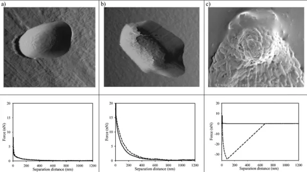
Figure 1.3. AFM topographs and associated force-distance curves of Klebsiella
terrigena ATCC33527 cells following attachment to (a) Isopore polycarbonate membranes, (b) poly-L-lysine-coated glass slides and (c) AFM cantilevers, showing that fixation method greatly alters both the morphology and elastic moduli of bacterial cells. Replicated with permission from Vadillo-Rodríguez et al.28
Both air and liquid imaging have been performed for mechanical investigations; however, biological materials may display large changes in elastic behavior depending on environmental humidity. As such, samples in air tend to have much larger Young’s moduli compared to samples imaged in liquids (e.g. a 10-fold difference was observed in between the elastic moduli of air-dried and rehydrated murine sacculi from Escherichia coli23), while fixation and cell immobilization can likewise have a major
10
impact on elasticity. As previously mentioned, it is usually preferable to compare results within studies rather than assuming a given stiffness value will apply under other experimental conditions, as AFM results of cells and tissues will by necessity reflect inherent differences in measurement techniques and sample preparation methods, as well as the natural variance in the material properties of bacterial cells and their secretions.
Table 1.1. Mechanical characterization of single-celled organisms by AFM.
Sample Tip properties Imaging
conditions Elastic properties Reference
Sulfate-reducing bacteria
Silicon nitride, k = 0.06 N/m
(nominal)
Contact mode, air, sample on mica
(Adhesion) −3.9 to −4.3 nN at cell surface, −5.1 to −5.9 nN at cell-substrate boundary, −6.5 to −6.8 nN at cell-cell boundary 24 Enteroaggregative Escherichia coli, wild-type and dispersin mutant Silicon, k = 2.8 N/m (nominal) Contact mode, liquid (distilled water) and air, sample on
gelatin-treated mica
(F-d slope) 0.133 for wild-type strain on agar, 0.069 for wild-type strain in broth, 0.81 for dispersin mutant on agar, 0.78 for dispersin
mutant in broth 25 Bacillus subtilis, Micrococcus luteus, Pseudomonas putida, two strains
of Escherichia coli Silicon nitride, k = 0.32 N/m (nominal) Contact mode, liquid (HM buffer), sample on APTES coverslip
(Spring constant) varies between 0.16 ± 0.01 to 0.41 ± 0.01, higher in Gram-positive cells
26 Pseudomonas aeruginosa Silicon nitride and silicon nitride with silicon oxide tips, k = 0.07 ± 0.01 (calibrated by the thermal method) Contact mode, liquid (milliQ water), sample on poly-L-lysine-coated glass
(Spring constant) 0.044 ± 0.002 N/m for unfixed, 0.11 ± 0.03 N/m for glutaraldehyde-fixed cells;
creep deformation behavior also investigated
27 Klebsiella terrigena Silicon nitride, k = 0.06 N/m (nominal) Contact mode, liquid (potassium phosphate buffer at pH 6.8), sample on polycarbonate membrane filter/poly-L-lysine-coated glass/immobilized on tip by gluteraldehyde fixation
(Adhesion) −0.26 ± 0.05 nN for membrane filter, −0.5 ± 0.2 for poly-L-lysine, −35 ± 2 nN for gluteraldehyde fixation, other adhesion parameters
also measured
11 Two Streptococcus salivarius strains Silicon nitride, k = 0.03 N/m (nominal) Contact mode, liquid (deionized water or 0.1 M KCl solution), sample on polycarbonate membrane filter
(Adhesion and repulsion) Fibrillated strain shows a larger repulsion range, interpreted to reflect the layer of fibrils; retraction results in three adhesion forces potentially corresponding to three different lengths of fibrils observed by electron microscopy
29 Desulfovibrio desulfuricans, Pseudomonas sp. and an unidentified local marine isolate Silicon nitride, k = 0.12 ± 0.02 N/m (calibrated by the thermal method) Contact mode, liquid (artificial seawater), sample
coated on tip and brought into interaction with
metals
(Adhesion) All three isolates adhere to aluminum better than mild steel, stainless steel 316 and copper; Desulfovibrio and Pseudomonas adhere
better than the marine isolate.
30 B. mycoides Silicon, k = 0.064 N/m and 0.4 N/m Contact mode (constant height), liquid (0.145 M NaCl), sample coated on tip and
brought into interaction with
glass
(Adhesion) 7.4 ± 3.7 nN of adhesion to hydrophilic glass surface, 49.5 ± 14.42 nN of adhesion to hydrophobic-coated glass surface
31 Marine bacterial depositions Silicon, k = 45.7 N/m (calibrated by the added mass
method)
Tapping mode, air, sample deposited on fluoridated and non-fluoridated
polyurethane
(Young's modulus) between 1.5 and 2.2 GPa 32
Bacterial depositions, suspected to be extracellular polymeric substances Silicon nitride, k varies from 0.03 to 0.5 N/m (nominal) Contact mode, liquid (MilliQ water), sample deposited on polystrene
(Adhesion) Forces of 0.8 ± 0.2 nN observed over bare polystyrene, as opposed to 0.2 ± 0.2 nN after
cell attachment 33 P. aeruginosa pili Silicon nitride, 0.008 ± 0.004 N/m Contact mode, liquid (water), sample attached to poly-L-lysine-coated tips and
brought into interaction with
mica surface
(Adhesion) Rupture forces of 95 pN during retraction. 34 E. coli biofilms Silicon nitride, k = 0.07-0.4 N/m
Contact mode, air, sample deposited
on glass
(Adhesion) Pull-off forces of 122.65 pN for cell-tip interaction and 51.79 pN for glass-cell-tip
interaction 35 Bacterial cellulose fibers Silicon nitride k = 1.03 ± 0.05 N/m (calibrated by the thermal method, nominal k of 0.5 N/m not used due to large discrepancy)
Contact mode, air, sample on silicon
nitride-coated silicon grating
12 E. coli and E. coli
spheroplasts Silicon nitride, k = 0.1 and 0.01 N/m (nominal, actual spring constants calibrated by the thermal method) Contact mode, liquid (TBS2 buffer), sample on APTES/Glut mica
(Spring constant) 0.194 N/m for intact cells, 0.571 N/m for fixed spheroplasts
37 Saccharomyces cerevisiae Silicon nitride, k = 0.008 ± 0.4 N/m (calibrated by the thermal method) Contact mode, liquid (milliQ water), sample on polycarbonate membrane filter
(Young's modulus) 6.1 ± 2.4 MPa on bud scar, 0.6 ± 0.4 MPa on surrounding cell wall
38 Aspergillus nidulans Silicon nitride, k = 0.47 ± 0.06 N/m (calibrated by the thermal method) Contact mode, liquid (PBS), sample on poly-L-lysine-coated glass
(Spring constant and Young's modulus) 0.29 ± 0.02 N/m and 110 ± 10 MPa for wild-type hyphae
in complete medium, decreases for mutant strain lacking a chitin synthesis gene, as well as in the
presence of 0.6 M KCl.
39
1.2.1.1.
Bacterial cell surfaces
Unlike many vertebrate cell lines, bacterial cells are not dependent on a highly specific set of environmental conditions to survive, and can tolerate extended AFM imaging sessions without detrimental effects40, 41. Their ease of procurement, non-demanding growth conditions and the fact that many laboratory species either are, or serve as models for, common pathogens make bacteria popular targets for AFM imaging. Bacteria must be immobilized prior to imaging in liquid media, as their mobility otherwise makes it impossible to image. Even sessile bacteria can be laterally pushed by the AFM tip42. Immobilization can be performed by drying and rehydrating, electrostatic binding to a positively charged surface (e.g. gelatin or poly-L-lysine), entrapment in adhesive proteins, covalent binding to amino- or carboxyl-functionalized surfaces or by physical confinement in microwells or porous membranes42, 43. It should be kept in mind that the immobilization method may modify the surface properties of the entrapped cells, e.g. by directly altering the bacterial
13
surface chemistry or triggering a defense mechanism against the environmental stresses associated with the immobilization technique. Due to the small sizes of bacteria, it is also possible to functionalize the AFM tips with bacterial cells, which can then be used to test the interaction between the bacterium and material surfaces. Ting et al., for example, used such tips to show that the Gram-negative bacteria Massilia timonae and Pseudomonas aeruginosa adhere better to stainless steel surfaces than does the Gram-positive Bacillus subtilis44.
AFM studies of bacteria frequently focus on the mechanisms by which certain molecules inhibit the growth of pathogens. Many antibiotics (e.g. beta-lactam antibiotics, polymyxins and glycopeptide antibiotics) act by inhibiting the synthesis of bacterial cell walls or membranes, and thereby alter the membrane integrity of the affected bacteria. Other antibiotic-mediated effects, such as membrane thinning45 or pore formation46, can also be observed by AFM, and the effects of less conventional antibiotics, such as plant extracts47 and peptide sequences48, 49, can also be assessed. The antimicrobial peptide PGLa has been demonstrated to lower the stiffness of Escherichia coli membranes and create micelle-like structures around cell membranes prior to their eventual rupture, while garlic extract was also associated with membrane disruption.
14
Figure 1.4. The effect of an antimicrobial peptide (CM15) on E. coli cell walls, as
observed by high-speed AFM. (a) Disruptions begin to appear on cell surfaces as early as t = 13 s and increase in severity with time. (b, c) Although some bacteria resist the effects of the peptide, these individuals nevertheless react to prolonged treatment (d, at t = ∼30 min). Replicated with permission from Fantner et al.50
Chitosan and chitooligosaccharides were also tested for their antimicrobial effect on E. coli and Staphylococcus aureus, and the thick peptidoglycan wall of S. aureus was found to allow this bacterium to better retain its overall morphology, despite experiencing a significant decrease in cell rigidity51. While the effects of antibacterial
15
molecules may be apparent even under dry imaging, it is also possible to quantify the resulting changes in a time-dependent manner by characterizing the mechanical properties of bacterial cells in liquid before and after introducing the antibiotic in question into the medium6. This technique has the advantage of ensuring that the changes observed are fully due to the antibiotic, as opposed to a combination of it and the drying process, as bacterial cell walls may greatly alter their mechanical properties in response to relative humidity52.
A variety of subcellular elements can also be identified on membrane surfaces using AFM. Both bacterial and eukaryotic cells can be used in these efforts, and lipid bilayers can be substituted as simplified models of cell membranes53, 54. The structure and function of membrane proteins53, 55, 56, pore complexes57, gap junctions58, amyloid
aggregates59 and a variety of membrane-component lipids60, as well as the mode of action of cholera toxin61, were investigated by atomic force microscopy. Of particular interest are the recent developments in AFM-based rapid, high-resolution imaging methods, which have granted substantial insight into the nature of small surface elements. Processes such as the self-assembly of bacterial S-layer proteins62, protein
folding63, misfolding64 and crystallization65 events, the motion of “walking” proteins66, and drug-membrane interactions67 have been the subject of real-time imaging studies.
1.2.1.2.
Bacterial secretions, exudates and biofilms
It is well-known that cells in multicellular organisms enhance their survival and coordination through the production of an extracellular matrix (ECM); however, this property is not unique to higher eukaryotes. Bacteria also secrete proteins,
16
polysaccharides and quorum sensing molecules that relay information between conspecific cells and serve as a buffer against environmental stresses. In addition, the surface attachment of bacteria is also mediated by extracellular depositions, the adhesion capacities of which can be measured through AFM. Colanic acid production and surface lipopolysaccharide lengths, for example, were previously demonstrated to determine the strength of attachment of E. coli cells, as colanic acid-overproducing mutants were found to exhibit stronger attachment while shorter surface lipolysaccharides were associated with a lack of adhesive capacity68.
Bacterial biofilms are also of special interest with regards to AFM characterization. Biofilms are extracellular polysaccharides that are secreted to facilitate the attachment of bacteria (or other unicellular organisms) to a surface, and protect the adhering cells against hostile environmental factors such as antibiotics, detergents and heavy metals69. Biofilms are undesirable elements in many settings, and suitable means to inhibit their formation is necessary to prevent potential health hazards in food, agricultural and medical industries. As such, the mechanical properties of biofilms, as well as the mechanisms by which antifouling molecules act against biofilm production, have been investigated by using AFM and other mechanical characterization techniques70, 71. Corrosion damage and adhesive capacity of biofilms on a variety of metal surfaces have been detailed in the literature: Holden et al. report that unsaturated and liquid-grown biofilms of Pseudomonas putida respond differently to drying, suggesting that the biofilm composition is altered for optimal growth in dry and wet environments72. In another report, Tay et al. detail the effect of silver ions on Staphylococcus epidermidis biofilms and propose a mechanism through which silver
17
destabilizes the biofilm structure by binding to the electron donor groups provided by the biofilm components, thereby weakening the hydrogen bonds that hold the biofilm matrix together73.
1.2.2. Use of AFM in the characterization of eukaryotic cells and
tissues
A selection of AFM studies on the stiffness characterization of eukaryotic cells and tissues is provided in Table 1.2. AFM of mammalian cells and tissues is often undertaken for disease diagnosis or stem cell differentiation studies, as diseased tissues often display mechanical characteristics distinct from their healthy counterparts and stem cells are widely known to alter their differentiation pathways and mechanical characteristics depending on external stimuli. Consequently, AFM of cancer and stem cells can provide greater insight into the pathways required for events such as metastasis and lineage commitment. However, measurement of these samples is more difficult than single-cellular organisms or non-living biological materials such as biofilms and other secretions, as mammalian cells require a specific set of environmental conditions to survive and environmental stress can have a major impact on their mechanical properties. Nonetheless, cell culture conditions can be replicated to some extent within the liquid cell of an AFM, and a large number of successful investigations have been made regarding the mechanical character of living mammalian cells and tissues, either cultured ex vivo or collected immediately prior to imaging.
18
Sample Tip properties Imaging conditions Elastic properties Reference
NIH3T3 fibroblasts Silicon nitride, k = 0.018 N/m (calibrated by thermal method)
Contact mode, liquid (DMEM containing D-glucose (1000 mg/L) and
10% fetal bovine serum, fresh, warmed Ringer's solution used as medium replenishment), sample on
fibronectin-coated glass
(Young's modulus) 4 to 100 kPa over the cell surface, lower around the nucleus than in the
periphery 74 Breast cancer lines (MCF-10A and MCF7) Silicon nitride, k = 0.01 N/m (nominal) modified with a 4.5 μm diameter polystyrene bead
Contact mode, liquid (culture medium), sample
on glass
(Young's modulus) 0.2 to 1.2 kPa range, malignant cell line (MCF7) 1.4 to 1.8 times
softer than benign cell line (MCF-10A)
75
Osteoblasts, mesenchymal stem cells and osteosarcoma cells Silicon nitride, k ~ 20 N/m (calibrated using thermal method)
Contact mode, liquid (25 mM HEPES), fixed cells on polystyrene, glass or collagen-coated glass
(Young's modulus) 0.7 ± 0.1 to 2.6 ± 0.7 kPa range, lower Young's modulus for MG63
osteosarcoma cells on collagen
76 Zonal articular chondrocytes Spherical gold-coated borosilicate bead (5 μm diameter), k ~ 0.065 N/m (calibrated by thermal method)
Contact mode, liquid (DMEM), sample on
poly-L-lysine-coated glass
(Young's modulus) Instantaneous moduli at 0.55 ± 0.23 kPa for superficial, 0.29 ± 0.14 kPa
for middle/deep cells; relaxed moduli at 0.31 ± 0.15 kPa for superficial, 0.17 ± 0.09 kPa for middle/deep cells; apparent viscosities at 1.15 ±
0.66 kPa s for superficial, 0.61 ± 0.69 kPa s for middle/deep cells 77 Cardiac muscle, skeletal muscle and endothelial cells Silicon nitride, k = 0.03 to 0.05 N/m (calibrated using thermal method)
Contact mode, liquid (growth medium), sample
on glass slide
(Young's modulus) Moduli of 100.3±10.7 kPa for cardiac muscle, 24.7±3.5 kPa for skeletal muscle and 1.4±0.1 to 6.8±0.4 kPa depending
on the region tested for epithelial cells
78 Cardiac myocytes Silicon nitride, k = 0.06 N/m (nominal)
Contact mode, liquid (culture medium), sample
on laminin-coated petri dish
(Young's modulus) 35.1 ± 0.7 kPa for cardiomyocytes from 4-months old rats, 42.5 ± 1.0 kPa for cardiomyocytes from 30-months old
rats. 79 LLC-PK1 and MDCK kidney epithelial cell lines Silicon nitride, k = 0.12 N/m (nominal)
Contact mode, liquid (artificial urine)
(Young's modulus) 1.5 ± 0.8 MPa for LLC-PK1 cells, 5 ± 1.5 MPa for MDCK cells, oxalate treatment decreases Young's modulus to 1.2 ±
MPa for LLC-PK1 cells (other stiffness parameters also measured)
80 Neuronal growth cones Silicon nitride, k = 0.006 N/m (nominal)
Contact and dynamic modes, liquid (L15/ASW medium), sample on poly-L-lysine-coated glass
(Young's modulus) 3-7 kPa for the C domain, 7-23 kPa for the T domain, 10-40 kPa for the P
domain
19 Healthy and pathological erythrocytes Silicon nitride, k = 0.03 (nominal) Contact, liquid (PBS), sample on poly-L-lysine-coated glass and fixed by
glutaraldehyde
(Young's modulus) Moduli of 26 ± 7 kPa for healthy erythrocytes, 43 ± 21 kPa for hereditary
spherocytosis, 40 ± 24 kPa for thalassemia and 90 ± 20 kPa for G6PD deficiency samples
82 Liver endothelial cells Silicon nitride, k = 0.032 N/m
Contact, liquid (serum-free endothelial cell
medium), cells on collagen-coated petri dishes with and without glutaraldehyde fixation
(Young's modulus) Moduli of 2 kPa for living cells and over 100 kPa for fixed cells
83 Oral squamous cell carcinoma, normal and malignant lines Silicon nitride tip, k = 0.01 to 0.1 N/m; APTES-modified silicon oxide sphere tip, k ~ 0.5 N/m (calibrated using Sader method)
Contact, air, sample pre-fixed with 2% PFA and fixed with 3.7% PFA
(Young’s modulus) Median values of 6.75 MPa for “normal” and 4.36 MPa for metastatic cancer cells. Elasticity measurements taken
using sphere-modified tips.
84 PC-3 prostate cancer cells Silicon nitride tip, k = 0.012 N/m (calibrated using thermal method)
Contact, liquid (culture medium), samples treated
with anticancer drugs or DMSO control for 24 h
prior to analysis.
(Young’s modulus) c. 3 kPa for untreated cells, increased to c. 6-12 kPa in a dose-dependent manner following drug treatment. Frequency-dependency of the elastic modulus was also tested and found to change significantly for Celebrex, BAY, Totamine, TPA and VPA treatment, but not for DSF, MK and Taxol. This
effect is linked to the fact that the former drugs may alter crosslinking rates of cytoskeletal filaments, while the latter only change fiber
length and thickness.
85 Normal and cancerous bladder epithelium cells Silicon nitride tip, k = 0.011 to 0.018 (calibrated using thermal method)
Contact, liquid (culture medium), sample on glass
(Adhesion energy) average of 8.17 x 10⁻¹⁶ J for normal, 26.95 x 10⁻¹⁶ J for cancer cells (Young’s modulus) average of 27.57 kPa for
normal, 2.46 kPa for cancer cells
86 Porcine articular cartilage Silicon nitride, k = 0.06 N/m (nominal) and borosilicate glass beads with r = 2.5 μm, k = 0.06 and 13 N/m (nominal) Contact, liquid (PBS), tissues on poly-l-lysine-coated glass
(Dynamic elastic modulus) On the order of 2.6 MPa for borosilicate glass beads, about
100-fold lower for sharp tips
87 Mouse articular cartilage of arthritic Col9a1−/− knockout mice Silicon nitride, k = 0.06 N/m (nominal) and microspheres, k = 10 and 12 N/m Contact, liquid (PBS), bulk tissues glued on a round Teflon disk
(Dynamic elastic modulus) 1.3 ± 0.4 MPa for microspheres (no change recorded between ages) and 22.3 ± 1.5 kPa to 50.9 ± 4.7 kPa for
sharp tips (gradual increase with age)
20
Aortic intima of rats
Not listed, tip mounted on a custom platform for in vivo AFM imaging Anaesthetized living animals
(Young’s modulus) 0.4 – 0.5 MPa range for blood vessels without drug influence, raised to
c. 1.0 MPa in the presence of nitroglycerin and
decreased back to c. 0.3 MPa in the presence of norepinephrine 89 Anterior human corneal stroma Phosphorus-doped silicon, k = 25 and 33 N/m (calibrated by an optical method, as described by Sader et al.) Contact, liquid (15% dextran), bulk tissue placed on Teflon cell without attachment
(Young’s modulus) Between 1.14 and 2.63 MPa, consistent across the indentation depths
(between 1.0 and 2.7 μm) 90, 91 Human corneal basement membrane Borosilicate glass, k = 0.06 N/m (nominal) Contact, liquid (PBS), a 3 x 3 mm tissue piece dissected and glued onto a
well on steel disk
(Young’s modulus) 2 to 15 KPa, mean of 7.5 ± 4.2 kPa average for the anterior basement
membrane; 20 to 80 KPa, 50 ± 17.8 kPa average for the Descemet’s membrane
92 Monkey lenses Gold-coated tip, k = 0.01 N/m (nominal), calibrated by the relationship between applied voltage and cantilever deflection Contact, liquid (BSS), lenses dissected and placed on Teflon slide
(Young’s modulus) 1.720 ± 0.88 kPa 93
Human bone Various Various
(Young’s modulus) 16.6 ± 1.1 to 27.1 ± 1.7 for dry adult tibiae (lower for children), 13.4 ± 2.0 to 22.7 ± 3.1 for dry adult vertebrae (lower for wet samples), 16.58 ± 0.32 to 26.6 ± 2.1 for dry
adult femoral midshaft (lower for wet samples and in the femoral neck), other bone measurements and tissue hardnesses also noted
94 Bovine ocular tendon fibers Silicon nitride tip, k = 0.02 N/m (calibrated using thermal method)
Contact, air (sampling chamber kept at 100% humidity), sample glued
on glass petri dish
(Young’s modulus) 60 ± 2.69 MPa for lateral rectus, 59.69 ± 5.34 MPa for inferior rectus, 56.92 ± 1.91 MPa for medial rectus, 59.66 ± 2.64 MPa for superior rectus, 57.7 ± 1.36 MPa
for inferior oblique and 59.15 ± 2.03 for superior oblique tendons. Differences between
tendon elasticities are not statistically significant. 95 Breast tissue sections Borosilicate glass tip, k = 0.06 N/m (nominal), individual tips calibrated using thermal method Contact, liquid (PBS supplied with protease inhibitors and propidium iodide), samples sectioned
by cryomicrotomy
(Young’s modulus) c. 400 Pa in healthy and non-invasive tumor regions, 4-fold increase in
average stiffness in invasive tumor front. Higher average stiffness in the invasive front is
caused by highly stiff (>5 kPa) regions in this area. Aggressive breast cancer subtypes are also
found to exhibit higher Young’s moduli, quantified in terms of upper 10% stiffness.
21 Benign and aggressive prostate tumors Silicon nitride tip, k = 0.06 N/m (nominal), individual tips calibrated using thermal method Contact, liquid (physiological buffer), sample sliced by razor and
glued on glass
(Young’s modulus) 3.03 ± 0.64 kPa for benign, 1.72 7 ± 1.22 for cancerous tissues. The average Young’s modulus for cancer samples with Gleason scores in the 2-7 range was 2.07 ± 1.30, this value was 1.39 ± 0.48 for samples
with Gleason scores in the 8-10 range. In addition, metastatic tumors had an average modulus of 1.06 ± 0.58, while non-metastatic cancer tissue had an average elasticity value of
1.99 ± 1.24. These values reflect the tissue microenvironment and contrast macro-scale elastography results, in which more aggressive
cancers are stiffer.
97
1.2.2.1.
Cancer diagnosis and characterization
While medical advances have led to significant decreases in the incidences of many cancers, cancer still remains to be one of the most important diseases in recent history. It is well-established that cancer cells exhibit marked differences in stiffness and elasticity compared to their healthy counterparts98; however, diagnostic applications of mechanical characterization methods are currently limited. Effective diagnostic techniques must ascertain the presence (or absence) of disease with high confidence and using minimal amounts of sample tissue and time, and despite the ability of mechanical measurements to effectively distinguish between healthy and tumor cells, it is unlikely that a biopsy at an early stage of disease would yield tumor cells in numbers necessary to perform diagnosis based purely on mechanical data. Nonetheless, once the presence of a tumor is confirmed using more conventional methods, AFM may serve as a valuable tool for its characterization: Laidler et al., for example, report the possibility of utilizing AFM to determine whether a suspected breast or prostate tumor is malignant on the basis of its elastic properties99. In addition, AFM cantilevers can be functionalized with antibodies for disease-specific markers and used in the detection of cancer and other disorders100; however, this technique
22
effectively converts the AFM into a biosensor or sorting system instead of relying on its capacity for mechanical characterization.
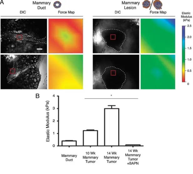
Figure 1.5. Changes associated with mammary tumor formation, as quantified by
AFM. Normal mammary duct tissue is less stiff compared to tumor tissue (a), and the tissue elastic modulus increases with tumor age (b). The inhibition of collagen cross-linking by BAPN prevents the tumor-associated increase in stiffness, suggesting that ECM modifications are involved. Replicated with permission from Lopez et al.101
Greater utility lies in the characterization of tumor cells and their interactions by AFM, which may further the current understanding of cancer biology and allow the design
23
and evaluation of novel cancer drugs. Discrepancies between the elasticities of cancer and non-malignant cells, for example, have been recorded in human lung, breast, pancreas, fibroblast, prostate, adenocarcinoma and other cell lines102, 103. These changes are suspected to increase the mobility of malignant cells during metastasis, and decreases in cell stiffness appear to be progressive, with more malignant cells expressing lower Young’s moduli. Fuhrmann et al. reported that the dysplastic Barrett’s esophagus cell lines are less rigid compared to metaplastic cells, which in turn are softer than their healthy counterparts104. Other changes in cell morphology may also occur to facilitate metastasis, and these too can be quantified by AFM. Sokolov et al., for example, have determined that cancerosus human cervical epithelial cells display two brush layers, in contrast to the single-length molecular brush of non-malignant cervical epithelium, which may also assist in invasion attempts by these malignant cells105. In addition, the tumor microenvironment also appears to contribute significantly to the alterations in tumor elasticity: Instead of the expected decrease in Young’s moduli, Weaver et al. observed a time-dependent increase in tumor stiffness during mammary tumor development in PyMT mice, and this increase in rigidity was also reflected in epithelial cells taken from the tumor site. However, cells taken out of their native environment and grown in vitro had lower Young’s moduli compared to their in vivo counterparts, and the inhibition of the ECM-crosslinking enzyme lysyl oxidase mitigated the gradual increase in the elastic modulus of mammary gland tumors101. As such, tissue culture samples and in vivo tumor cells may not necessarily
agree in elastic properties, as the stiffness of the former depends on the cytoskeletal properties of the cells, while that of the latter is largely mediated by the tissue microenvironment.
24
Drug responses of cancer cells can also be quantified by AFM. Zhang et al. observed that HeLa, HepG2 and C6 cells experience dose-dependent morphological changes on their cell membranes following treatment with colchicine or cytarabine. Surface roughness increased and pores appeared on the cell membrane after drug administration and before MTT-quantifiable decreases in viability could be observed, suggesting that the surface alterations represent an early response to drug presence106.
Such differences may be monitored to evaluate the effectiveness of drug candidates. Changes associated with gene deletion or restoration can also be observed through AFM: Zhou et al. reported that the expression of BRMS1 (restoring breast cancer metastasis suppressor 1) induces increases in cell adhesion capacity, cellular spring constant and Young’s modulus, which supports the idea that BRMS1 expression is associated with cytoskeletal rearrangements that are unfavorable for metastatic activity. Cell morphology and roughness were also altered following BRMS1 expression, possibly as a consequence of cytoskeletal modifications107.
1.2.2.2.
Diagnosis of other diseases
While cancer-associated changes in cell elasticity are particularly drastic, cancer is by no means the only condition to alter the mechanical properties of affected tissues. A variety of other conditions, including malaria, sickle cell anemia, hepatic fibrosis, cardiovascular disease, renal stiffness, muscular dystrophies and bone disorders, have also been associated with notable changes in the elastic properties of the affected tissues108-112. Mechanical diagnosis methods have been devised for some of such conditions, and AFM can be utilized to study the structural and mechanical properties of affected tissues; but it must be noted that, as with cancer, AFM-based diagnostic
25
methods for these diseases currently appear to be more suited towards supporting conventional diagnosis. However, the high-resolution imaging and mechanical probing capacity of AFM is ideal for the determination of the causes underlying the progression of the diseases in question.
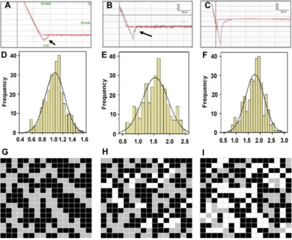
Figure 1.6. Young’s modulus measurements of erythrocytes from young and healthy
(YHP; a, d and g); old and healthy (OHP; b, e and h) and old and type-II diabetic (ODP; c, f and i) individuals. Erythrocytes exhibited higher stiffness in ODP individuals compared to YHP or OHP (1.78 ± 0.39 × 10⁵ N/m2 vs. 1.04 ± 0.19 × 10⁵
N/m2 and 1.53 ± 0.41 × 10⁵ N/m2). In addition, OHP and ODP erythrocytes exhibited higher adhesion compared to YHP erythrocytes (420 ± 25 pN and 510 ± 63 pN v. 200 ± 38 pN, respectively). Replicated with permission from Jin et al.113
26
Several reports on AFM-based disease characterization exist in the literature. Szymoński et al., for example, demonstrate that red blood cells belonging to patients with hereditary blood diseases generally bear a higher Young’s modulus compared to healthy erythrocytes82. In particular, gluteraldehyde-fixed, poly-L-lysine-immobilized erythrocytes of patients with hereditary spherocytosis, thalassemia or G6PD deficiency were stiffer compared to normal cells, while patients with anisocytosis displayed two distinct peaks in their histogram of Young’s moduli, corresponding to healthy and diseased populations of cells. In patients with hereditary spherocytosis, changes in cell morphology were also observed. Similarly, Vatner et al. have investigated the effects of aging on cardiac myocytes by AFM indentation, comparing 4 months- and 30 months-old rats in order to assess whether myocyte health influences the aging-associated diastolic dysfunction of the left ventricule110. Myocytes were found to significantly increase in stiffness with age, suggesting that the observed dysfunction can be linked at least in part to the malfunction of individual myocytes. The effect of aging on cellular Young’s moduli was also noted by Sokolov et al., who have shown that older human epithelial cells are more rigid than their younger counterparts, and have further demonstrated that an increase in the density of cytoskeletal elements is responsible for the age-related increase in stiffness114, 115. The age-related progression of osteoarthritis was also quantified on the articular cartilage of normal and arthritic mice, and nanometer-scale tips (but not microbeads) were shown to be able to resolve the gradual changes in cartilage stiffness88.
Another interesting frontier in AFM-based disease characterization is the investigation of malfunctioning proteins that are involved in the pathogenesis of neurodegenerative
27
disorders such as Alzheimer’s disease, Parkinson’s disease, Huntington’s disease and amyotrophic lateral sclerosis. These disorders severely decrease the quality of life, are exceptionally common among the elderly and have no definitive cures, which renders it crucial to gain further insight into the nature of their causative agents. As such, the formation of amyloid plaques has been investigated in detail using a variety of biological, chemical and material characterization methods, including several AFM-based studies. Lansbury et al., for example, reported on the in vitro formation of metastable Aβ amyloid fibril precursors that may later develop into complete amyloid assemblies, and suggest that the halting of this mechanism may prevent the onset of Alzheimer’s disease by retaining the precursor fibrils (“protofibrils”) in their benign intermediate form4. In addition, Kowalewski and Holtzman demonstrated that the size,
shape and production kinetics of Aβ aggregates were altered depending on the surface on which the aggregation occurs116 (i.e. particulate assemblies were generated on the hydrophilic mica surface, while β-sheets formed on the hydrophobic graphite), and suggested that the β-sheet forming behavior of Aβ on graphite may yield useful information on how protein folding occurs in vivo. The ability of Aβ peptides to form ion-channel like structures without interacting with other membrane components was also confirmed using AFM, with trimeric, tetrameric, pentameric and hexameric pore structures being identified in topographic images. Both D- and L-enantiomers of Aβ peptides were able to form these channels, suggesting that the pore formation mechanism is not stereospecific59.
28
1.2.2.3.
Stem cell differentiation
Fluctuations in mechanical properties do not necessarily suggest aging-related deterioration or a disease state, as healthy cells may also respond to environmental signals by altering their membrane integrity. This is most obviously observed in stem cells, as differentiation induces fundamental changes not only in cell morphology and expression patterns, but also in membrane content and stiffness117-119. Since the
maintenance, recruitment and differentiation of stem cells are intimately linked to the mechanical properties of their immediate environment, it is feasible to use AFM measurements in order to determine the factors that drive the differentiation process in these cells. Such factors are relatively clear in some cases, such as the mesenchymal stem cell differentiation into myogenic, chondrogenic or osteogenic lineages, but the mechanical triggers behind the differentiation of other cells are less understood. In addition to stem cell differentiation, changes in the microenvironmental conditions of tissues can be assessed using AFM or other mechanical characterization methods, especially in situations involving the slow recovery of a damaged system, as in the cases of bone fracture healing. Other processes, such as the local remodeling and ECM secretion of cells in a tissue culture environment, or the capacity of biomimetic materials to imitate the mechanical environment of their tissue model, also fall within the purview of AFM.
The differences between stem and derived cells have also been investigated by AFM. Guilak et al. have confirmed that chondrocytes, osteoblasts and adipocytes, the primary differentiation products of mesenchymal stem cells, display different rigidities, and determined that undifferentiated mesenchymal stem cells are similar to