MOLECULAR MECHANISMS OF ADENOSINE
REGULATION OF HELPER T CELL
RESPONSES
A THESIS SUBMITTED TO
THE GRADUATE SCHOOL OF ENGINEERING AND SCIENCE OF BILKENT UNIVERSITY
IN PARTIAL FULFILLMENT OF THE REQUIREMENTS FOR THE DEGREE OF
MASTER OF SCIENCE IN
MOLECULAR BIOLOGY AND GENETICS
By Altay KOYAŞ
MOLECULAR MECHANISMS OF ADENOSINE REGULATION OF HELPER T CELL RESPONSES
By Altay KOYAŞ May 2017
We certify that we have read this thesis and that in our opinion it is fully adequate, in scope and in quality, as a thesis for the degree of Master of Science.
_____________________________________ Çağlar ÇEKİÇ (Advisor)
_____________________________________ İhsan GÜRSEL
_____________________________________ Güneş ESENDAĞLI
Approved for the Graduate School of Engineering and Science:
____________________________________ Ezhan KARAŞAN
Abstract
MOLECULAR MECHANISMS OF ADENOSINE
REGULATION OF HELPER T CELL RESPONSES
Altay KOYAŞ
M.Sc. in Molecular Biology and Genetics Advisor: Çağlar ÇEKİÇ
May, 2017
Polarizations of helper T cells into different functional subsets is important in order to influence the progression of immune-related diseases. One of the major outcomes in immune cell activation and generation of immunogenic response followed by TCR stimulation, is elevation of extracellular adenosine and upregulation of adenosine A2A
receptors. Adenosine A2A receptor stimulation elevates intracellular cAMP to regulate
helper T cell responses., Intracellular receptors of cAMP, PKA and EPAC proteins regulate cellular responses downstream of cAMP. In this study, we showed that adenosine differentially suppresses Th1 polarization rather than polarization of other functional T cell subsets. Adenosine signaling strongly decreased T cell accumulation in all the polarizing conditions except for Th2 condition. Adenosine-mediated decrease in T cell accumulation is associated with decreased proliferation and survival. PI3K-AKT pathway by targeting the Akt phospho-activation is one of the essential factors for regulation of immune
response. One of the targets for Akt is Foxo1, which is inhibited by Akt phosphorylation. Foxo1 is known to suppress T cell proliferation and important in T cell trafficking and survival. Mechanistic studies have shown that adenosine signaling decreases the phosphorylation of Akt and Foxo1 molecules downstream of TCR. A Foxo1 inhibitor, AS1842856, reverses the reduction in T cell accumulation after adenosine receptor stimulation in particularly Th1 and Th17 conditions by increasing T cell survival in these conditions rather than T cell proliferation. Further studies using PKA and EPAC specific analogs indicated that both pathways may be required for adenosine mediated suppression of Th1 polarization; however, PKA pathway alone is largely responsible from inhibition of T cell proliferation. Results of this study have major implications to understand potential cell-intrinsic effects of one of the major immunoregulatory pathways.
Özet
ADENOZİNİN YARDIMCI T HÜCRESİ
YANITLARININ REGÜLASYONUNDAKİ
MOLEKÜLER MEKANİZMALARI
Altay KOYAŞ
Moleküler Biyoloji ve Genetik, Yüksek Lisans Tez Yöneticisi: Çağlar ÇEKİÇ
Mayıs, 2017
Yardımcı T hücrelerinin, farklı fonksiyonel alt gruplara polarizasyonu, immün kökenli hastalıkların ilerlemesine olan etkileri nedeniyle önem teşkil etmektedir. T hücresi reseptör uyarımını takiben oluşan hücre aktivasyonu ve immünojenik yanıtın ortaya çıkardığı en büyük etkilerden biri, ekstrasellüler adenozin miktarının artışı ve adenozin reseptörü A2A
nın up-regülasyonudur. Adenozin A2A reseptör uyarımı, yardımcı T hücre cevaplarının
düzenlenmesi için hücre içi cAMP miktarını arttırır. cAMP nin hücre içi reseptörleri, PKA ve EPAC proteinleri, cAMP nin alt yolağı ile oluşan hücresel cevapları düzenler. Bu çalışmada, adenozinin, Th1 polarizasyonunu, diğer fonksiyonel T hücresi altgruplarından farklı olarak baskıladığını göstermekteyiz. Adenozin sinyali, Th2 dışındaki bütün polarizasyon koşullarında T hücresi birikimini kuvvetli bir biçimde azaltmaktadır. Adenozin aracılı T hücresi birikimindeki azalış, azalmış proliferasyon ve sağkalım ile ilişkilidir. PI3K-AKT yolağı, Akt fosfo-aktivasyonunu hedefleyerek, immün yanıtın düzenlenmesindeki en temel faktörlerden biridir. Akt nin hedeflerinden biri olan Foxo1, Akt fosforilasyonuyla inhibe edilmektedir. Foxo1’in T hücresi proliferasyonunu baskıladığı ve T hücresi göçü ve sağkalımında önemli olduğu bilinmektedir. Mekanistik çalışmalar göstermektedir ki, adenozin sinyali, TCR alt yolağı olan Akt ve Foxo1
fosforilasyonunu azaltmaktadır. Foxo1 inhibitörü olan AS1842856, adenozin reseptör uyarımı sonrası azalan T hücresi birikimini, özellikle Th1 ve Th17 koşullarında, proliferasyonu etkilemektense T hücresi sağkalımını arttırarak geri çevirmektedir. PKA ve EPAC spesifik analoglar ile yapılan daha ileri çalışmalar, her iki yolağın da Th1 polarizasyonunun baskılanması için gerekli olabileceğini göstermektedir; bununla beraber, T hücresi proliferasyonunun inhibisyonundan ağırlıklı olarak PKA yolağı sorumludur. Bu çalışmanın sonuçları, başlıca immünoregülatör yolaklarının potansiyel hücre içi etkilerinin anlaşılmasında önemli çıkarımlara sahiptir.
Acknowledgement
First, I would like to thank to my supervisor Asst. Prof. Çağlar Çekiç for his support and patience during my MSc. Under his supervision, I was able to improve my academic background as well as technical skills and I become more qualified. None of them would happen without his guidance. I also would like to specially thank to Prof. İhsan Gürsel and Assoc. Prof. Güneş Esendağlı for evaluating my thesis and being in my jury. Their assessments about my thesis are really important in improving myself with their valuable knowledge. Also, I was very lucky to work with Çekiç grup members, special thanks to Ali Can Savaş, Merve Kayhan, İmran Akdemir. Thank you all for your support and friendship during this years. Not just Çekiç members, the rest of Bilkent MBG members were also really helpful during my education. I also want to thank you all, and specifically, thanks to Gürsel group for their help, as well as Aslı Dilber Yıldırım, Seniye Targen, and Başak Özgür for their friendship.
Of course, my friends from Hacettepe University, Dept. of Biology, without their support in my hard times, I couldn’t come to an end. You are very special for me, that is because I thank you Eda Öğüt, Pelin Deniz, Cansu Sevik, Duru Sancar, Ebru Sağ, Gülçin Günal Karataş, Atilla Mehmet Güler, and other members of Biology Dept. Furthermore, I also thank to Emre Ekinci for his support. Last but not least, my dearest family, thank you for everything, especially my siblings, Sümeray Koyaş Şahin and Atılay Koyaş, for their supports and companions.
This project was financially supported by The Scientific and Technological Research Council of Turkey (TUBITAK) (115Z170) together with European Molecular Biology Organization (EMBO) and Turkish Academy of Sciences (Çekiç, TÜBA-GEBİP 2015). I was financially supported by Bilkent University Department of Molecular Biology and Genetics and TUBITAK for my graduate studies.
Table of Contents
Abstract ... iii
Özet ... v
Acknowledgement ... viii
Table of Contents ... ix
List of Figures ... xii
List of Tables ... xiv
Abbreviations ... xv
Chapter 1 ... 1
Introduction ... 1
1.1 THE IMMUNE SYSTEM ... 1
1.1.1 INNATE IMMUNE SYSTEM ... 2
1.1.2 ADAPTIVE IMMUNE SYSTEM ... 2
1.1.2.1 B LYMPHOCYTES ... 3
1.1.2.2 T LYMPHOCYTES ... 5
1.2 PURINERGIC SIGNALING ... 8
1.2.1 ADENOSINE and ADENOSINE RECEPTORS ... 10
1.2.2 ADENOSINE RECEPTOR SIGNALING in IMMUNE SYSTEM ... 14
1.2.2.1 ADENOSINE SIGNALING in T LYMPHOCYTES ... 15
1.3 FOXO SIGNALING ... 16
1.3.1 FOXO1 SIGNALING in T LYMPHOCYTES ... 17
1.4 AIM OF THE STUDY ... 19
Chapter 2 ... 21
Materials ... 21
2.1.1 General Laboratory & Cell Culture Reagents and Materials ... 21
2.1.2 Cytokines & Antibodies & Reagents ... 21
2.1.3 Mice ... 26
2.2 BUFFERS and SOLUTIONS ... 26
2.2.1 Flow Cytometry Buffers ... 26
2.2.2 Western Blot Buffers ... 26
2.2.3 Chemicals for cAMP Assay ... 28
Chapter 3 ... 29
Methods ... 29
3.1 CELL CULTURE EXPERIMENTS ... 29
3.1.1 CD4+ T Cell Isolation & Enrichment ... 29
3.1.2 Carboxyfluorescein Succinimidyl Ester (CFSE) Labeling ... 30
3.1.3 T cell activation for Western Blotting ... 31
3.1.4 CD4+ Th Cell Differentiation ... 31 3.1.4.1 Day:0 ... 31 3.1.4.2 Day: 2 ... 32 3.1.4.3 Day: 4 ... 33 3.2 FLOW CYTOMETRY ... 34 3.3 WESTERN BLOTTING ... 35 3.3.1 Protein Isolation ... 35
3.3.2 Sodium Dodecyl Sulfate Polyacrylamide Gel Electrophoresis (SDS-PAGE) ... 35
3.3.3 Western Blotting ... 35
3.4 cAMP ASSAY ... 36
Chapter 4 ... 37
Results ... 37
4.1 Adenosine Receptor Signaling Preferentially Inhibits Th1 Polarization ... 37
4.2 Adenosine Receptor Signaling inhibits accumulation of Th1, Th17 and Th9 cells but not Th2 cells ... 39
4.3 Reduced T Cell Accumulation after Adenosine Receptor Signaling Correlates with Decreased Proliferation and Survival ... 40
4.4 Adenosine receptor signaling through A2AR causes activation of downstream cAMP signaling. ... 43
4.5 Both PKA and EPAC pathways are important for inhibition of T cell
activation by adenosine receptor signaling ... 45
4.6 Adenosine Receptor Signaling Reduces the Phosphorylation of Akt and Its Downstream Target Foxo1 ... 47
4.7 Foxo1 inhibition reverses decrease in T cell accumulation by adenosine receptor signaling in Th1 and Th17 conditions. ... 48
4.8 Foxo1 does not change anti-proliferative effect of adenosine receptor signaling in T cells ... 49
4.9 Foxo1 inhibition promotes survival of Th1 and Th17 cells in the presence of adenosine receptor signaling ... 50
Chapter 5 ... 52
Discussion ... 52
Bibliography ... 56
APPENDIX ... 69
List of Figures
Figure 1.1: The generation and maturation processes of B lymphocytes. ... 4
Figure 1.2: The interaction between T cell & APC during T cell activation. ... 6 Figure 1.3: CD4+ T cell subsets and their functions in adaptive immunity. ... 7
Figure 1.4: Purinergic signaling mechanism in naïve,effector and regulatory T
lymphocytes. ... 10
Figure 1.5: General mechanism of adenosine signaling. ... 12
Figure 1.6: The interaction between adenosine receptors in cellular mechanism. ... 14
Figure 1.7: General effect of foxo1 signaling in T lymphocytes. ... 18
Figure 4.1: Adenosine receptor stimulation reduces the IFNg production by inhibiting Th1
polarization. ... 39
Figure 4.2: Absolute CD4+ T cell numbers are diminished by adenosine receptor stimulation in CD4+ Th conditions except for Th2 ... 40 Figure 4.3: Adenosine receptor signaling functions as anti-inflammatory in CD4+ T cells
effecting via proliferation level together with survival. ... 41
Figure 4.4: Adenosine signaling activates cAMP signaling and its downstream targets.
... 44
Figure 4.5: Both PKA- and EPAC- dependent pathways are important in regulating the
Adenosine/cAMP signaling during inhibition of T cell activation. ... 45
Figure 4.6: AKT phosphorylation and its downstream target Foxo1 are diminished due to
adenosine receptor signaling. ... 47
Figure 4.7: Diminished T cell accummulation caused by NECA can be reversed by Foxo1
Inhibitor ... 48
Figure 4.8: AS1842856 is failed to reverse the anti-proliferative effect caused by NECA in
CD4+ Th subsets. ... 49
Figure 4.9: Decreased survival in Th1 and Th17 by NECA is reversed in the presence of
AS1842856. ... 51
Appendix Figure 1: Main gating strategy to target CD4+ Th subsets in flow cytometry.
Appendix Figure 2: Gating strategy for Th1 polarization in flow cytometry. ... 70
Appendix Figure 3: Gating strategy for Th2 polarization in flow cytometry. ... 71
Appendix Figure 4: Gating strategy for Th9 polarization in flow cytometry. ... 72
Appendix Figure 5: Gating strategy for Th17 polarization in flow cytometry. ... 73
Appendix Figure 6: Gating strategy for Treg polarization in flow cytometry. ... 74
Appendix Figure 7: The effects of adenosine analog and Foxo1 inhibitor on naïve T cell phenotype………... 74
List of Tables
Table 1.1: The list of the cell culture media components and buffers ... 22
Table 2.1: The list of the antibodies used to generate CD4+ T cell subsets ... 22
Table 2.2: The list of the cytokines used to generate CD4+ T cell subsets ... 23
Table 2.3: The list of enzyme/ligands used to generate CD4+ T cell subsets ... 23
Table 2.4: The list of other reagents used during signaling experiments ... 24
Table 2.5: The list of flow cytometry chemicals and antibodies ... 24
Table 2.6: The list of western blot antibodies and chemicals ... 25
Table 3.1: Reagent cocktails used to generate Th cell subset differentiation (Day:0) ... 32
Table 3.2: The feeding conditions material list (Day:2) ... 33
Abbreviations
Ab Antibody
ABB ADA
Annexin Binding Buffer Adenosine deaminase
ADP Adenosine diphosphate
AMP Adenosine monophosphate
APC Antigen presenting cell
AR Adenosine receptor
ATP Adenosine triphosphate
BCR B cell receptor
BCL2 B cell lymphoma 2
cAMP cyclic Adenosine monophosphate
CD Cluster of differentiation
CFSE Carboxyfluorescein succinimidyl ester
CREB cAMP response element binding protein
CTLA-4 DAMP
Cytotoxic T lymphocyte antigen 4 Damage-associated molecular pattern
DC Dendritic cell
ddH2O Double-distilled water
dH2O Distilled water
FOX Forkhead box
EPAC Exchange protein directly activated by cAMP
ERK Extracellular signal-regulated kinase
HBSS Hank's balanced salt solution
IBD Inflammatory bowel disease
IgA Immunoglobulin A
IgD Immunoglobulin D
IgG Immunoglobulin G
IL Interleukin
iNKT Invariant natural killer T cell
MHC Major histocompatibility complex
MS Multiple sclerosis
NK Natural killer
PAMP Pathogen associated molecular pattern
PD-1 Programmed death 1
PI Propidium Iodide
PI3K PKB
Phosphoinositide 3-kinase
Protein kinase B (also known as Akt)
RPMI Roswell Park Memorial Institute
SA Streptavidin
SCID Severe combined immunodeficiency
Tfh Follicular helper T cells
TGF Transforming growth factor
Th T helper cell
Chapter 1
Introduction
1.1 THE IMMUNE SYSTEM
Immune system is responsible for protecting the host from pathogens and from cancer formation. In general, during this process, by discriminating self from non-self or modified, misplaced self (pathogens, mutated endogenous proteins or intracellular components); a unique, complex network between the members of immunity and other physiological systems takes role to maintain the homeostasis for healthy lifespan. Although there are various types of microorganisms and molecules that might be harmful for the host, the immunogenic response can be divided into two parts depending on their
1.1.1 INNATE IMMUNE SYSTEM
Innate immunity forms the first line of defense mechanism of the host. When the physical/chemical barriers of the host (e.g. epithelia, mucosal surfaces) are breached by the pathogen; a quick, vigorous immunogenic response occurs encoded by germline, without showing any-specificity and without providing long-lasting protection. One of the key aspects of innate immunity, it is developed earlier during evolution and the response is not changed depending on the source of infection, but it is effective against wide-range of pathogens at the same level even after many encounters with the same type of them, which is caused by the absence of memory against the infectious agent. This branch of immune system consists of phagocytes (monocytes, macrophages, neutrophils, dendritic cells), cells responsible for secretion of inflammatory mediators (basophils, eosinophils and mast cells) together with other elements (complement system, acute-phase proteins, cytokines) and NK cells.
In this system, all cells are originated from pluripotent hematopoietic stem cells and a differentiation process occurred through myeloid lineage predominantly. The discrimination between the self and non-self molecules, which is essential for appropriate immunogenic response, relies on the recognition of PAMPs (Pathogen-associated molecular patterns) of pathogens through their PRRs (Pattern Recognition Receptors). PAMPs are not found on the vertebrates but shared commonly by many pathogens and toxins. Any problem in that detection process can cause the initiation of immunogenic response against host’s self-antigens which might result in pathological conditions such as allergy, auto-immune disorders [1-3]. One of functions of the innate immunity is activate cellular adaptive immunity, which is activated as a second line of more specific defense mechanism that is capable of forming immunological memory.
1.1.2 ADAPTIVE IMMUNE SYSTEM
The most characteristic feature of the adaptive immunity is the generation of antigen specific response occurred by the members in that branch of the immune system. Tightly regulated selection mechanisms allow to create lymphocytes that have no immunogenic response against self-molecules of the host but effective to foreign antigens by their specific receptors. In contrast to innate immunity, generation of effector response takes
days rather than hours to develop and provides a long-lasting immunity against subsequent exposures with the same antigen by initiating more rapid and vigorous immune response provided by memory cells. In general, adaptive immunity relies on two fundamental steps; it starts with the recognition/presentation of the antigen, which gives rise to cell activation, differentiation and proliferation of lymphocytes. This step provides the selection of lymphocytes bearing appropriate receptor and multiplying of them under the control of “clonal selection” process, followed by the effector responses either by B lymphocytes via release of antibodies into body fluids and tissues, or by T lymphocytes via orchestrating the immune response and initiating the cytotoxic activity.
All lymphocytes are originated from hematopoietic stem cells within the bone marrow. The maturation process of B cells also occurs within bone marrow, whereas T cells travel to the thymus for this step. After the maturation, they migrate to secondary lymphoid organs of the host and maintain as “naïve” cells. Maturated lymphocytes bear the receptors specific to antigen after the process named as “gene-rearrangement” early in the development which allows for generation of more than 108 antigen receptors and antibody types [4]. Four types of genetic fractions, V (Variable), D (Diversity), J (Joining), and C (Constant), play the key roles for this process. A unique recombination [5] of genes of each within these segments responsible for unique antigen receptor production together with the inaccuracy in splicing and an enzyme “deoxyribonucleotidyltransferase” capable of inserting nucleotides around segments, further mount up the receptor generation [6]. Whole procedure is conducted under control of enzymes produced by RAG (Recombination Activating Genes)1-2. Defects in mechanism of RAG genes lead to severe immunodeficiency caused by the absence of lymphocytes bearing appropriate receptors [7].
1.1.2.1 B LYMPHOCYTES
They are responsible for producing antibodies which are important in toxin-neutralizing, blocking pathogen-adhesion, and opsonizing bacteria for favoring phagocytosis [1]. There are two main sites on antibody molecule structure; constant region determines the function of antibody whereas the variable region is responsible for antigen binding. During
adhesion and signaling system. At slightly later stages on ontogenesis, they undergo a process ended up with the lack of expression of CD5 molecule and form as “B2” cells. IgM and IgD are co-expressed on mature B2 cells before they encounter with an antigen. Within germinal centers, where B cell response occurs, modification processes can be initiated; V gene segment rearrangement allows to “receptor editing” [8] and “class switching” in immunoglobulins, defining as the VDJ unit of antibody amend to different constant region [9] together with the “somatic hypermutation” for adjusting the binding strength; ensures for production of fine-tuned IgA, IgE, or IgD. Before the final response arises, they leave the germinal center as “plasma cells” and initiate antibody secretion within secondary lymphoid tissues [3]. General diagram about B cell development is shown in Figure 1.1
Figure 1.1: The generation and maturation of B cells. [10]
B-cell responses can be initiated by either T-cell dependent or independent manner. In B and T cell interaction, processed antigen is presented to CD4+ T cell, complexed with MHC-II molecule leading to the secretion of cytokines by T cells, necessary for B cell that ended up with B cell division & maturation [1]. On the other hand, repetitive parts of the antigen where the antigen is recognized and bound to, can initiate the short-lived, less specific B cell response without T cells, leading to direct secretion of IgM without class-switching and formation of memory [11].
1.1.2.2 T LYMPHOCYTES
Unlike B cells, T lymphocytes are not capable of recognizing antigen directly, but instead, they are activated after the antigen is presented to them by MHC complex. Antigen is loaded onto MHC complex in two different ways; It might be generated as a result of infected/dysfunctional cells and expressed on the cell surface by MHC I or taken up from the extracellular microenvironment via endocytosis by APCs. All nucleated cells express MHC I complex and capable of presenting the antigen for elimination by CD8+ T cells via cytotoxic effect. Antigen presenting cells are responsible for taking up the antigens, processing into short peptides and transferring them to the secondary lymph nodes where they encounter with naïve T cells and antigen presentation process occurs [1]. Migration of immature T cells from bone marrow to thymus occurs throughout lifespan of the individual [12].
The structure of T cell receptors based on the constant and variable regions similar to B cell receptors, however, they are found as membrane-anchored form. The a and b chains are mostly found among TCR types which recognize the MHC-antigen complex [13, 14] whereas under the 10% of T cells [1] bear g and d chains, which are thought to have functions for mucosal defense [3]. T cells bearing a/b TCRs remain in the thymus and undergo various selection process [15] for appropriate immunogenic response to prevent the self-destruction. The cells which are capable of interacting with MHC complex, but not showing any affinity against self-antigens and MHC complex are specifically selected during this process [16, 17]. Furthermore, in thymic regulation process, many various expressed molecules on the surface of T cells are switched depending on their selection. Two major classes of T cells occur after this procedure; T cells that express CD4+ molecule which can interact with MHC II molecule and respond to orchestrate immunogenic functions by secreting cytokines and taking role in B cell activation and the ones that express CD8+ molecule, which are able to recognize MHC I complexed peptides. CD8+ T cells are effective in cytotoxic activity against intracellular pathogens. They also get help from CD4 T cells for better expansion and effector differentiation after activation. After the activation, some T cells still remain in the lymph nodes as “memory cells”, ready to initiate faster secondary responses against the same pathogen exposures. Discrimination
Naïve T cells express CD45RA whereas CD45RO is found on memory T cells [1, 3]. In activation process, recognition of MHC-antigen complex is not enough for appropriate immune response. The interaction between MHC-antigen complex and CD4 or CD8 molecules together with CD3 only, results in the formation of unresponsive T cells (anergic). Co-stimulatory molecules are also essential for T cell response. Communication between co-stimulatory molecules, e.g. CD80 (B7-1), CD86 (B7-2), CD40 expressed on the APCs and CD28 expressed on the T cell is also essential for activation [1]. A general diagram explaining T cell activation by APCs is shown in Figure 1.2.
.
Figure 1.2: The interaction between T cell and APC during T cell activation. [18]
CD8+ T cells and their cytotoxic innate counterpart, NK cells use similar mechanism to kill the target cells. They bind to cells, creating pores on the targeted cell membrane by insertion of perforins followed by the transmission of granzyme-containing cytoplasmic granules to targeted cell’s cytoplasm through the pores and manipulate the cell’s metabolism by activating caspase enzymes, thereby inducing cell apoptosis for elimination of infection. To activate apoptosis, cytotoxic T cells can also bind to “Fas” molecules on the surface of targeted cells via their Fas ligand “FasL” [1].
CD4+ T cells can be sub-grouped based on cytokine production profile they produce [19]. In early stages of T cell activation, helper (CD4+) T cells defined as “Th0” which mainly responsible for IL-2 production participate in T cell proliferation and stimulation of CD8+ T cells [1]. When the activation signals still continue, they further start to respond these
signals and begin to polarize into different subsets (Th1, Th2, Th17, Th9 and Tfh) relying on the condition at infected zone, responsible for secretion of various cytokines for appropriate immune response [20, 21]. General information about CD4+ T cell subsets is shown in Figure 1.3.
Figure 1.3: CD4+ T cell subsets and their functions in adaptive immunity.[22]
Specific cytokines are responsible for certain differentiation processes; IL-12, as a result of production by NK cells or macrophages enhances Th1 differentiation whereas IL-4 is the key factor in Th2 differentiation process which is secreted by basophils and mast cells as well as Th2 cells themselves. Also, IL-6 and TGF-b together induce the polarization of Th17 whereas TGF-b and IL-4 are responsible in Th9 differentiation. After the polarization process, polarized Th cells begin to express their characteristic proteins to secrete their main cytokines. IL-2 and IFNg production is a result of Th1 polarization leading to contribution to cell-mediated immune response. Th2 cells are capable of secreting various cytokines; IL-4, IL-5, IL-10 and IL-13, effective in humoral response as well as allergic responses. IL-17, IL-21 and IL-22 production is generated by Th17 cells
that take part in elimination of extracellular pathogens. The Th9 cells are responsible for production of IL-9 predominantly [23-25].
Some part of CD4+ cells have a functional role in downregulating immune responses. These cells are called as “Regulatory T cells” (Treg) which show their characteristic forms by expressing forkhead transcription factor (FoxP3) together with CD25. The regulatory activity comes from the secretion of IL-10 and TGF-b [26]. Some of these cells are generated in thymus known as “natural Treg” (nTreg) whereas some Tregs are transformed from naïve CD4+ T cells at the early stage of activation in the presence of IL-10 and TGF-b in periphery, which are called as “induced Tregs” (iTreg). However, CD25 or FoxP3 expression pattern can be slightly different in this phenotype [27].
A subgroup of T lymphocytes named as “invariant NK T cells (iNKT)” provide rapid response against infection in innate immunity. These cells express, invariant ab T cell receptor together with natural killer cell markers (e.g. NK1.1) and responsible in recognizing the lipids instead of peptides coming from either the pathogen or damaged cell itself which is presented by APCs through their MHC-class I related molecule CD1d [28, 29].
1.2 PURINERGIC SIGNALING
Among various bio-molecules, ATP is the fundamental energy currency for living cells. It is a member of purine family and consists of an “adenine” base and “ribose” bound to three ionized phosphate groups [30]. It can be generated as a result of different biochemical processes e.g., glycolysis, TCA or Krebs cycle as well as oxidative phosphorylation [31]. Beside the major function of ATP in energy supply, it can also takes part of regulation in the various physiological processes such as cardiovascular system [32], visceral organs, e.g. gut, bladder, vagina [33, 34]. Together with its degradation products, AMP or adenosine, ATP is also vital for activation of inflammasome, key molecule for the secretion of pro-inflammatory interleukin-1b (IL-1b). Therefore, ATP is considered to be one of the endogenous immunostimulatory damage-associated molecular patterns (DAMPs), which will be discussed later [35]. In general, purinergic receptors can
be branched into three groups based on their pharmacological activities and their structural properties [36].
P2X class has a function as ATP-gated ion channels. They specifically respond to ATP with various ligand affinities [37]. After ligand binding, the response is seen as the influx of extracellular cations, including calcium [38]. Among these types of receptors, P2X7, reveals its ion channel function under the condition of micromolar ATP concentrations whereas at higher ATP levels (milimolar range), P2X7 form pores and cause apoptosis [37].
P2Y subclass of purinergic receptors belong to G-protein coupled receptor group shows affinity to ATP, ADP, UTP, UDP, UDP-Glucose whereas P1 subclass recognizes adenosine [38].
Another important function of ATP after its secretion from damaged tissues, stressed or dying cells as “danger signal” is to initiate the appropriate immunogenic response through its recognition by purinergic receptors [39-41]. Extracellular ATP accumulation is predominantly occurrs through Pannexin 1 channels which are members of purinergic receptors. That hemi-channel type, together with connexins are believed to allow intercellular communications [38]. It is also known that TCR stimulation, as well as osmotic stress is responsible for accumulation of ATP through Pannexin 1 [42-44]. Main feature of ATP in immune response can be detected as dual effects. High concentrations of ATP enhance the activation of immune cells and pro-inflammatory response when secreted acutely by the activated cells and when acting in autocrine/paracrine fashion. Nevertheless, when the generation of extracellular ATP is chronically available at lower levels, it may cause an anti-inflammatory response [41, 45, 46]. These effects which are regulated by P1 and P2 receptors, are confirmed in CD4+ T lymphocytes as well as monocytes, macrophages, DCs and neutrophils. In Figure 1.4. general mechanisms of purinergic signaling in T cell types is shown.
Figure 1.4: Purinergic signaling mechanism in a) Naïve T cells, b) Effector T cells c) Regulatory T cells [47]
1.2.1 ADENOSINE and ADENOSINE RECEPTORS
Adenosine, a nucleoside, member of purine family which is a ligand for P1 receptors has many different roles in physiological systems. It shows various effects in nervous system (such as dementia, sleep, pain, Parkinson’s disease), renal system, cardiovascular diseases, pulmonary disorders, cancer, and inflammation [48]. It is responsible in modulating the responses against tissue-disturbing events to provide tissue protection. After stressful conditions occur, such as hypoxia, ischemia, trauma, inflammation; adenosine functions as a tissue protective molecule [49] by increasing the oxygen supply to the tissue, inducing anti-inflammatory responses and stimulating angiogenesis [50]. The concentration of adenosine is low at extracellular area when there is no stressful environment, however, it can be rapidly elevated to regain homeostasis after pathophysiological conditions [29]. ATP level elevation in the extracellular space as “danger signal” causes initiation of a
hydrolyzing process by ectonucleotidases that converts ATP/ADP to AMP via CD39 (also known as; ectonucleoside triphosphate diphosphohydrolase-1) , and from AMP to adenosine via CD73 (also known as ecto-5’-nucleotidase) [51] to terminate P2 receptor activation and preventing from pro-inflammatory effects of ATP. In addition to that, extracellular adenosine can also be found followed by transportation from intracellular area through bidirectional nucleoside transporters [31].
For termination of adenosine signaling, equilibrative nucleoside transporters (ENTs) and concentrative nucleoside transporters (CNTs) remove extracellular adenosine from the extracellular space [52, 53]. Furthermore, adenosine deaminase (ADA), an enzyme responsible for converting adenosine into inosine, is also effective for terminating adenosine signaling process [54] whereas adenosine kinase is responsible for conversion of adenosine into AMP [55]. Two different isoforms of ADA; ADA and CECR1 (also known as ADA2) which are responsible for enhancing the co-stimulatory signaling during immune response followed by T-cell proliferation and type 1 cytokine production [56, 57] are found on various cell types including lymphocytes [58].
Several studies point out the importance of ADA, CD39 and CD73 in physiological regulation mechanism by adenosine signaling. Polymorphisms in CD39 is responsible for the elevation of ATP/ADP and lowering of Adenosine levels, cause the predisposition to inflammatory bowel disease (IBD) as well as multiple sclerosis (MS), whereas loss-of-function of CD73 in humans causes arterial calcifications [59-62].
Adenosine exerts its effects by binding to four types of receptors; A1, A2A, A2B and A3;
which are members of G-protein coupled receptor family (GPCRs). Adenosine receptor signaling regulates cellular events mainly by changing the intracellular level of cAMP by activating or inhibiting adenylyl cyclase activity [29]. A1, A2A, and A2B are highly
homologous whereas A3 varies among species [63]. A1 and A3 are coupled with Gi, Go,
Gq, and decrease intracellular cAMP level by inhibiting adenylyl cyclase, while A2A and
A2B are coupled with Gs, Gq, and Golf increase intracellular cAMP level following by
activating adenylyl cyclase [64, 65]. The effect of adenosine signaling is determined by the repertoire of receptors expressed on tissue. Adenosine signaling is described briefly in Figure 1.5.
Figure 1.5: General mechanism of adenosine signaling [65]
A1 activation via pertussis-toxin sensitive G protein, leading to adenylyl cyclase inhibition
[66]. Furthermore, release of Ca2+ ions from intracellular stores by phospholipase C (PLC)
activation is another effect of A1 receptor activation. Elevated intracellular Ca2+ level is
capable of activating various enzymes; such as phospholipase A2 (PLA2), phospholipase D
(PLD), protein kinase C (PKC) to generate an appropriate physiological response [36, 48]. Briefly, activation of A1 does not create a consensus response; as a matter of fact, both pro-
and anti-inflammatory responses can be seen following by the activation. These dual effects of A1 are seen depending on the tissue/organ and injury stage/progression together
with the cell type and other signaling mechanisms for optimum physiological response [31]. It enhances pro-inflammatory response in human neutrophils by fomenting chemotaxis and adherence of these cells to epithelium under the condition when adenosine levels at sub-micromolar concentrations [67, 68]. Moreover, A1 antagonism is shown to
have protective effects in lungs [69] while agonists of A1 may provide a protection in renal
effects which come up with upregulation of cyto-protective genes, together with increased heat-shock protein 27 (HSP27) phosphorylation, which has a role in preventing the denaturation and aggregation of cellular proteins [70] [71]. Absence of A1 may causes
pro-inflammatory effect in the lungs, demyelination in central nervous system (CNS), induced level of pro-inflammatory cytokine secretion in sepsis-induced mice [72-74]. According to one study, A1 and A3 are elevating G-CSF production which is important in stimulation of
bone marrow cells, therefore can be used against chemotherapy to reverse its effect by inducing the number of leukocytes after the treatment [75].
A2A and A2B are responsible for elevating the intracellular cAMP level after stimulation of
adenylyl cyclase via GS and Golf and specifically, A2B is capable of activating PLC through
Gq [64]. In A2A signaling, elevated cAMP levels lead to regulation of various pathways
including Ca2+, K+ channels, CREB, PLC, MAPK activation [76, 77]. The structure of these two receptors seem quite related, however, A2B consists of the longest extracellular
loop 2 (ECL2) among all adenosine receptors which has a major role in ligand binding, together with the highest number of cysteine residues found among all GPCRs. The length of ECL2 in A2B, may partially contribute the decreased affinity of that receptor to ligands
compared to A2A [78, 79]. Correspondingly, there are few numbers of A2B agonists which
are useful, e.g NECA (Non-selective adenosine receptor agonist) [80], and highly A2B
selective agonist, BAY60-6583 [81]. In A2A-dependent signaling by agonists pointed out
the several protective effects against myocardial infarction, lung as well as spinal cord [82-84] whereas A2B agonists show their effects in the lungs during hypoxia in relation with
HIF-1 transcriptional induction [85]. Furthermore A2B is essential for IL-4 production in
human mast cells [29]. More importantly, A2A receptor have also major role in the
regulation of immune response together with A2B.
A3 is shown as the most variable receptor type of adenosine among species. It causes
inhibition of adenylyl cyclase and decreasing the intracellular cAMP level, as well as stimulation of PLC and calcium mobilization [48, 86, 87]. Besides its cardioprotective effects through KATP channels, it is also effective in regulation of various cellular
functions, such as cell growth, survival and differentiation [87]. Moreover, an agonist of A3, IB-MECA is shown to inhibit tumor growth [48, 87] and A3 is shown to mediate the
allergic response in mice; e.g histamine release in mast cells [88]. Molecular interactions within adenosine receptors is shown in Figure 1.6.
Figure 1.6: The interaction between adenosine receptors in cellular mechanism [48] 1.2.2 ADENOSINE RECEPTOR SIGNALING in IMMUNE SYSTEM
In general, adenosine dependent regulation of immune system based on the repertoire of adenosine receptor and bio-availability of adenosine. During inflammation, neutrophils are the first recruited class of immune cells. In neutrophil response, adenosine decreases neutrophil activation as well as neutrophil migration and release of pro-inflammatory cytokines. Moreover, expression of adhesion molecules is reduced mainly by A2A signaling
followed by elevated cAMP which leads to PKA activation [89]. On the other hand A3
takes part as well in the affecting neutrophil migration and inhibition of superoxide anion generation [90, 91].
In macrophages, A2A, together with A2B reduce the secretion of pro-inflammatory
cytokines and provide the accumulation of anti-inflammatory cytokine IL-10 [92, 93]. More importantly, adenosine, together with inosine are shown to inhibit M1-macrophage activation, which has pro-inflammatory response, and direct the M2-like phenotype, which favors angiogenesis and tissue protection [94, 95].
Dendritic cells are also essential for immune system. A2A activation on mature DCs,
changes their profiles into anti-inflammatory by reducing TNFa, IL-6, IL-12 production, and inducing the augmentation of IL-10. Moreover, in the presence of adenosine, DCs have limited ability to induce Th1 polarization from naïve CD4+ T cells [96-98]. Stimulation of A2B results in the development of incorrect DCs, functioning as promoting
the tumor growth; [99] that are characterized by both expression of low level DC marker, “CD1a” and CD14, a monocyte marker which is not normally expressed on the surface of DC.
1.2.2.1 ADENOSINE SIGNALING in T LYMPHOCYTES
Among all four types of adenosine receptors, A2A is the predominantly expressed subtype
in T lymphocytes and is induced after TCR stimulation; [100, 101] thereby primary effects of adenosine occur through A2A receptors. In general, adenosine can inhibit T cell
activation, proliferation, secretion of pro-inflammatory cytokines, and cytotoxic activity in an A2A dependent manner [29].
Two downstream molecules of cAMP signaling, PKA and EPAC are responsible for mediating the adenosine signaling response on T cells and most of the effects by cAMP are mediated by PKA [102]. After adenosine binding to A2A, elevation of cAMP level through
Gs followed by the protein kinase A (PKA) activation restricts TCR activation [102].
cAMP mediated signaling is directly targets the cAMP response element binding protein (CREB) and phosphorylates nuclear factor of activated T cells (NF-AT) as well as inhibiting the NF-kB [103].
The adenosine dependent, restricted TCR activity, decreases the generation of various processes essential for appropriate immune response; e.g., inhibition of TCR-mediated pro-inflammatory cytokine secretion, such as IFNg, IL-2, IL-12 and TNFa; CD25 and CD69 expression, and proliferation together with cytotoxic activity by regulating granular exocytosis from both CD4+ and CD8+ T lymphocytes; whereas favor the immunosuppressive molecules, such as cytotoxic T-lymphocyte associated protein 4 (CTLA-4), programmed cell death protein 1 (PD-1), IL-10, TGF-b [101, 104, 105]. In
addition to reduce of Th1 and to lesser extent, Th2 polarization, A2A has also ability to
limit IL-17 generation by blocking the IL-6 production; which has essential role in Th17 differentiation and IL-17 secretion, together with TGF-b. Moreover, this process also induces the FoxP3 expression, resulted in favoring the “inducible” Treg differentiation [106]. CD39 and CD73 are co-expressed on Tregs, and different studies show that, adenosine-mediated immunosuppression can also be generated via CD39/CD73 expressed on Treg cells [107, 108].
Extracellular adenosine also affects the maintenance and survival of naïve T cells. Specifically, downregulation of IL-7Ra (CD127) expression after the TCR stimulation is prevented by AKT phosphorylation as a downstream of PI3K signaling, as a result of PKA-dependent adenosine signaling mechanism [109].
1.3 FOXO SIGNALING
Foxo is a subclass of Fox (Forkhead box) transcription factor family which is named after studying of fkh (Fork head) gene in D. melanogaster by monitoring a spiked-head in adults after the mutation on this gene occurs [110]. They are responsible in regulating the cellular responses and regulated by post-transcriptional mechanisms resulted in the controlling of their cellular localization [111]. Its genetic characterization has studied more specifically via its homologue DAF-16 in C. elegans. Activation of DAF-16 resulted in longer lifespan compared to normal nematodes [112]. Together with other transcriptional factors, they provide appropriate responses under the effect of environmental cues. The “O” subclass of Fox family has 4 members: Foxo1, Foxo3, Foxo4 and Foxo6. One of the most enquired kinase that is essential for regulating the Foxo response is Akt, and its phosphorylation function on Foxo is controlled by PI3K in response to various cell stimuli [111]. Nevertheless, according to studies, Akt phosphorylation of Foxo proteins has no direct effects on their functions, yet, it regulates binding for Foxo proteins onto 14-3-3 proteins [113].
14-3-3 proteins are modulators on various cellular processes. After binding to their targets, 14-3-3 proteins can regulate their stabilization, cellular localization, interaction with other
complexes and their enzymatic activity [114-116]. Phosphorylation of Foxo proteins by Akt direct them to accumulate in the cytoplasm after their inactivation [117].
Foxo proteins are responsible in the regulation of cell cycle progress and apoptosis [118]. It has been shown that, in breast cancer, overexpression of Foxo proteins reduce the tumor progression in vitro and and tumor size in vivo [119, 120]. Cellular localization of them is essential in tumoral response; cytoplasmic localization cause lower survival in breast cancer patients where nuclear localization, under the effect of its response, elevating apoptosis while reducing angiogenesis [119-122]. After finding the importance of this transcription factor family in cancer progression, it was studied among other cancer types, e.g. glioblastoma [123], leukemia [124]; which brings the field of investigation the effect of foxo proteins in immunity.
1.3.1 FOXO1 SIGNALING in T LYMPHOCYTES
Foxo1 proteins are important in maintaining the T cell homeostasis. Among other Foxo members, Foxo1 is upregulated specifically during the T cell maturation [125]. When there is no activation, T cell survival and homeostatic proliferation is predominantly dependent in IL-7 signaling [126]. The physiological effect of IL-7 in T cells generate after binding its receptor which consists of a-chain (IL-7Ra, also known as CD127) and common-g-chain following by the activation of STAT5 transcriptional factor [127]. IL-7R expression is strongly down-regulated after TCR-activation, however it is re-expressed in memory T cells [128].
Akt-mediated phosphorylation as a result of TCR-activation, influence of CD28 co-stimulatory signaling and cytokine influence, initiates the nuclear export Foxo1 for inactivation in T cells whereas in resting conditions, Foxo1 proteins stay in the nucleus [129, 130]. It has been proposed that Foxo1 inactivation is essential for T cells to enter cell proliferation process based on a study in which the Akt-insensitive Foxo1 mutant expression resulted in the suppression of proliferation [122, 131]. Foxo1-deficiency causes the generation of T cells with activated phenotype and increases the differentiation to effector T cells. In addition to importance of Foxo1 in maintaining the naïve T cells, Th1 and Th17 polarization is also reduced under the influence of Foxo1 [128]. Furthermore, the
genes responsible for stimulatory molecules are upregulated in the absence of Foxo1 [132-134]. General information about Foxo1 signaling in T cells is shown in Figure 1.7.
Figure 1.7: General effect of Foxo1 signaling in T cells [111]
There is a balanced mechanism between IL-7R expression and Foxo1 signaling to provide the optimal maintenance and trafficking of T cells. In the absence of Foxo1 or inactivation caused by the TCR triggered AKT phosphorylation following by PI3K signaling, there is a diminished expression of IL-7R; which is also effective in screening the activated phenotype [128, 135]. Thymic exiting and migration of peripheral lymphoid organs, depend on various trafficking molecules; e.g. sphingosine-1-phosphate receptor 1 (S1P1,
Edg1), CCR7, and CD62L [136]. Migration of T cells into secondary lymphoid organs is
reduced due to the low levels of expression of these molecules caused by Krupple-like 2 (KLF2) deficiency, which is a transcription factor selectively expressed on mature T lymphocytes, leading to thymic accumulation of T cells [137]. Foxo1 overexpression resulted in the induced levels of KLF2, whereas absence of Foxo1 leads to reduction of it, correlated with S1P1, CCR7 and CD62L expressions [135, 138]. In addition to the importance of maintenance and tolerance, Foxo1 is also important in Treg differentiation process. There are some binding docks identified for Foxo proteins in FoxP3 promoter region [139], and it has been pointed out that TGF-b responsiveness in CD4+ T cells is under the influence of Foxo1 [140].
1.4 AIM OF THE STUDY
CD4+ T cells have been studied to understand their effects in different pathological conditions because they not only help CD8+ T cells and B cells but also shape the course of inflammatory responses. CD4+ T cells can polarize into different functional subsets to regulate the immunogenic response. Therefore, these cells can be targeted to cure immune related disorders. Before doing so, it is important to identify how these cells are regulated in different microenvironments. It is also important to know by what molecular mechanisms their responses are regulated. Adenosine is one of the important components of different inflammatory conditions. Also by being a GPCR, it functions similarly by other extracellular regulators of inflammation targeting their corresponding GPCRs. One example for such molecules is prostaglandins. Therefore, the main aim of the study was a thorough analysis of the effect of adenosine receptor signaling in helper T cells by using highly enriched naïve CD4+ T cells instead of testing cytokines from supernatants without doing intracellular cytokine staining and measuring true polarization of T cells. [101, 141, 142].
Our second aim was to identify potential molecular mechanisms for adenosine regulation of helper T cells. Because we observed an anti-proliferative and apoptotic effect of adenosine signaling in certain Th polarizing conditions we focused our attention on Foxo1 transcription factors. This is because Foxo1 transcription factors can inhibit survival and proliferation of T cells. TCR activation causes Akt phospho-activation and Foxo1 is inhibited after being phosphorylated by Akt. Our previous studies and others have shown that adenosine can inhibit Akt activation downstream of TCR. To address the effect of Foxo1 inhibition on T cell accumulation in the presence or absence of adenosine receptor stimulation we used a highly selective Foxo1 inhibitor, AS1842856.
Finally, our last aim was to understand which arm of cAMP pathway, namely PKA or EPAC pathway, is more responsible from suppressive effects of adenosine receptor signaling on T cells. To address this question, we used PKA vs. EPAC specific cAMP analogs
To our knowledge this is the first study testing the effects of adenosine receptor signaling in all major polarizing conditions, testing not only cytokine responses but also T cell accumulation. We also first time identified sustained activation of Foxo1 by reduced phosphorylation of Akt by adenosine may particularly suppress T cell survival rather than proliferation. Another novel finding was the combinatorial targeting of both EPAC and PKA pathways was required to phenocopy the effects of adenosine on IFNg production/Th1 differentiation. Results of this study has important implications for mechanistic understanding of how helper T cell responses can be shaped in different cytokine and metabokine milieu.
Chapter 2
Materials
2.1 MATERIALS
2.1.1 General Laboratory & Cell Culture Reagents and Materials
All the 96-well cell culture plates were bought from Greiner Bio One, Austria. All pipettes were bought from Gilson, USA and Rainin, United Kingdom. BD FalconTM 5 ml polystyrene round-bottom tubes were used in cell enrichment procedure. CD4+ enrichment procedure was performed via EasySep MouseTM Cd4+ T cell Enrichment Kit (Cat. No: 19752) and for magnetic sorting of CD4+ cells, EasySepTM Immunomagnetic Cell Separation equipment (Cat. No: 18000) was used. CFSE, a dye for monitoring the cell proliferation was kindly gifted from Thor Lab (Bilkent, Ankara). For flow cytometry, CytoFlex was used from Beckman Coulter. Raw data were analyzed by GraphPad Prism 6. All equipment that used in western blot experiments were bought from Biorad, Hercules,
LANCEâ Ultra cAMP kit (Cat. No: TRF0262, Perkin Elmer) was used. ½ Area 96-well plates for cAMP assay, were bought from Perkin Elmer. The list of general chemicals and buffers during the cell culture experiments can be seen in table 1.1
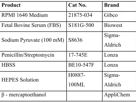
Table 1.1: The list of the cell culture media components and buffers
Product Cat No. Brand
RPMI 1640 Medium 21875-034 Gibco
Fetal Bovine Serum (FBS) S181G-500 Biowest Sodium Pyruvate (100 mM) S8636
Sigma-Aldrich Penicillin/Streptomycin 17-745E Lonza
HBSS BE10-547F Lonza HEPES Solution H0887-100ML Sigma-Aldrich β - mercaptoethanol AppliChem
2.1.2 Cytokines & Antibodies & Reagents
The list of the antibodies, cytokines, enzyme/ligands, and other reagents that had been used during CD4+ cell subset differentiation experiments can be seen in Table 2.1, 2.2, 2.3 and 2.4 respectively.
Table 2.1: The list of the antibodies used to generate CD4+ T cell subsets
Product Cat No. Brand
Goat IgG Fraction to Hamster IgG 55397 MP
LEAF™ Purified Anti-mouse CD3ε 100314 BioLegend
LEAF™ Purified Anti-mouse CD28 102112 BioLegend
LEAF™ Purified Anti-mouse IFN-γ 505812 BioLegend
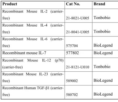
Table 2.2: The list of the cytokines used to generate CD4+ T cell subsets
Product Cat No. Brand
Recombinant Mouse IL-2
(carrier-free) 21-8021-U005 Tonbobio
Recombinant Mouse IL-4
(carrier-free) 21-8041-U005 Tonbobio
Recombinant Mouse IL-6
(carrier-free) 575704 BioLegend
Recombinant mouse IL-7 577802 BioLegend
Recombinant Mouse IL-12 (p70)
(carrier-free) 21-8121-U010 Tonbobio
Recombinant Mouse IL-23
(carrier-free) 589002 BioLegend
Recombinant Human TGF-β1
(carrier-free) 580702 BioLegend
Table 2.3: The list of the enzyme & ligands used during signaling experiments
Product Cat No. Brand
Adenosine Deaminase (ADA) 10102105001 Roche NECA 35920-39-9 Tocris SCH 58261 (A2A Antagonist) 160098-96-4 Tocris 8-pCPT-2-O-Me-cAMP-AM
(cAMP analog-Epac specific) 4853 Tocris
6-Bnz-cAMP sodium salt
(cAMP analog-PKA specific) 5255 Tocris
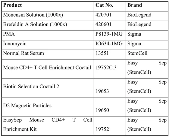
Table 2.4: The list of other reagents used to generate CD4+ T cell subsets
Product Cat No. Brand
Monensin Solution (1000x) 420701 BioLegend
Brefeldin A Solution (1000x) 420601 BioLegend
PMA P8139-1MG Sigma
Ionomycin IO634-1MG Sigma
Normal Rat Serum 13551 StemCell
Mouse CD4+ T Cell Enrichment Coctail 19752C.3 Easy Sep (StemCell)
Biotin Selection Coctail 2
19653 Easy Sep (StemCell) D2 Magnetic Particles 19650 Easy Sep (StemCell) EasySep Mouse CD4+ T Cell
Enrichment Kit 19752
Easy Sep
(StemCell)
All chemicals and antibodies for flow cytometry and western blot experiments can be seen in table 2.5 and 2.6 respectively.
Table 2.5: The list of flow cytometry chemicals & antibodies
Product Cat No. Brand
FoxP3 Transcription Factor Staining Buffer Set
TNB-
0607-KIT Tonbobio
16% Formaldehyde Stock Solution
(Methanol-free) 28908 Thermo Scientific
LIVE/DEAD® Fixable Green Dead
Cell Stain Kit (For 488 nm Excitation) L349780
Life
Technologies
LIVE/DEAD® Fixable Aqua Dead
Cell Stain Kit (For 405 nm Excitation) L34957
Life
Technologies
Saponin 84510 Fluka
CD16/32 (Fc Block) 14-061-85 eBiosicences
BCL-2 PE 633508 BioLegend
CD4 PE 12-0041-83 eBiosciences
CD4 PerCpCy5.5 65-0041-U100 Tonbobio
CD4 PE-Cyanine 7 60-0042-U100 Tonbobio
CD44 Biotin – Rat Monoclonal Atb 10232 StemCell
CD45 Alexa Fluor® 700 103128 BioLegend
CD8a PerCpCy5.5 65-1886-U100 Tonbobio
CD8a APC-Cyanine7 25-0081-U100 Tonbobio
FoxP3 AF647 126407 BioLegend
IFNɣ PE-Cyanine7 60-7311-U100 Tonbobio
IL - 17 APC 506916 BioLegend
IL-4 PE 50-7041-U100 TonboBio
IL - 4 Brilliant Violet 421™ 504127 BioLegend
IL - 9 PE 514104 BioLegend
Streptavidin - PE S866 Invitrogen
Streptavidin - APC 17-4317-82 eBiosciences
Streptavidin – BV510 563261 BD
Table 2.6: The List of Western Blot Antibodies & Chemicals
Product Cat No. Brand
p-Akt (s473) rabbit mAb 9271 Cell Signalling
p-FOXO1 (s256) rabbit Ab 9466 Cell Signalling
p-CREB (Ser133) 9198S Cell Signalling
p-ERK ½ 9461 Cell Signalling
AKT 9272 Cell Signalling
FOXO1 9454 Cell Signalling
CREB 4820S Cell Signalling
β-Actin Antibody (C4) sc-47778 Santa Cruz
Phosphatase Inhibitor 04 906 837 001 Roche
Goat a-rabbit IgG HRP Conjugate 1706515 Biorad
HRP Goat a-mouse IgG 405306 BioLegend
Acrylamide / Bisacrylamide BA004 InroBio
Clarity Western ECL Substrate 1705060 BioRad
TEMED 161-0801 BioRad
BCA Kit 23227 Thermo Fisher
PageRuler™ Plus Prestained Protein Ladder, 10 to 250 kDa
26620 Thermo
Scientific
2.1.3 Mice
All mice that have been used for experiments were generated at Animal Research Facility in Bilkent University, Dept. of Molecular Biology and Genetics. During the experiments, wild-type C57BL/6 (For Th2 differentiation, Balb/c mice was used as well.) 10-15 weeks old mice were used. All ethic permissions were taken and all procedures are done by following the Experimental Animals Regulations.
2.2 Buffers and Solutions 2.2.1 Flow Cytometry Buffers
- FACS Buffer: 2% FBS and 0,05% NaN3 in HBSS
- Fixation Buffer: 4% Formaldehyde in HBSS
- Permeabilization Buffer: 1% FBS + 0.1% NaN3 and 0.1% Saponin in HBSS
2.2.2 Western Blot Buffers
5x Sample Buffer - 10 g SDS - 25 mL b - mercaptoethanol - 50 mL Glycerol - 0.25 g Bromophenol blue - 3.94 g Tris – HCl in 25 ml dH2O (store at -200C)
10x Running Buffer - 10.08 g SDS - 30.3 g Tris - 144 g Glycine in 1L dH2O Transfer Buffer - 6.06 g Tris Base - 28.8 g Glycine in 1.2L dH2O - 400 ml MetOH Adjust to 2L with dH2O
RIPA Buffer (for 10 ml)
- 750 ul 2M NaCl (2M NaCl: 1.16 g NaCl in 10 ml ddH2O)
- 1 ml 10% NP-40 (10% NP-40: 1 ml NP-40 in 9 ml ddH2O)
- 500 ul 10% Sodium DOC (10% Sodium DOC : 1 g Sodium DOC in 10 ml ddH2O)
- 100 ul 10% SDS (10% SDS: 1 g in 10 ml ddH2O) - 250 ul 2M Tris HCl (2M Tris HCl: 3.14 g in 10 ml ddH2O, ph:8) - 500 ul 1M NaF - 100 ul 100mM NaVO4 in 6.5 ml ddH2O. Stripping Buffer - 15 g Glycine - 1 g SDS - 10 ml Tween 20 in 800 ml dH2O (Adjust pH to 2.2) Adjust to 1L with dH2O TBS-T: %0.1 Tween in TBS (1 mL tween in TBS) 5% NFDM: 2.5 g NFDM in 50 mL TBS-T
10% APS: 5g APS in 50 mL dH2O 10% SDS: 5 g SDS in 50 mL dH2O
1 M Tris pH 6.8: 6.057 g Trizma base in 20 mL dH2O, adjust pH to 6.8 then adjust
volume to 50 mL with dH2O
1M Tris pH 8.8: 6.057 g Trizma base in 20 mL dH2O, adjust pH to 8.8 then adjust
volume to 50 mL with dH2O
2.2.3 Chemicals for cAMP Assay
- 0.6M Perchloric acid (2.14 ml 70% aqueous solution into 48 ml dH2O)
Chapter 3
Methods
3.1 Cell Culture Experiments
3.1.1 CD4+ T Cell Isolation & Enrichment
Spleens were isolated from C57BL/6 and Balb/c mice and transferred into HBSS including 10% FBS (v/v) on ice. The organ was mashed and strained through Falcon™ 40 µM Cell Strainer. (Fisher Scientific) (Cat. No: 08-771-1). Centrifugation was performed at 1200 rpm/min for 5 minutes. After the removal of supernatant, cells were resuspended in HBSS with 10% FBS at the appropriate volume according to the number of desired cell numbers for experiments. After the resuspension, cells were transferred into the 5 ml polystyrene round-bottom tubes for CD4+ T cells enrichment, which is necessary for fitting into the magnetic sorter. After the transfer; “Normal Rat Serum” was added to the suspension at 50 ul/ml of the cells (e.g. for 2 ml of cells, 100 µl of cocktail is added) together with the 3 ul of CD44 Biotin antibody; then, “CD4+ T cell Enrichment Cocktail” was added to mix at 50 µl/ml. Suspension was mixed well and incubated in the refrigerator (2-80C) for 15
minutes. “Biotin Selection Cocktail 2” was added after the 15 min of incubation at the level of 100 ul/ml. Cells were mixed well by pipetting again and incubated in the refrigerator (2-80C) for another 15 minutes. For binding of the magnetic particles to non-CD4+ T cells, “D2 Magnetic Particles” was added into the mix at 100 ul/ml. It is important that the magnetic particles have to be vortexed vigorously for at least 30 seconds to
dissolve any aggregates and to optimize the binding affinity. After the addition of magnetic particles, and mixing, cells were incubated in refrigerator (2-80C) for 5 minutes. For
optimum magnetic separation, total volume of cells was brought to 2.5 ml by adding HBSS with 10% FBS and gently mixed by pipetting slowly 2-3 times. Tube was placed into the magnet without the cap and waited for 5 minutes. The magnet and the tube was inverted in one continuous motion into the new 5 ml polystyrene tube to collect only CD4+ cells, and get rid of the particles, other materials in the suspension. Shaking the tube and collecting any remaining drops should not be done. CD4+ Enriched cells were counted by hemocytometer and after the calculation the exact number of cells need for the experiment, the suspension was centrifuged, then resuspended in the condition medium (RPMI 1640, with 10% FBS, 10 mM HEPES and 50 µM b-mercaptoethanol). ADA (Adenosine Deaminase) was added to the media at 1U as final concentration, and cells were preincubated for 30 minutes at room temperature to get rid of the effects of endogenous adenosine and creating a new starting point for the experiments.
3.1.2 Carboxyfluorescein Succinimidyl Ester (CFSE) Labeling
In CFSE Labeling, after the counting of the cells, centrifugation was performed at 1200 rpm for 5 minutes than they were resuspended in 1ml HBSS. On the other hand, CFSE Stock Solution was prepared with HBSS in another falcon tube in 2x concentrations (10 uM) at 1 ml. Two pipettes were used for mixing process, cell suspension and CFSE solution were taken through different p1000 pipettes and put them into another 15 ml falcon tube simultaneously. Falcon was gently inverted 1-2 times and waited for 7 minutes at room temperature. It is important that CFSE is a fluorescent reactive dye, so the environment should be as dark as possible while working with it. At the end of the 7 minutes, rest of the falcon was filled with (10% FBS) HBSS quickly, centrifuged at 1200 rpm for 5 minutes. After resuspension of the cells with condition medium include ADA and preincubating for 30 minutes at room temperature in dark, cells were ready for the differentiation processes. During CFSE experiments, 6 x10⁴ cells/well were used for CFSE experiments and they were not restimulated with PMA and Ionomycin at the end of the differentiation process. After this period, cells were collected and flow cytometry was performed directly.
3.1.3 T cell activation for Western Blotting
Lymph nodes were harvested from Black/6 mice and mashed, followed by filtering through Falcon™ 40 µM cell strainer. Cells were counted and after centrifugation at 1200 rpm for 5 min, they were resuspended with RPMI 1640, containing 10% FBS, 10 mM HEPES and 50 µM b-mercaptoethanol together with 1x ADA at desired volume according to experiments. For western blotting, 3x10⁶ lymphocytes/90 ul were used. Cells were splitted to 1.5 ml Eppendorf tubes in 90 ul and incubated in +40C for 2 hours. After that, NECA (1µM), SCH 58261 (1µM), EPAC (3µM), PKA (30µM) analogs together with 1µg/ml α-CD3e and α-CD28 were added as final concentrations in 10 µl/well from 10x stock solutions. Antagonists were added 10 mins before than agonists and analogs. After 30 min incubation at +40C, hamster IgG was added to tubes as 50 µg/ml final concentration. Later, tubes were incubated at 370C in thermoshaker. After 10 min, cells were moved onto ice to stop the reaction and protein isolation protocol was performed followed by western blot protocol.
3.1.4 CD4+ Th Cell Differentiation 3.1.4.1 Day:0
The 96-well plate was coated with goat IgG fraction to hamster IgG. 50 µM of the IgG was prepared with HBSS and added to wells 50µl at 50µg/ml concentration. Plates were coated with IgG on shaker for 2 hours, at the low-level shaking (≈100-200 rpm/min). At the end of the coating stage, plate was washed with HBSS once, and blocked with RPMI 1640 media for 10-15 min. Later, the media was removed, and the treatments were added to coated wells. Waiting for another 10 – 15 minutes is necessary for the strengthening the interaction between the ligands and antibodies. The cells which preincubated in the medium with ADA were later added to the wells. Before the start of the incubation, plates were centrifuged at 1000 rpm for 5 minutes on low brake. In general, for Th differentiation experiments, 3x10⁴ CD4+ T cells/well were used during the experiments at the 150 µl medium/well. In “Uncoated Groups” cells were plated on well without IgG-coating. Depending on the type of the experiment, medium of uncoated group was included IL-7 (5ng/ml) as well as HEPES (10mM) and b-mercaptoethanol (50 µM). During the experiments, cells stimulated with 1µg/ml α-CD3e and α-CD28 only were used as control groups.
Table 3.1 Reagent cocktails used to generate Th cell subsets differentiation (Day:0)
Th POLARIZATIONS (Final Concentrations)
Th1 Th9 Th17 Th2 Treg
HEPES (10mM) & b-mercaptoethanol (50 µM) α-CD3ε/α-CD28 (1µg/ml) IL-12 (10ng/ml) TGF-β1 (10ng/ml) TGF-β1 (1ng/ml) IL-2 (20ng/ml) TGF-β1 (5ng/ml) α-IL4 (10µg/ml) IL-4 (10ng/ml) IL-6 (50ng/ml) IL-4 (100ng/ml) IL-2 (5ng/ml) α-IFNγ (10µg/ml) IL-23 (5ng/ml) α-IFNγ (10ng/ml) α-IL-4 (10µg/ml) α-IFNγ (5µg/ml) 3.1.4.2 Day: 2
Plates were centrifuged at 1000 rpm for 5 minutes on low brake, removed 120 ul/well of old medium slowly and added 140 ul/well of freshly prepared medium on top of the existing medium included ligands. It is important that the new medium should include HEPES (10mM), b-mercaptoethanol (50 µM) and 1U of ADA. The materials in medium for feeding procedures are listed below.
![Figure 1.1: The generation and maturation of B cells. [10]](https://thumb-eu.123doks.com/thumbv2/9libnet/5885628.121604/20.892.137.721.477.704/figure-generation-maturation-b-cells.webp)
![Figure 1.2: The interaction between T cell and APC during T cell activation. [18]](https://thumb-eu.123doks.com/thumbv2/9libnet/5885628.121604/22.892.284.630.394.711/figure-interaction-t-cell-apc-t-cell-activation.webp)
![Figure 1.3: CD4+ T cell subsets and their functions in adaptive immunity.[22]](https://thumb-eu.123doks.com/thumbv2/9libnet/5885628.121604/23.892.227.692.279.711/figure-cd-t-cell-subsets-functions-adaptive-immunity.webp)
![Figure 1.4: Purinergic signaling mechanism in a) Naïve T cells, b) Effector T cells c) Regulatory T cells [47]](https://thumb-eu.123doks.com/thumbv2/9libnet/5885628.121604/26.892.139.712.118.642/figure-purinergic-signaling-mechanism-naïve-cells-effector-regulatory.webp)
![Figure 1.5: General mechanism of adenosine signaling [65]](https://thumb-eu.123doks.com/thumbv2/9libnet/5885628.121604/28.892.271.652.134.544/figure-general-mechanism-of-adenosine-signaling.webp)
![Figure 1.6: The interaction between adenosine receptors in cellular mechanism [48]](https://thumb-eu.123doks.com/thumbv2/9libnet/5885628.121604/30.892.157.747.119.534/figure-interaction-adenosine-receptors-cellular-mechanism.webp)
![Figure 1.7: General effect of Foxo1 signaling in T cells [111]](https://thumb-eu.123doks.com/thumbv2/9libnet/5885628.121604/34.892.135.746.123.415/figure-general-effect-foxo-signaling-t-cells.webp)