POSTERS
after 2, 6, or 16 hours. Expression of CA markers (iNOS and TNF-a), AA markers (Fizz-1 and TGF-b), and matrix metalloproteinase-9 (MMP-9) were analyzed by real-time PCR. The role of b1-integrins was investigated by culturing in the presence or absence of integrin b1 blocking antibodies.
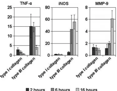
Results: AA macrophages cultured on type III collagen significantly (P < 0.05) upregulated both the AA associated genes Fizz-1 and TGF-b and the CA associated genes TNF-a and iNOS; in contrast, those on type I collagen only reinforced the AA associated genes without effect on CA associated genes. Furthermore, AA macrophages on type III collagen significantly (P < 0.05) upregulated MMP-9, but this did not occur in any of the CA cultures. On type III collagen, TNF-a was maximally upregulated after 2 hours, whereas iNOS and MMP-9 showed the highest upregulation after 16 hours of culture. Inhibition of b1 integrin signalling resulted in downregulation of TNF-a, iNOS and MMP-9, although levels were still raised compared to culture on type I collagen.
Conclusions: Type III collagen results in switching AA macrophages into an intermediate phenotype that has both AA and CA features, but also expresses MMP-9, which is neither a specifically described feature of CA or AA macrophages. The increased type III collagen production generally seen in early fibrosis may provide macrophages with such a phenotype in order to remove cellular and ECM debris. This phenotype is b1-integrin driven.
Figure 1. Regulation of CA genes upon culturing AA macrophages onto type I or type III collagen.
405
TLR3 INDUCED EXPRESSION OF RANTES MODULATES THE FUNCTION OF ACTIVATED PRIMARY HEPATIC STELLATE CELLS B. Wang1,2, J.F. Schlaak1.1University Hospital of Essen, Essen,
Germany;2Tongji Hospital, Wuhan, China
E-mail: [email protected]
Background/aims: Chronic hepatitis C virus (HCV) infection is one of the most common entities leading to liver fibrosis. Recently, RANTES was described as a central mediator of liver fibrogenesis. However, the interaction between TLR3, which is activated by HCV, and RANTES and its role in the activation of HSCs is not well understood.
Methods: Wild-type and TLR3−/− mice were treated with Poly I:C. The expression of RANTES and IFN-b in liver samples was detected by quantitative real time RT-PCR (qrt-PCR). Primary HSCs were isolated from human liver tissue or C57BL/6J wild-type mice. HSCs were stimulated with different TLR1–9 ligands or TLR3 ligands alone for 20 h. RANTES expression was estimated by qrt-PCR, while the production of RANTES was detected by ELISA. Activated HSC
were stimulated with RANTES, supernatants were collected and co-incubated with MH1 cell lines, and the expression of HCV in MH1 cells, as well fibrosis-related genes (TGFb, PDGF, CTGF, RhoA, MMP13) and TLR signaling pathway-related genes (TLR3, MyD88, TIRAP, RIG-I) of HSCs were estimated by qrt-PCR. Functional assays included cell proliferation and migration transwell assays. Results: Only TLR3 stimulation could induce the secretion of RANTES by primary HSC in a time- and dose-dependent manner which could be blocked by in vitro transfection of TLR3 siRNA. TLR3 could stimulate the proliferation and migration of HSC via the RANTES signaling pathway, but had little effect upon their phenotypic transformation, which was associated with the down-regulated expression of TGF-b. There was a weak antiviral effect of RANTES, while the expression of TLR signaling pathway-related genes was induced in a time-dependent manner.
Conclusions: These data show that TLR3/HCV is a potent inductor of RANTES in HSC which leads to up-regulation of fibrosis-related genes. Therapies that are aimed at suppression of RANTES expression may provide a useful strategy to inhibit HCV-induced liver fibrosis when HCV eradication can not be achieved.
04d. MOLECULAR AND CELLULAR
BIOLOGY: LIVER REGENERATION
406
NANOPARTICLE LABELING: A NEW ERA IN VIVO TRACING THE CELLS IN LIVER STUDIES
E. Akhan1, M.M. Aydin2, V. Ibrahimova3, E. Bugdayci1, D. Tuncel3,
K.C. Akcali2.1Bilkent University,2Molecular Biology and Genetics, 3Chemistry, Bilkent University, Ankara, Turkey
E-mail: [email protected]
Background and Aim: Tracking molecules in vivo has always been challenging and there is increasing demand for tools that allow researchers to trace especially during cell-based therapies. Conjugated polymer based water-dispersible nanoparticles (NP) represent a new class of probes for cell imaging and tracking because they offer high brightness, improved photostability, high fluorescent quantum yield and non-cytotoxicity comparing to conventional dyes. We aimed to develop a novel approach by using fluorescein emitting NP to label Mesenchymal Stem Cells (MSCs) and HUH7 cell lines and assess their fate in vivo in liver regeneration, liver fibrosis and xenografts.
Methods: Copolymer of fluorene-benzothiodiazole derivatives containing cross-linkable allyl groups attached to fluorene monomer has been converted into water-dispersible green-light-emitting NPs using reprecipitation method. Liver regeneration model was achieved by partial hepatectomy in Sprague-Dawley rats. Liver fibrosis was induced by treating C57BL/6 mice with CCl4 for 4 weeks. MSCs were labeled with NP and injected from tail vein of host animal and their fate were examined. Liver xenograft model was generated by injecting NP-labeled HUH7 cell line into the CD1 nude mouse. Viability of MSCs and HUH7 cells after NP treatment was measured by MTT assay. NP-labeled MSCs and HUH7 cells were examined on tissue sections under fluorescein microscopy. Results: We showed that NP labeled MSCs have ability to migrate to the site of injury in liver regeneration and fibrosis as evidenced by the presence of intense fluorescein staining in the liver suggesting NP labeling did not interfere with the physiology of MSC. We did not observe any staining in the absence of tissue injury or when non-labeled MSCs were injected. After NP treatment, significant number of cells, 68% of the labeled cells, were found viable as
POSTERS
revealed by MTT. In vivo xenograft results showed the presence ofNP-labeled HUH7 cells as long as 8 weeks after the injection. Conclusions: The utilization of NP labeling is a promising tool for the tracking of cells in vivo and could have many applications. This approach clearly can be used in elucidating the molecular mechanisms of many physiological and pathological conditions of liver as well as cell-based therapies.
407
ANTI-b1-INTEGRIN ANTIBODIES IMPROVE SURVIVAL OF ISOLATED HUMAN HEPATOCYTES SIGNIFICANTLY INCREASING ADHESION TO HEPATIC SINUSOIDAL ENDOTHELIUM UNDER FLOW AND ENGRAFTMENT IN MURINE LIVER FOLLOWING TRANSPLANTATION
D.C. Bartlett1,2, V.S. Aldridge1, A. Wilhelm1, N. Davies1, J. Youster1,
D.H. Adams1,2, P.N. Newsome1,2.1NIHR Biomedical Unit and Centre
for Liver Research, University of Birmingham,2Liver Unit, Queen
Elizabeth Hospital Birmingham, Birmingham, UK E-mail: [email protected]
Background: Hepatocyte transplantation offers a potential alternative to orthotopic liver transplantation but is limited by poor survival of transplanted cells. This may be partly due to apoptosis of isolated hepatocytes following their detachment from extracellular matrix with loss of b1-integrin-mediated survival signals. Anti-b1-integrin antibodies have been shown to reduce apoptosis of rat hepatocytes and improve their survival in allogeneic transplantation. The purpose of this study was to determine the effect of b1-integrin blocking antibodies on the survival and initial engraftment of transplanted human hepatocytes.
Methods: Primary human hepatocytes were isolated from liver tissue obtained with ethical approval from Queen Elizabeth Hospital Birmingham. Integrin surface expression was confirmed using flow cytometry. Cells were incubated in suspension with anti-b1-integrin blocking antibodies or isotype matched control for 1 hour. Viability and caspase 3 activity were assessed using flow cytometry and ELISA respectively. A modified flow adhesion assay was used to investigate the resistance to flow of cells adherent to sinusoidal endothelium (HSEC). An FC blocking agent was used to exclude the possibility of treated cells binding to HSEC via antibody-FC receptor interactions. 1×10⁶ fluorescently labelled cells were
injected into C57BL/6 mice via the portal vein under general anaesthesia and the mice culled after 15 minutes. The livers were immediately frozen and sectioned and the number of fluorescent cells per field of view counted.
Results: Mean surface expression of the b1-integrin subunit on human hepatocytes was 86.8% (MFI 46.8). Hepatocytes treated with b1-integrin antibodies showed increased viability (85.4% vs. 79.0% p = 0.02) and reduced caspase 3 activity. b1-integrin blockade significantly increased the mean percentage of cells remaining adherent to HSEC under flow compared to IgG control (30.6% vs. 12.7%, p = 0.03) and significantly increased the number of cells per field of view in the livers of mice following transplantation (1.6 vs. 0.7, p = 0.017).
Conclusion: b1-integrin blocking antibodies increase the survival of isolated hepatocytes and improve their ability to remain adherent to HSEC under flow resulting in increased initial engraftment of transplanted human hepatocytes in the mouse liver. The use of b1-integrin blocking antibodies may provide a means to increase the efficacy of human hepatocyte transplantation.
408
CD133+ STEM CELLS FOR THE TREATMENT OF END STAGE LIVER DISEASE
L. Brodosi1, L. Catani2, S. Lorenzini1, R. Giordano3, V. Giudice4,
D. Sollazzo2, S. Gitto1, M. Baccarani2, M. Bernardi1, R.M. Lemoli2,
P. Andreone1. 1Clinical Medicine,2Departement of Hematology,
University of Bologna, Bologna,3Departement of Regenerative
Medicine, Fondazione Ospedale Maggiore, Mangiagalli e Regina Elena, University of Milan, Milan,4Immunohematology Service and Blood
Bank, University of Bologna, Bologna, Italy E-mail: [email protected]
Previous studies showed that bone marrow (BM) cells contribute to liver regeneration after tissue injury. The main objective of the present study is to evaluate the feasibility and the safety of the purification and intrahepatic reinfusion of increasing numbers of autologous BM-derived G-CSF-mobilized CD133+ stem cells (SCs) in patients with end-stage liver disease (ESLD). For this purpose, G-CSF at 7.5 mg/Kg/b.i.d. is administered subcutaneously (sc) from day 1 until the completion of peripheral blood stem cells (PBSC) collection. Collection of PBSC begin on day + 5 only if the concentration of CD133+ cells is >8/mL. CliniMacs device is used for the positive selection of CD133+ SCs (under GMP conditions) from PB of mobilized standard-volume leukapheresis. At least 4 weeks after SC mobilization, collection and cryopreservation, highly purified autologous G-CSF-mobilized CD133+ cells are re-infused through the hepatic artery by transfemoral or transbrachial arteriography. CD133+ cells are administered to patients starting from 5×10⁴/Kg patient’s body weight and increased every 3 patients
up to 1×10⁶/kg. G-CSF at 5 mg/Kg/day is administered sc for
3 days after the reinfusion of SCs for their expansion and to induce a selective proliferative advantage of reinfused cells in vivo. Biological assays (phenotype of circulating SCs, clonogenic assays, serum cytokines) were done during the mobilization and re-infusion phases together with the phenotypic characterization of the isolated CD133+ SCs. The clinical trial is ongoing. Up to date, 9 patients have been successfully mobilized with G-CSF and highly purified autologous CD133+ SCs have been re-infused in 7 cases. Based on preliminary data, we suggest the feasibility and safety of intrahepatic reinfusion of highly purified CD133+ stem cells in patients with ESLD. Biological studies show that:
1. circulating hematopoietic and endothelial progenitors are increased after G-CSF treatment;
2. highly purified CD133+ cells express hematopoietic and endothelial markers;
3. serum concentration of HGF, SDF-1, VEGF and MMP9 and clonogenic capability of hematopoietic progenitors are increased during the mobilization and re-infusion phases;
4. clonogenic potential of endothelial progenitors shows variable expression.
409
IMPLICATION OF NADPH OXIDASES (NOXES) IN LIVER REGENERATION
E. Crosas-Molist1, A. S ´anchez2, M. Fern ´andez2, P. Sancho1,
E. Bertran1, I. Fabregat1,3.1Bellvitge Biomedical Research Institute
(IDIBELL), L’Hospitalet de Llobregat,2Dep. of Biochemistry and
Molecular Biology II, School of Pharmacy, Complutense University, Madrid,3Dep. of Physiological Sciences II, School of Medicine,
University of Barcelona, Barcelona, Spain E-mail: [email protected]
Background/aims: Reactive oxygen species (ROS) function as signaling molecules at physiological concentrations mediating numerous biological processes. The NADPH oxidase (NOX) family main function is the production of ROS. Previous results from our group have demonstrated that different members of the NOX family