i
NOVEL MONOCLONAL ANTIBODIES TARGETING
CONFORMATIONAL ERBB2 EPITOPES
A THESIS SUBMITTED TO
THE DEPARTMENT OF MOLECULAR BIOLOGY AND GENETICS AND THE GRADUATE SCHOOL OF ENGINEERING AND SCIENCE
OF BILKENT UNIVERSITY
IN PARTIAL FULFILLMENT OF THE REQUIREMENTS FOR THE DEGREE OF
DOCTOR OF PHILOSOPHY
OVER PAGE
1.1COVER PAGE
BY
CEYHAN CERAN
OCTOBER 2012
ii 1.2 DEDICATION PAGE
To my loving, understanding, caring, self-sacrificing,
smart, wise, imaginative, helpful and inspiring mother, Reyhan
iii 1.3 SIGNATURE PAGE
I certify that I have read this thesis and that in my opinion it is fully adequate, in scope and in quality, as a thesis for the degree of Doctor of Philosophy.
____________________ Prof. Dr. Mehmet Öztürk
I certify that I have read this thesis and that in my opinion it is fully adequate, in scope, and in quality, as a thesis for the degree of Doctor of Philosophy.
____________________ Prof. Dr. Tamer Yağcı I certify that I have read this thesis and that in my opinion it is fully adequate, in scope, and in quality, as a thesis for the degree of Doctor of Philosophy.
____________________ Assoc. Prof. Dr. Işık Yuluğ
I certify that I have read this thesis and that in my opinion it is fully adequate, in scope, and in quality, as a thesis for the degree of Doctor of Philosophy.
____________________ Assoc. Prof. Dr. İhsan Gürsel
I certify that I have read this thesis and that in my opinion it is fully adequate, in scope, and in quality, as a thesis for the degree of Doctor of Philosophy.
____________________ Assist. Prof. Dr. Elif Erson Bensan
Approved for the Graduate School of Engineering and Science
____________________ Prof. Dr. Levent Onural Director of the Graduate School of Engineering and Science
iv
ABSTRACT
NOVEL MONOCLONAL ANTIBODIES TARGETING
CONFORMATIONAL ERBB2 EPITOPES
1.4ABSTRACT
Ceyhan Ceran
Ph.D. in Molecular Biology and Genetics Supervisor: Prof. Dr. Mehmet Öztürk
October 2012, 233 pages
ERBB2 is a tyrosine kinase receptor which can act as homodimers or heterodimers with other members of the ERBB family. Nearly 30% of breast cancers overexpress ERBB2, which can be effectively targeted by anti-ERBB2 monoclonal antibodies. Trastuzumab directed against an epitope on subdomain IV of the extracellular domain (ECD) of ERBB2 is a clinically used therapeutics but the response rate is poor and acquired resistance is frequent. Pertuzumab that binds to subdomain II and inhibits receptor dimerization is another promising therapeutics under clinical trials. Anti-ERBB2 antibodies directed to novel epitopes are potentially useful tools for replacement and combinatorial therapies. We produced five new anti-ERBB2 antibodies, all directed against epitope(s) present only on the native ECD. They performed selective growth inhibitory effects depending on the level of ERBB2 expression and cellular background. When used alone, novel anti-ERBB2 antibodies displayed modest but significant growth inhibition on SK-BR-3, BT-474 and MDA-MB-361 cells with ERBB2 overexpression; while no detectable inhibition was observed on MCF-7 and T47D cells lacking ERBB2 amplification. When the antibodies were tested in combination with TNF-α, they acted synergistically on SK-BR-3 cells, producing upto 80% growth inhibition; but performed antagonistically on BT-474 cells. Detailed investigation of a representative antibody indicated G1-arrest as the main mechanism of the anti-proliferative effects exerted on SK-BR-3 cells. Antibody treatment induced permanent inhibition of DNA synthesis, leading to accumulation of cells at G1-phase; an effect which was accelerated in the presence of TNF-α. In addition, treated SK-BR-3 cells displayed inhibition of Akt and ERK1/2 phosphorylation leading to cyclin D1 accumulation and growth arrest, independently from TNF-α. Novel antibodies against conformational epitopes present on the extracellular domain of ERBB2 receptor may serve as new analytical and diagnostic tools, in addition to being potent anti-cancer bioactive molecules. Cell-dependent synergy and antagonism between anti-ERBB2 antibodies and TNF-α provide evidence for a complex interplay between ERBB2 and TNF-α signaling pathways. Such complexity may drastically affect the outcome of ERBB2-directed therapeutic interventions.
v
ÖZET
KONFORMASYONAL ERBB2 EPİTOPLARINI HEDEF ALAN
ÖZGÜN MONOKLONAL ANTİKORLAR
1.5 ÖZET
Ceyhan Ceran
Moleküler Biyoloji ve Genetik Doktora Tezi Tez Yöneticisi: Prof. Dr. Mehmet Öztürk
Ekim 2012, 233 sayfa
Bir tirozin kinaz almacı olan ERBB2, gerek homodimer yapıda, gerekse ERBB ailesinin diğer üyeleriyle heterodimer oluşturarak etki gösterir. Meme kanserlerinin % 30’unu oluşturan ve ERBB2 almaç ifadesi yüksek olan vakalar, hedefe yönelik tedavi kapsamında anti-ERBB2 antikorlarıyla başarılı bir şekilde tedavi edilebilmektedir. ERBB2’nin hücredışı bölgesindeki IV. alt-bölgeye karşı geliştirilmiş Trastuzumab klinikte kullanılan bir terapötik olmasına karşın, elde edilen başarılı yanıt oranı zayıftır ve hastalarda sıklıkla tedaviye karşı bağışıklık gelişir. II. alt-bölgeye bağlanarak almaç dimerizasyonunu engelleyen Pertuzumab, klinik çalışmaları yürütülmekte olan diğer bir ümit verici terapötiktir. Yeni epitoplara karşı geliştirilen anti-ERBB2 antikorları, replasman ve kombine tedavilerde kullanılabilecek etkin araçlardır. ERBB2 almaçının yalnızca doğal haldeki hücredışı bölgesine bağlanan beş adet özgün anti-ERBB2 antikoru ürettik. Bunlar, hücredeki ERBB2 ifade düzeyine ve hücrenin yapısal özelliklerine bağlı olmak üzere değişik oranda çoğalmayı baskılayıcı özellik gösterdiler. Tek başlarına kullanıldıklarında, SK-BR-3, BT-474 ve MDA-MB-361 gibi ERBB2 ifadesi yüksek hücrelerin çoğalmasını mütevazi ancak anlamlı ölçüde baskılarken, MCF-7 ve T47D gibi normal ifade düzeyine sahip hücrelerde belirgin etki göstermediler. Antikorlar TNF-α ile birlikte kullanıldıklarında, SK-BR-3 hücrelerinde sinerjik bir etki göstererek hücre çoğalmasını % 80’e varan oranlarda baskılarken, BT-474 hücrelerinde antagonistic etki gösterdiler. Antikorlardan seçilen bir tanesiyle yapılan detaylı inceleme, SK-BR-3 hücrelerinde görülen çoğalma baskılayıcı etkinin ana mekanizmasının hücre döngüsünün G1-evresinde durma ile ilintili olduğunu ortaya koymuştur. TNF-α ile birlikte kullanıldığında artan antikor etkisi, DNA sentezini kalıcı olarak engelleyerek hücrelerin G1-evresinde birikmesini sağlamıştır. Buna ek olarak, antikor tedavisine tâbi tutulan SK-BR-3 hücrelerinde TNF-α’dan bağımsız olarak Akt ve Erk1/2 proteinlerinin fosforlanmasındaki azalmayı takiben Cyclin D1 birikmesi gözlenmiş ve hücre çoğalması engellenmiştir. ERBB2 almacının hücredışı bölgesindeki üç boyutlu yapıya bağlı oluşan epitoplara karşı geliştirilen özgün antikorlar, anti-kanser özellik taşıyan biyoaktif ajan olma potansiyellerinin yanı sıra, yeni analitik ve tanısal araçlar olarak da kullanılabilir. Anti-ERBB2 antikorları ve TNF-α arasında hücre yapısına bağlı olarak görülen sinerji ve antagonism, ERBB2 ve TNF-α arasında karmaşık bir etkileşim olduğunu göstermektedir. Böyle karmaşık etkileşimlerin, ERBB2 hedefli tedavilere gösterilen yanıtı ciddi ölçüde etkilemesi mümkündür.
vi
ACKNOWLEDGEMENTS
1.6ACKNOWLEDGEMEN
I owe my deepest gratitude to Prof. Tamer Yağcı and Prof. Mehmet Öztürk for helping me learn the scientific way of thinking, enlightening my route with invaluable advises and giving me the opportunity to become one of the numerous scientists they have raised. In addition to being great mentors, I would like to thank them for always believing in me since 2001, helping me find my way when I was lost, allowing and more importantly encouraging me to stand on my own feet when they knew I was experienced enough.
I am indepted to the members of the supervisory committee, Assoc. Prof. Işık Yuluğ, Asist. Prof Elif Erson Bensan and Assoc. Prof. İhsan Gürsel for their priceless advice on vital aspects that enabled me save precious time and effort.
I would like to express my very great appreciation to all Bilkent MBG faculty members who were more than mere instructors for me. I especially would like to thank Assoc. Prof. Dr. Rengül Atalay and Assoc. Prof. Işık Yuluğ for the fruitful discussions and guidance they have generously provided whenever I knocked their door in great despair. I also want to thank Asist. Prof. Can Akçalı not only for the scientific knowledge I have gained in his enjoyable and fruitful lectures, but also for his friendship throughout which he has thought me to keep patient when need be and gave hope when I needed most.
I am grateful to all Gürsel group, especially Assoc. Prof. İhsan Gürsel, Tamer Kahraman, Gizem Tinçer König and Fuat Yağcı in addition to Tülin Erşahin and Assoc. Prof. Mayda Gürsel, without whose knowledge and assistance the flow cytometric analysis in this study would not have been successful.
I want to send my special thanks to all past and present members of Yağcı and Öztürk groups; especially Nuri Öztürk, Mine Mumcuoğlu, Şerif Şentürk, Haluk Yüzügüllü, Özge Gürsoy Yüzügüllü, Hani Alotaibi, Ayça Arslan Ergül, Sevgi Bağışlar, Ender Avcı, Emin Öztaş, Fatih Semerci, Eylül Harputlugil, Mustafa Yılmaz, Pelin Gülay, Çiğdem Özen, Gökhan Yıldız, Dilek Çevik, Ayşegül Örs and Derya Soner for all material, knowledge and experience exchange that made research easy, enjoyable and fruitful throughout my PhD life; but above everything, for absolute friendship.
vii
I would like to thank all graduate friends at Bilkent MBG, especially Ebru Bilget Güven, Emre Onat and Işıl Nalbant Çevik for sharing invaluable knowledge and experience.
I am especially grateful to Şükrü Atakan, Nilüfer Sayar, Gurbet Karahan, Sinem Yılmaz Özcan, Kerem Mert Şenses, İrem Gürbüz, Muammer Üçal, Damla Gözen and Derya Soner for being wonderful friends who were always there in good and bad times both academically and non-academically. It is a pleasure for me to have them all as life-long friends.
I cannot thank Bilge Kılıç enough for she was always helpful, generous, understanding, sympathetic, kind and thoughtful. I am honoured to have her as one of my best friends in life.
I would like to express my gratefulness to Sevim Baran and Füsun Elvan who were always supporting and encouraging even at times of shortest notice, Tülay Arayıcı, Abdullah Ünnü, Turan Daştandır and Yavuz Ceylan who were always there to provide the perfect research environment.
And above all, I would like to thank my parents Reyhan Ceran and Abdullah Ceran for their continuous uphold, patience, care, sympathy, unconditional and endless love at all times.
This work was supported grants from both TUBITAK and State Planning Office.
viii
TABLE OF CONTENTS
1.7
TABLE OF CONTENTS
COVER PAGE ... i
DEDICATION PAGE ... ii
SIGNATURE PAGE ... iii
ABSTRACT ... iv
ÖZET... v
ACKNOWLEDGEMEN ... vi
TABLE OF CONTENTS ... viii
LIST OF TABLES ... xiii
LIST OF FIGURES ... xiv
ABBREVIATIONS ... xvii
1 INTRODUCTION ... 1
1.1 Breast Cancer ... 1
1.2 ERBB2 ... 1
1.3 Anti-ERBB2 Antibody Based Targeted Therapy of ERBB2 Overexpressing Breast Cancers... 10
1.3.1 Mechanism of Action of anti-ERBB2 Antibody Treatment ... 12
1.3.1.1 Increased Autophosphorylation, Internalization, Degradation and Down-regulation of ERBB2 ... 13
1.3.1.2 Growth Inhibition Modulated Through PI3K and MAPK Pathways14 1.3.1.3 Inhibition of ERBB2 Shedding ... 14
1.3.1.4 ADCC ... 15
1.3.1.5 Inhibition of DNA Repair (after chemotherapy or radiation) ... 16
1.3.1.6 Inhibition of Angiogenesis... 16
1.3.2 Response to Trastuzumab ... 17
1.3.3 Mechanisms of Trastuzumab Resistance ... 18
1.3.3.1 Variations in ERBB2-ECD ... 20
1.3.3.2 Activating Mutations on ERBB2 Transmembrane and Kinase Domain………. ... 21
1.3.3.3 Inhibition of Receptor Downregulation ... 22
ix
1.3.3.5 Crosstalk Among Other ERBB Family Members ... 23
1.3.3.6 Constitutive Activation of Effector Proteins in PI3K Pathway .... 24
1.3.3.7 Increasing Signalling From Other Receptors... 26
1.3.3.8 Fc Receptor Status ... 27
1.3.3.9 DNA Methylations ... 27
1.3.3.10 Other Factors... 27
1.3.3.11 Complexity Accompanying ERBB2 gene amplifications ... 28
1.4 Alternative Therapeutics for Treatment of ERBB2 Overexpressing Breast Cancer... ... 28
1.5 TNF-α and ERBB2... 31
2 OBJECTIVES AND RATIONALE ... 35
2.1 Aim of the Study ... 35
2.2 Rationale ... 35
3 MATERIALS AND METHODS ... 37
3.1 MATERIALS ... 37
3.1.1 General Chemicals ... 37
3.1.2 Instruments ... 39
3.1.3 Cell Lines and Tissue Culture Reagents ... 40
3.1.4 Animal Experiments... 44
3.2 SOLUTIONS AND MEDIA ... 45
3.2.1 Cell Culture Solutions ... 45
3.2.2 General Solutions ... 46
3.2.3 Cell Lysis Buffers ... 49
3.2.4 SDS-PAGE Solutions ... 50
3.2.4.1 Conventional SDS-PAGE Gel Buffers ... 50
3.2.5 Solutions for Staining the Proteins on SDS-Polyacrylamide Gels .... 51
3.2.6 Solutions for Staining Total Proteins on PVDF Membranes ... 52
3.2.7 Western Blotting Solutions ... 52
3.2.8 ELISA Solutions ... 52
3.2.9 Immunofluorescence (IF) Solutions ... 53
3.2.10 Agarose Gel Electrophoresis Solutions ... 53
3.2.11 Bacterial Culture Media and Solutions ... 54
x
3.2.13 Antibody Purification Buffers using Protein G Columns ... 55
3.2.14 Flow Cytometry and Cell Cycle Analysis Buffers ... 56
3.2.15 SAβ-Gal Staining Solutions ... 57
3.3 METHODS ... 59
3.3.1 Cell Culture ... 59
3.3.1.1 Thawing Cell Lines ... 59
3.3.1.2 Expansion of Adherent Cell Lines ... 60
3.3.1.3 Expansion of Suspension Cell Lines ... 60
3.3.1.4 Freezing Cell Lines ... 61
3.3.1.5 Transient Transfection of Huh-7 Cells ... 61
3.3.2 Generation of anti-ERBB2 Monoclonal Antibodies ... 63
3.3.2.1 Immunization of BALB/c mice against human ERBB2 ... 63
3.3.2.2 Harvesting splenocytes and preparation of cells... 63
3.3.2.3 Preparation of Hybridoma Clones and Subcloning ... 64
3.3.3 Indirect ELISA ... 67
3.3.4 Antibody Isotyping and Purification of anti-ERBB2 Antibodies ... 68
3.3.4.1 Collecting Antibodies from Hybridoma Supernatants... 68
3.3.4.2 Preparation and Collection of Ascites from Mice ... 69
3.3.4.3 Isotyping of Monocolonal Antibodies ... 69
3.3.4.4 Purification of Monoclonal Antibodies by FPLC using ProteinG Columns………… ... 70
3.3.5 Cloning ERBB2-deletion Constructs ... 71
3.3.5.1 Preparation of ERBB2-deletion Constructs ... 71
3.3.5.2 PCR-Amplification of human ERBB2 regions ... 78
3.3.5.3 Restriction Digestion of PCR Products ... 79
3.3.5.4 Ligation of Digested Fragments ... 80
3.3.5.5 Preparation of Competent DH5α E. coli Cells ... 80
3.3.5.6 Transformation of Ligated Plasmids to DH5α... 81
3.3.5.7 Plasmid Isolation from transformed DH5α... 82
3.3.5.8 Selection of Clones by Diagnostic Digest ... 82
3.3.5.9 Sequence Verification of Constructed Plasmids ... 83
3.3.6 Cell Lysis and Protein Quantification ... 83
xi
3.3.8 SDS-PAGE ... 85
3.3.8.1 Conventional SDS-PAGE ... 85
3.3.8.2 SDS-PAGE using Commercial NuPAGE™ Bis-Tris System ... 88
3.3.8.3 Coommassie Staining of Total Proteins Resolved by Polyacrylamide Gel Electrophoresis ... 89
3.3.9 Western Blotting ... 89
3.3.10 Immunofluorescence (IF) ... 91
3.3.11 Sulforhodamine (SRB) Based Growth Assay ... 92
3.3.12 Flow Cytometry ... 94
3.3.13 Long-term bromodeoxyuridine (BrdU) incorporation assay ... 95
3.3.14 Cell Cycle Analysis by Flow Cytometry ... 97
3.3.15 Senescence Associated β-Gal (SAβ-gal) Assay ... 103
3.3.16 Analyses of Multiple Drug Interactions ... 105
3.3.17 Statistical Analyses ... 106
4 RESULTS ... 107
4.1 Generation of Novel Monoclonal Antibodies Directed to Extracellular Domain of Human ERBB2 ... 107
4.2 Characterization of Novel anti-ERBB2 Monoclonal Antibodies... 108
4.2.1 Novel Antibodies React Selectively With Human ERBB2 ECD .... 108
4.2.2 Comparison of Binding Strengths of Novel Antibodies for Human ERBB2……… ... 108
4.2.3 Comparison of Affinities for Recombinant ERBB2-ECD by ELISA110 4.2.4 Comparison of Affinities for Cellular ERBB2-ECD by Flow Cytometry………….. ... 111
4.2.5 Comparison of Affinities for Cellular ERBB2-ECD by Indirect Immunofluorescence ... 114
4.3 Human ERBB2 Epitope(s) Recognized by BH-antibodies ... 116
4.3.1 The Role of Structural Integrity of ERBB2 in Antibody Binding ... 116
4.3.2 BH-antibodies Do Not Compete for The ERBB2 Epitope Recognized by Trastuzumab ... 119
4.3.3 Identification of Epitopes Recognized by BH-antibodies ... 122
4.4 Biological Activity of anti-ERBB2 Antibodies ... 129
xii
4.4.2 Effect of BH-antibodies in Combinatorial Treatments ... 136
4.4.3 Mechanism of Growth Inhibition Induced by BH-antibodies on SK-BR-3 Cells……….. ... 145
5 DISCUSSION AND CONCLUSION ... 155
5.1 Novel anti-ERBB2 Antibodies Recognize Conformational Epitope(s) Restricted to Intact ERBB2-ECD... 155
5.2 Novel anti-ERBB2 Antibodies Perform Growth Inhibitory Effects on ERBB2-overexpressing Breast Cancer Cells and Influenced by TNF-α in a Cell Type Dependent Manner ... 157
5.3 BH-antibodies Induce G0/G1 Arrest on ERBB2-overexpressing SK-BR-3 Cells……. ... 161
6 FUTURE PERSPECTIVES ... 167
7 REFERENCES ... 169
8 APPENDIX ... 199
8.1 Permission for Table 1.1 ... 199
xiii
LIST OF TABLES
1.8 LIST OF TABLES
Table 1.1 Relationship between ERBB2 status and breast pathology.. ... 6 Table 3.1 List of specific chemicals, enzymes and commercial kits used during
the project. ... 38 Table 3.2 List of constructs, recombinant proteins, protein purification and
analysis reagents used during the project. ... 39 Table 3.3 List of instruments used during the project. ... 40 Table 3.4 High-amplitude amplifications and deletions of breast cell lines... 43 Table 3.5 List of cell culture solutions and reagents used during the project. ... 44 Table 3.6 Oligonucleotide primers used during cloning of ERBB2 constructs. . 75 Table 3.7 Production of fragments used for contruction various ERBB2
constructs. ... 76 Table 3.8 Fragment components of each ERBB2 construct. ... 76 Table 3.9 Oligonucleotides used for sequence verification of the ERBB2
constructs.. ... 77 Table 3.10 Antibodies and assay conditions used for immunoblotting. ... 90 Table 3.11 Antibodies and assay conditions used for immunofluorescence. ... 92 Table 5.1 Summary of biological activities of BH1, TNF-α and their
xiv
LIST OF FIGURES
1.9 LIST OF FIGURES
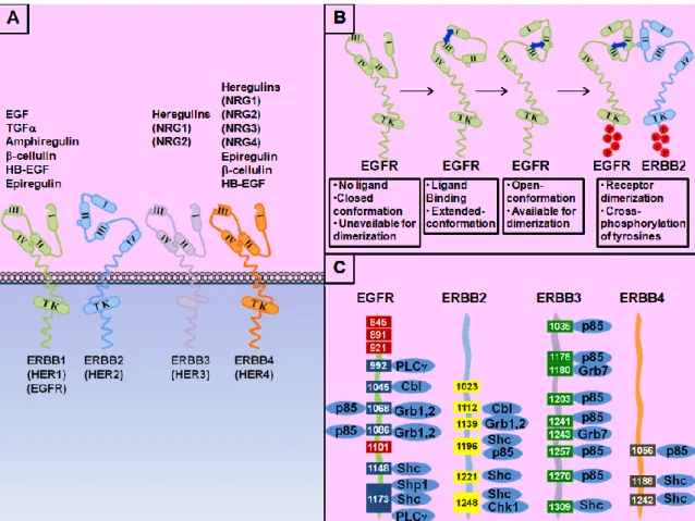
Figure 1.1 ERRB family of tyrosine kinase receptors.. ... 4
Figure 1.2 ERBB2 signaling.. ... 5
Figure 1.3 Common ERBB2 mutations observed on human cancers. ... 9
Figure 1.4 Mechanism of Action of anti-ERBB2 treatment.. ... 13
Figure 1.5 Mechanisms of Trastuzumab Resistance.. ... 19
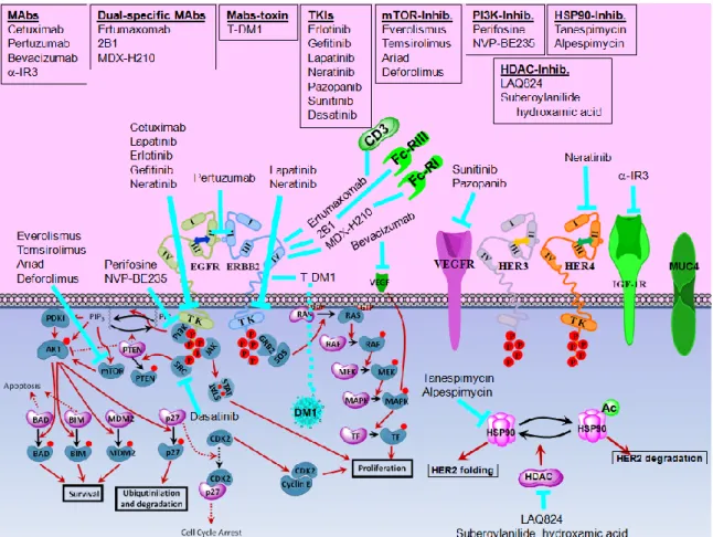
Figure 1.6 Alternative Therapeutics. ... 29
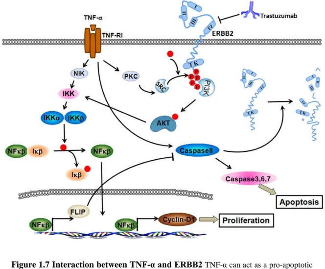
Figure 1.7 Interaction between TNF-α and ERBB2. ... 34
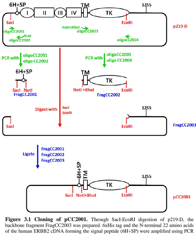
Figure 3.1 Cloning of pCC2001... 73
Figure 3.2 Cloning of pCC2002... 74
Figure 3.3 Sequence verification of ERBB2 constructs.. ... 77
Figure 3.4 Resolving gel for SDS-PAGE ... 87
Figure 3.5 Stacking gel for SDS-PAGE ... 87
Figure 3.6 Typical read out of a PI stained cell suspension ... 102
Figure 4.1 Structure of recombinant human ERBB2 ECD/Fc fusion protein used for immunization and ELISA assays.. ... 107
Figure 4.2 Pairwise alignment of EGFR and ERBB2 amino acid sequences. .. 109
Figure 4.3 ERBB2 and EGFR reactivities of novel anti-ERBB2 monoclonal antibodies. ... 110
Figure 4.4 BH-antibodies purified by Protein-G affinity chromatography... 110
Figure 4.5 Analysis of BH-antibody affinities for recombinant ERBB2 by ELISA. ... 111
Figure 4.6 Analysis of BH-antibody affinities for cellular ERBB2 by Flow Cytometry. ... 113
Figure 4.7 Analysis of BH-antibody affinities for cellular ERBB2 by Flow Cytometry. ... 113
Figure 4.8 Analysis of cellular ERBB2 by indirect immunofluorescence using BH-antibodies.. ... 115
Figure 4.9 Western blot analysis of endogenous ERBB2 expression on different breast cancer cell lines. ... 116
xv
Figure 4.10 Analysis of antibody binding to endogenously expressed ERBB2 denatured under reducing conditions.. ... 117 Figure 4.11 Immunoprecipitation of endogenous ERBB2 by BH-antibodies. .. 118 Figure 4.12 Trastuzumab does not compete with BH-antibodies for ERBB2-ECD binding on ELISA assays. ... 120 Figure 4.13 BH-antibodies do not compete with Trastuzumab for ERBB2-ECD
binding on ELISA assays.. ... 120 Figure 4.14 BH-antibodies and Trastuzumab do not share the same epitope on
cellular ERBB2 protein. ... 121 Figure 4.15 Human ERBB2 mRNA and protein domains; and the
corresponding nucleotides on ERBB2 mRNA. ... 123 Figure 4.16 ERBB2 deletion constructs expressing different ECD-subdomains.
... 124
Figure 4.17 Epitope mapping of anti-ERBB2 antibodies using indirect immunofluorescence.. ... 127 Figure 4.18 Verification of protein products of ERBB2 constructs. ... 127 Figure 4.19 Epitope mapping of anti-ERBB2 antibodies using combined
immunoprecipitation-western blot assay. ... 129 Figure 4.20 Effects of BH-antibodies on SK-BR-3 cell growth. ... 130 Figure 4.21 Dose effect of BH-antibodies on SK-BR-3 cell growth. ... 131 Figure 4.22 Effects of BH-antibodies on breast cancer cell panel with different
levels of ERBB2 expression. ... 131 Figure 4.23 Effects of BH-antibodies together with TNF-α on SK-BR-3 cell
growth.. ... 132 Figure 4.24 Dose effect of BH-antibodies on SK-BR-3 cell growth in the
presence of 1000 Unit/ml TNF-α.. ... 133 Figure 4.25 Dose effect of TNF-α on SK-BR-3 cell growth in the presence of 5
µg/ml BH-antibodies. ... 134 Figure 4.26 Effects of BH-antibodies together with TNF-α on breast cancer cell
panel with different levels of ERBB2 expression. ... 136 Figure 4.27 Effect of paired BH-antibody combinations on SK-BR-3 cell growth.
xvi
Figure 4.28 Effects of drug interactions between BH-antibodies and Trastuzumab on SK-BR-3 cell growth. ... 138 Figure 4.29 Effects of BH-antibodies on SK-BR-3 cell growth. ... 140 Figure 4.30 Interaction between BH1 and TNF-α on different breast cancer cell
lines. ... 143 Figure 4.31 Linear analyses of interaction between BH1 and TNF-α on different
breast cancer cell lines. ... 144 Figure 4.32 Effects of BH-antibodies on SK-BR-3 DNA synthesis in the presence
or absence of TNF-α. ... 147 Figure 4.33 Analysis of senescence induction upon BH1 treatment in the
presence or absence of TNF-α. ... 148 Figure 4.34 Effects of BH-antibodies on SK-BR-3 cell cycle.. ... 149 Figure 4.35 Change in cell cycle distribution and BrdU incorporation of SK-BR-3 cells upon BH1 treatment with or without TNF-α. ... 151 Figure 4.36 Effects of BH1±TNF-α treatment on molecular responses
xvii
ABBREVIATIONS
1.10 ABBREVIATIONS
Amp Ampicillin
AP Alkaline Phosphatase
APS Ammonium persulphate
BrdU Bromodeoxyuridine
BSA Bovine serum albumin
CaCl2 Calcium Chloride
cDNA complementary DNA
c-Met Mesenchymal-epithelial transition factor
CO2 Carbon dioxide
CNS Central nervous system
CTD Carboxy terminal domain
CTF Carboxy terminal fragment
DAPI 4',6-diamidino-2-phenylindole
ddH2O Double distilled water
DMEM Dulbecco’s Modified Eagle’s Medium
DMSO Dimethyl sulfoxide
DNA Deoxyribonucleic Acid
dsDNA ECD
Double stranded DNA Extracellular domain
ECL Enhanced chemiluminescence
EDTA Ethylenediaminetetraacetic acid
EGF Epidermal growth factor
EGFR Epidermal growth factor receptor
ELISA Enzyme linked immunosorbent assay
ErbB Epidermal growth factor receptor
ErbB1 Epidermal growth factor receptor 1
ERBB2 Epidermal growth factor receptor 2
ErbB3 Epidermal growth factor receptor 3
ErbB4 Epidermal growth factor receptor 4
xviii
FBS Fetal Bovine Serum
FITC FPLC
Fluorescein Isothiocyanate
Fast protein liquid chromatography
g gram
HRP Horseradish peroxidase
Ig Immunoglobulin
IgG Immunoglobulin G isotype
K Potassium
Kan Kanamycin
Kb Kilobase
kDa Kilodalton
lt Litre
MAb Monoclonal antibody
MAPK Mitogen activated protein kinase
µg microgram mg miligram MgCl2 Magnesium chloride min minute µl microliter ml mililiter µm micrometer μM micromolar mM milimolar
mRNA messenger RNA
MUC4 Mucin4
N-terminal Amino terminal
Na Sodium
NaCl Sodium chloride
Na-DOC NaOH
Sodium deoxycholate Sodium hydroxide
NEAA Non-essential amino acid
ng Nanogram
xix
NTD N-terminal domain
PBS Phosphate buffered saline
PBS-T Phosphate buffered saline with Tween-20
PCR Polymerase chain reaction
PI Protease Inhibitor Cocktail
PI3K Phosphotidylinositol-3-kinase
pmol Picomole
PNPP p-Nitrophenyl phosphate disodium salt
P/S Penicilline/Streptomicine
PTEN Protein tyrosine phosphatase
PVDF Polivinilidene flouride
RNA Ribonucleic acid
RNAse A Ribonuclease A
rpm Revolutions per minute
RIPA RPMI
Radioimmunoprecipitation assay Roswell Park Memorial Institute RT
SD
Room temperature Standard deviation
SDS Sodium dodecyl sulphate
SDS-PAGE SDS polyacrylamide gel electrophoresis
sec Second
SFM Serum free media
SRB Sulforhodamine B
TBS Tris buffered saline
TBS-T Tris-buffered saline with Tween-20
TCA Trichloroacetic acid
TF Transcription factor
TGF-α Transforming growth factor alpha
TGF-β1 Transforming growth factor beta
TK Tyrosine kinase domain
TNF-α Tumor necrosis factor alpha
Tris Tris (Hydroxymethyl)- Methylamine
xx
U Unit
UV Ultraviolet
v/v Volume/volume
VEGF Vascular endothelial growth factor
1
1 INTRODUCTION
1.1 Breast Cancer
With a lifetime risk of ~12%, breast cancer is the most common cancer in women worldwide [1], ranking second overall. Although the survival rates were increased due to the recent developments in breast cancer therapy, it is still the most frequent cause of cancer-related death in women worldwide (with 458,000 estimated deaths in 2008), and fifth overall [2]. More than 225,000 new cases of female-breast cancer are estimated to occur in 2012, in the United States alone, and 40,000 female deaths are expected as a result [3].
Breast cancers can be divided into two main subtypes according to their estrogen dependency. Estrogen-dependent breast cancers can further be divided into Luminal A and Luminal B types. Luminal A type represents 58% of all breast cancer cases [4]. Luminal B type which accounts for 16% of all breast cancer cases, overexpresses ERBB2 in addition to the common hormone receptors, shows greater proliferative potential and lower frequency of relapse-free survival [5]. Estrogen-independent breast cancers which account for ~26-30% of all breast cancer cases, are more aggressive than estrogen-dependent cases, tend to have poorly differentiated phenotypes, higher recurrence rate and decreased overall survival and can be further divided into basal-like and ERBB2-positive subtypes [5]. Of all breast cancer cases, ~16% are basal-like mostly with a triple-negative phenotype, where the expression of Estrogen Receptor (ER), Progesterone Receptor (PR) and ERBB2 is either low or absent. The rest, which comprise ~6% of all breast cancer cases have an estrogen-independent, ERBB2-positive phenotype [4]. Therefore ~25-30% of all breast cancer cases are associated with ERBB2 overexpression. [6, 7]
1.2 ERBB2
ERBB2 (also known as HER2) is a Receptor Tyrosine Kinase (RTK) identified in the early 1980s which is shown to be amplified in breast cancer cells [8]. The gene contains 27 exons spanning 38 kilobases and is located on chromosome 17q21 [9].
2
The 4.8 kb mRNA product is translated into a polypeptide of 1255 amino acids [10] and through extensive glycosylation, the precursor protein of 170 kDa is further processed and matured into 185 kDa glycoprotein [11]. In addition to the 185 kDa holoreceptor, breast cancer cell lines were documented to express two ERBB2 peptides of lower molecular mass [12]. The presence of the 68 kDa and 95 kDa peptides were very significant in the ERBB2-overexpressing cell line SK-BR-3 [12].
Being the human ortholog of the rat Neu protooncogene [10, 13], ERBB2 belongs to the ERBB transmembrane receptor family together with EGFR (HER1 or ERBB1), ERBB3 (HER3) and ERBB4 (HER4) [14]. Each family member contains four extracellular subdomains at their N-terminal, a transmembrane domain and an intracellular domain containing tyrosine residues that are used for signal transduction (Figure 1.1 A). In addition, except for ERRB3, all ERRB receptors have intracellular tyrosine kinase domains and intrinsic kinase activity. Extracellular subdomain I and III were documented to take role in the establishment of receptor conformations that allow dimerization through subdomain II [15] (Figure 1.1 B). In the absence of the ligand, the receptor is found on a closed-conformation, where subdomain II makes an intramolecular contact with subdomain IV and is not available for dimerization [16, 17]. Upon ligand binding by subdomains I and III, conformation of the receptor is changed exposing the dimerization domain [18, 19] and allowing dimerization of two receptors present in this open-conformation (Figure 1.1 B). ERBB2 is unique in the family in two ways; first it has no identified ligands upto date [20]. Second, the structure of its extracellular region is radically different from other family members; resembling the ligand-activated state of other ERRB receptors, it provides the receptor with a fixed open-conformation [17, 18], negating the requirement of ligand-induced activation. Due to its constitutive open-conformation, ERBB2 is permanently available for heterodimerization with other ligand-activated ERRB-family receptors and is frequently documented as their preferred partner [14] (Figure
1.1 B). Since their dimerization arm is permanently exposed, ERBB2 receptors
readily homodimerize and activate each-other when they are overexpressed. This is a key mechanism in cell proliferation observed both in ERBB2 overexpressing cells in vitro [21] and ERBB2-positive breast cancers [6, 22]. Recently, an autoinhibition mechanism was revealed, where the αC helix and β4 sheet in its kinase domain form a loop inactivating ERBB2 kinase. Since many kinase domain mutations observed on
3
human cancers were localized on this loop, the loss of this autoinhibition was suggested to cause oncogenic ERBB2 activation [23].
Upon dimerization, ERRB receptors cross-phosphorylate one another on multiple intracellular tyrosine residues and provide docking sites for adaptor proteins (Figure 1.1 C). In adition, via phosphorylating the adaptor proteins, they create additional docking sites available for the down-stream signalling transduction pathway and alter the activity of intracellular signalling proteins by phosphorylating them (Figure 1.2). Main auto- and trans-phosphorylation residues present on the intracellular region of ERRB receptors, and the adaptor molecules that interact with these docking sites are summarized on Figure 1.1 C [14]. Being the main autophosphorylation sites on ERBB2; Tyr-1221/1222 and Tyr-1248 interact with Shc through Src homology 2 (SH2) domain and participate in Ras-Raf-MAPK signal transduction pathway [24–26]. In addition, phosphorylated Tyr-1196 and Tyr-1248 interact with Shc through a phosphotyrosine binding (PTB) domain [24]. Also associated with ERBB2 overexpression, phosphorylation of Tyr-1139 induces GRB2 and GRB7 interaction through SH2 domain contributing to a subset of breast tumors [27]. Phosphorylation of Tyr-877 is performed by Src family kinases and was initially suggested to influence the intrinsic kinase activity of ERBB2 [28], however, Tyr-877Phe substitution of ERRB2 did not alter its biological and/or biochemical properties [25], refuting its role in ERBB2 activation [23]. Tyr-1028 on rat NEU corresponding to Tyr-1023 on human ERBB2, was shown to be a negative regulator of NEU-mediated transformation [29]. Upon EGF stimulation, EGFR heterodimerizes with ERBB2; PI3K interacts with phosphorylated Tyr-1196 on ERBB2 [30] and Ser-1113 is phosphorylated by CaMK II which serves as an indicator of poor prognosis [31]. In addition, neuregulin mediated heterodimerization of ERBB2 and ERBB3 induces phosphorylation of Thr-686 on ERBB2 by PKA; and in turn stimulates the crosstalk between PKA and cAMP leading to activation of PI3K-Akt and MEK-Erk pathways [32]. c-Cbl ubiquitin ligase is recruited to the phosphorylated Tyr-1112 on ERBB2 and induces poly-ubiquitination and proteolytic degradation of ERBB2 [33].
4
Figure 1.1 ERRB family of tyrosine kinase receptors. A) ERRB family of tyrosine kinase receptors are shown in their ligand-free conformation; while ERRB1, ERRB3 and ERRB4 are found in closed-conformation without a bound ligand, ERBB2 is constitutively present in the open-conformation. Each receptor contains four extracellular subdomains, a transmembrane domain and an intracellular C-terminal tail. All except ERRB3 also have an intracellular tyrosine kinase domain. Identified ligands of the receptors are listed above each receptor. B) Proposed mechanism of receptor dimerization is illustrated. Upon ligand binding (dark blue arrow), a conformational change occurring at the extracellular subdomains of EGFR exposes the previously buried subdomain II. ERBB2 and ligand-activated EGFR interact through their exposed subdomain II, and heterodimerize. C) Main auto- and trans-phosphorylation sites on the intracellular C-terminal tail of each ERRB receptor and the adaptor molecules interacting with the respective phosphorylated tyrosine residues are shown.
5
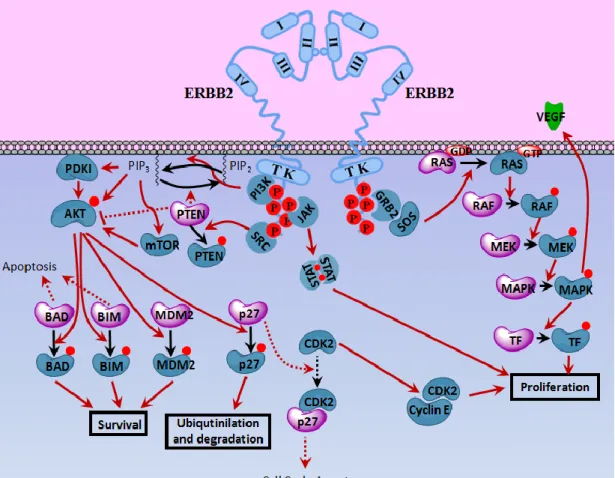
Figure 1.2 ERBB2 signaling. Upon homodimerization, tyrosine residues (in addition to few threonine and serine residues) on the cytosolic tail of ERBB2 are phosphorylated activating the downstream effectors mainly on PI3K-Akt, Ras-Raf-MAPK and JAK/STAT pathways. As a result, proliferative signals are received, pro-apoptotic signals are blocked and cell cycle arrest is inhibited leading to to cell survival and proliferation. Effectors shown in dark blue are the molecular forms induced upon ERBB2-dimerization and participate in proliferation. Effectors shown in pale purple are the molecular forms which participate in cell cycle arrest and apoptosis that are decreased upon ERBB2-dimerization. /: molecular transitions/signal transductions in action. : inhibited signal transductions. : phosphorylated residues. TK: Tyrosine kinase domain. TF: Transcription factor.
Upon dimerization, receptors transphosphorylate each other and recruit downstream effectors such as PI3K, Src, Grb2 [29, 34–36]. When Src is recruited to intracellular phospho-tyrosine residues on activated ERBB2, it phosphorylates PTEN downregulating its activity. Therefore PTEN can no longer antagonise PI3K pathway. Activated ERBB2 further recruits and activates PI3K, facilitating Akt activation through PI3K-mediated phosphorylation. Activated Akt further phosphorylates pro-apoptotic proteins Bad, Bim and MDM2, leading to cell-survival [37]. It also phosphorylates p27 leading its ubiquitinilation and degradation. Since p27 is no longer active to block CDK2, CDK2 interacts with CyclinE, leading to cell
6
proliferation [38, 39] (Figure 1.2). As an additional mechanism for silencing tumor suppression, ERBB2 was shown to interfere with TGFβ-induced cytostasis and oncogene-induced senescence by deregulating C/EBPβ isoform translation [40]. Cell culture conditions were reported to induce a switch between the two main signalling pathways downstream of ERBB2; PI3K and MAPK pathways [41].
It was shown that ERBB2-overexpression in NIH3T3 cells leads to malignant cellular transformation and tumorigenesis [21, 42]. Numerous reports have demonstrated that ERBB2 amplification and receptor overexpression was observed in several human cancers including breast [6, 22], ovarian [43], endometrial [44], gastric [45], lung [46], esophageal [47], oropharyngeal [48], bladder [49]. Relation between ERBB2 status and breast pathology is described by Ross et. al. and shown on Table 1.1 [50].
Table 1.1 Relationship between ERBB2 status and breast pathology. Reprinted with permission from "Ross JS, Slodkowska EA, Symmans WF, Pusztai L, Ravdin PM, Hortobagyi GN: The HER-2 Receptor and Breast Cancer: Ten Years of Targeted Anti–HER-2 Therapy and Personalized Medicine. The Oncologist Anti–HER-2009, 14:3Anti–HER-20–68." [50]. Copyright AlphaMed Press 2009.
7
A Val664Glu point mutation found on the transmembrane domain of rat ERBB2/NEU (corresponding to Val659Glu in humans) was shown to be sufficient to transform cells [51, 52], where the glutamic acid variant was suggested to stabilize dimerization leading to conversion of pro-oncogenic NEU to an oncogenic protein [53]. Through examination of transgenic mice overexpressing ERBB2/NEU, single point missense mutations and in-frame deletions were detected at the juxtamembrane domain; and the cysteine-rich juxtamembrane region was suggested to have an important role in regulation of NEU catalytic activity [54, 55]. While the human equivalent Val659Glu has been shown to constitutively activate ERBB2 in vitro [56], up to date it has never been observed in human breast cancers [57]. Through the computational analysis of the transmembrane regions of the ERBB2-homodimer, it was shown that Val664Glu mutation stabilizes the active conformation of the dimeric RTK, whereas Ile-655Val destabilizes the active conformation [58].
ERBB2 polymorphisms/mutations found on human tumors are summarized on
Figure 1.3. The common Ile655Val polymorphism that is suggested as a risk factor for breast cancer development is studied by various groups. While some groups reported increased risk of predisposition with Val-allele [59–63], many others observed no-association [64–68] or even decreased risk [69].
Screening kinase domain of ERBB2 in metastatic breast cancer patients, Gori et. al. have detected one insertion in exon 20 (ins780 (C)) and a missense mutation in exon 21 (Ser856Pro) [70]. Through the investigation of breast, gastric and colorectal tumors, missense mutations in exon 18 (Lys724Asn, Thr733Ile), exon 19 (Leu755Ser, Asp769His) and exon 20 (Val777Leu, Val777Met, Glu799Pro), exon 21 (Val842Ile, Leu869Glu), exon 22 (Arg896Cys) and one in-frame deletion on exon 19 (Leu755-Thr759del) was detected on ERBB2-kinase domain [71]. Another study analysing exon 18-24 of ERBB2 on non-small cell lung cancers (NSCLC) identified four main types of insertion at exon 20 (ins776(TyrValMetAla), ins781(GlySerPro), Gly-776Val + ins777(C), Gly-776Leu + ins777(C)) [72].
Screening ERBB2 in various tumour types, Stephens et. al. detected further somatic mutations and in frame insertions in NSCLC, glioblastoma, gastic and ovarian tumours. These ERBB2 mutations which were located on exon 19 (Leu755Pro), exon 20 (ins774(AlaTyrValMet), Gly776Ser, ins779(ValGlySer)),
8
exon 21 (Asn857Ser) and exon 23 (Glu914Lys) of the kinase domain were not associated with ERBB2 overexpression in these tumours [73]. Kinetic analysis demonstrated that, the catalytic activity of ERBB2 dramatically increases when Gly776 and Gly778 are mutated [23].
Point mutations at the transmembrane domain of ERBB2 was detected in malignant central nervous system (CNS) tumors [74]. As a rare event, His559Ala point-mutation located on the Trastuzumab-binding site was detected on the juxtamembrane domain of ERBB2 in invasive human breast cancers overexpressing ERBB2 [75].
In a recent study, Zito el. al. resequenced every individual amplicon in human breast cells with ERBB2 amplification; the high-resolution analysis of the total
ERRB2 coding sequence on five cell lines and two breast tumors did not reveal any
activating mutations on ERBB2 [76]. This suggests that, receptor overexpression and ERBB2 activating mutations may have mutually exclusive roles in breast cancer development.
9 Figu re 1 .3 Com m on E RBB 2 m u tat ion s ob se rve d on h u m an c an ce rs. Tw ent y seve n exon s fo rm ing the hu m an ERBB2 i sof o rm -a m R N A a re il lus tr at ed w it h num ber ed pi nk bo xes d rawn to sc al e; nu m be rs abo ve the bo xe s ind ica te the n ucl eot ide p osi ti on s su rr o undi ng ea ch exon. Pu tat ive pr ot ei n do m ai ns ar e il lu st rat ed w it h ye ll ow boxe s d rawn to sc al e ac cor d ing to the inf or m at ion pr es en ted on N C B I and i n di cat ed re fer enc es. B lue num be rs b el ow t he bo xes i ndi ca te the am ino aci d p os it ions at t he bou nda ri es of ea ch pu ta ti v e pr o te in do m ai n an d bl ac k nu m be rs abo ve the boxe s indi ca te the ir cor re spond in g nuc leo ti d e p o si ti o ns o n the fu ll l eng th m R N A se qu enc e. Mu ta ti on s ob se rved o n hu m an ER B B 2 rec ept or ar e ind ica ted i n red. T he put at ive V al 6 59 G lu su bst it ut ion on h um an ER B B 2 cor res p ondi ng t o t h e rat ho m ol o gue V al 66 4 G lu is i nd ica ted i n g ree n. : subs ti tu ti o n , ins : i n se rt ion, o r de l: d el et ion.
10
ERBB2 amplification and overexpression is tested either at gene level by fluorescence in-situ hybridization (FISH) or at protein level by immunohistochemistry (IHC) respectively. While IHC is easier to perform, faster and less expensive; lack of standardization in fixation and paraffin-embedding protocols, sensitivity of the antigen epitope, and the subjectivity in measurement of staining intensity may cause variation, leading to false-positive and false-negative results. On the contrary, FISH is a standardized procedure providing quantitative more reliable and reproducible results but more expensive, complex and slower than IHC. IHC based HercepTest (DakoCytomation Denmark A/S, Glostrup, Denmark), and FISH based PathVysion (Vysis Inc., Downers Grove, IL) kits are approved by FDA for the verification of HER2 status of breast tumors. Although there is strong correlation (upto 90%) between IHC and FISH results for strong positive cases (IHC +3), tumors with IHC scores of +2 are recommended to be confirmed by FISH analysis [45, 48, 77, 78].
While normal breast cells express ~20,000 ERBB2 receptors on their membrane (IHC score: 0), the protein expression in tumor cells was documented to reach as high as 2,000,000 ERBB2 molecules per cell (IHC score: +3) [79]. ERBB2 levels which were selectively much higher in cancer cells than the normal tissues were shown to strongly correlate with breast cancer prognosis and pathogenesis [22].
ERBB2 gene amplification is observed in 20–25% of invasive breast cancers and is highly correlated with protein expression [6, 22, 78, 80]. ERBB2 overexpression which was detected both in primary lesions and metastatic sites was found to be relatively homogeneous among ERBB2 overexpressing tumor cells [81]. In addition to above, being a cell surface receptor, ERBB2 was an ideal candidate target for monoclonal antibody (MAb) based breast cancer therapy [82]
1.3 Anti-ERBB2 Antibody Based Targeted Therapy of ERBB2
Overexpressing Breast Cancers
ERBB2 amplification alone was shown to cause cell transformation [21] and was linked to TNF-α/macrophage resistance in vitro which was reversed upon treatment by monoclonal antibodies (MAbs) targeting extracellular domain (ECD) of ERBB2 [83, 84]. In addition, 25% of all breast cancers have ERBB2 overexpression
11
which was linked with a more aggressive type of breast cancer [6, 22], therefore ERBB2 was acknowledged as a significant therapeutic target in breast cancer treatment. A large panel of MAbs targeting ERBB2-ECD was developed [85] and shown to inhibit monolayer proliferation and revert anchorage independent phenotype [86]. In vivo data also supported the findings that indicate anti-ERBB2 antibody therapy as a treatment regime for ERBB2-overexpressing tumors. Radiolabeled antibodies against ECD were localised to ERBB2-overexpressing mammary tumors in NEU-transgenic mice; suggesting a potential for a successful therapeutics [87].
More than 100 monoclonal antibodies were developed against extracellular region of human ERBB2 by various groups [88]. Among this wide panel of anti-ERBB2 antibodies, mouse monoclonal 4D5 was shown to have anti-tumor effects both in vitro and in vivo [84, 89–91].
In order to overcome the human anti-mouse antibody response that limits the therapeutic efficiency of murine monoclonal 4D5 (MuMAb4D5), the heavy and light chain variable regions (VH and VL) of the antibody are humanized by gene conversion mutagenesis. In addition, 5 aminoacids on framework region 3 (FR3) were changed by site-directed mutagenesis to increase the affinity and the therapeutic efficiency of the humanized antibody named as Trastuzumab (Herceptin®) [92]. Trastuzumab is the first FDA approved humanized MAb therapeutics targeting ERBB2. Having received first FDA approval in 1998, Trastuzumab is now used both for metastatic and for early-breast cancer treatment mostly in adjuvant settings [93]. When Trastuzumab was used as a single agent therapy for ERBB2-overexpressing, metastatic breast cancer (MBC), ~66-88% of the patients showed primary resistance [94–96]. While combined therapy was better than Trastuzumab monotherapy for MBCs, the majority of patients developed resistance within one year even if they had an initial response [82]. In addition, although Trastuzumab led to promising results in the treatment of ERBB2-overexpressing early-stage breast cancers, ~15% of the patients still developed metastasis [97], demonstrating the clinical limitations caused by the development of Trastuzumab-resistance in the long run.
Recently another monoclonal antibody (2C4) developed at the same time with 4D5 is being investigated as an alternative therapeutics for the treatment of ERBB2-overexpressing breast tumors. 2C4 which initially displayed growth inhibitory
12
activity weaker than 4D5 was later found promising due to its inhibitory effects on ERBB2 dimerization. The therapeutic activity of Pertuzumab, the humanized form of 2C4 is being tested in clinical trials [98].
Although numerous additional anti-ERBB2 antibodies were developed up to date, Trastuzumab and Pertuzumab are the two main antibodies extensively studied so far.
Through crystal structure analysis, Trastuzumab was found to interact with ERBB2 through three regions (557–561, 570–573, 593–603) on the ERBB2 extracellular domain IV, located juxtamembrane of the receptor [17]. The dimerization inhibitory antibody Pertuzumab binds to the dimerization domain on ECD-II [99].
1.3.1
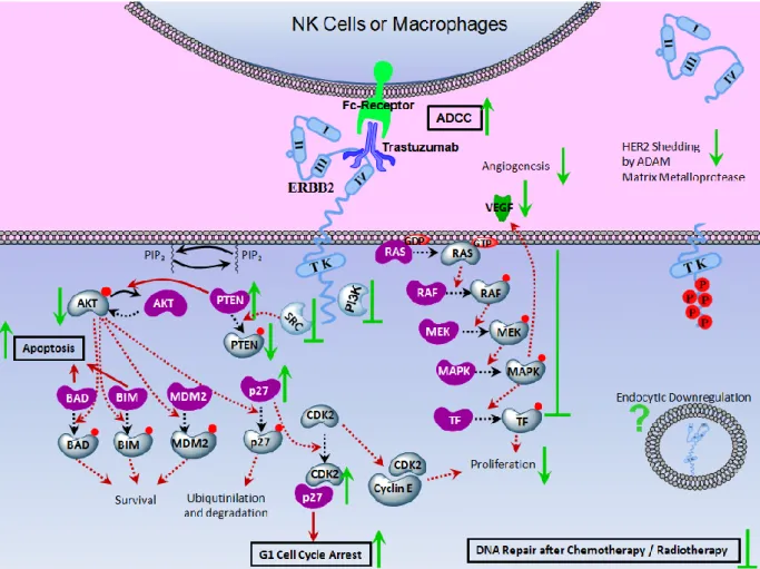
Mechanism of Action of anti-ERBB2 Antibody Treatment
Being the first monoclonal antibody developed for selective targeting of ERBB2 overexpressing tumors, Trastuzumab provided most of the information gathered on the mechanism of anti-ERBB2 treatment. As summarized on Figure 1.4, anti-ERBB2 treatment acts mainly by inhibiting growth inducing signals transduced through MAPK and PI3K pathways. Antibody treatment stimulates tumor cell elimination by further activating antibody dependent cellular cytotoxicity (ADCC) mechanism and apoptotic pathway. In addition, it was suggested that antibody interaction with ERBB2 receptor may decrease receptor shedding, influence endocytic receptor downregulation and enhance the chemotherapeutic response.
13
Figure 1.4 Mechanism of Action of anti-ERBB2 treatment. Molecular responses of ERBB2 overexpressing cells following anti-ERBB2 antibody treatment are shown. PI3K and MAPK pathways are downregulated. Effectors shown in dark purple are the molecular forms induced upon Trastuzumab treatment which participate in anti-proliferative pathways. Effectors shown in pale blue are the molecular forms which participate in proliferation and survival pathways and are decreased upon ERBB2-dimerization. /: molecular transitions/signal transductions in action. /: inhibited molecular transitions/signal transductions. : upregulation, activation or stabilization. : downregulation, inactivation or degradation. : inhibition.
?
: conflicting results : phosphorylated residues. TK: Tyrosine kinase domain.1.3.1.1 Increased Autophosphorylation, Internalization, Degradation and Down-regulation of ERBB2
Contradictory observations are documented regarding the effect of anti-ERBB2 treatment on ERBB2 phosphorylation. While some groups showed decrease of ERBB2 phosphorylation [100], others demonstrated induction of tyrosine phosphorylation of the receptor upon antibody treatment [33, 101, 102].
14
Phosphorylation of Tyr-1112 of ERBB2 induces recruitment of c-Cbl to the receptor [33]. Binding of anti-ERBB2 antibodies were shown to stimulate ubiquitinylation of the receptor by activating E3 ubiquitin ligase c-Cbl, leading to ERBB2 degradation [103]. While receptor internalization and degradation upon anti-ERBB2 treatment (mostly by Trastuzumab) is demonstrated by several groups as a means to explain the inhibition of ERBB2 kinase activity and downstream pathways [84, 101, 102, 104– 106], others failed to show a decrease in ERBB2 levels [107–110].
1.3.1.2 Growth Inhibition Modulated Through PI3K and MAPK Pathways
ERBB2 overexpressing cells induce Src which phosphorylates and deactivates PTEN. Following Trastuzumab treatment of ERBB2-overexpressing cells, Src is dissociated from ERBB2; PTEN which is left active upon Src-inhibition is translocated to the plasma membrane [111], where it dephosphorylates and deactivates Akt [108]. Since phosphorylation of the internal tyrosine residues on ERBB2 is reduced by anti-ERBB2 treatment [100], anti-ERBB2 treatment blocked PI3K-docking necessary to activate the kinase. Through inhibition of PI3K, the membrane localization of PTEN is increased and phosphorylation level of Akt is decreased leading to increased GSK3β stability, decreased Cyclin D1 expression and G1-arrest [111–114]. Inducing the association between p27 and CDK2, anti-ERBB2 treatment decreases CDK2/Cyclin E mediated cell cycle progression. The decrease in CDK2/Cylin E mediated phosphorylation and degradation of p27 further stabilizes p27 and leads to a negative feedback resulting in G1-arrest [39, 115, 116]. Cyclin G2 which belongs to the G-cyclins that are upregulated during cell cycle arrest and apoptosis is also induced by Trastuzumab [117]. Decreasing the phophorylated MAPK levels, the treatment also inhibited the MAPK pathway [113, 118].
1.3.1.3 Inhibition of ERBB2 Shedding
Truncated ERBB2 receptors lacking extracellular N-terminal region were detected in human breast tumors. Through cleavage by matrix metalloproteases, the
15
wild type 185kDa ERBB2 was reported to be cleaved into two fragments; 110 kDa ERBB2-ECD circulates in the blood [119, 120], while the 95 kDa C-terminal fragment (CTF) is left attached to the cell membrane being constitutively activated [56, 121–124]. Elevated levels of p110 circulating in the serum was shown to be associated with poor prognosis in advanced breast cancer patients [125–128]. CTFs are also detected in a subset of breast cancer patients that developed nodal metastasis, leading to worse prognosis and poor clinical outcome [129, 130].
Trastuzumab decreased ERBB2 shedding by inhibiting ADAM metalloproteinase activity; an effect which is not shared by Pertuzumab [119, 120]. Trastuzumab not only decreased serum ERBB2-ECD levels, but also the intact ERBB2 levels; suggesting the presence of another mechanism in addition to sheddase inhibition [131]. At least ~20% reduction in serum ERBB2 (p110) levels has been proposed as a marker of better prognosis upon Trastuzumab therapy (patients with elevated pre-treatment ECD levels had higher response rates to Trastuzumab) [132–134].
1.3.1.4 ADCC
Antibody dependent cellular cytotoxicity (ADCC) is one of the mechanisms of tumor elimination induced by antibody binding and conducted by natural killer (NK) cells, macrophages and monocytes expressing Fc receptors that recognize IgG molecules bound to target cells. Anti-ERBB2 antibodies were shown to elicit apoptosis in breast cancer cell lines through ADCC [86, 92] and significant lymphoid infiltration was observed in Trastuzumab treated patients [107]. Kute et. al. demonstrated that ERBB2 overexpressing breast cancer cells that developed resistance to anti-proliferative effects of anti-ERBB2 treatment in vitro were still sensitive to ADCC response and were successfully eliminated by the effector-mediated cytotoxicity; emphasizing the importance of ADCC mechanism on anti-ERBB2 dependent tumor elimination [135].
FcRIII receptor activates effector-mediated target cell lysis whilst FcRIIB performs as an ADCC inhibitory receptor. While mice engineered to express only the activating FcRIII receptors were capable of a successful cytotoxic response, those lacking the activating receptors were insufficient to arrest tumor growth in vivo. In
16
addition, anti-ERBB2 antibodies lacking the Fc region (Fab molecules) were unable to trigger significant anti-tumorigenic response; indicating ADCC as one of the main mechanism of anti-ERBB2 treatment in vivo [136, 137].
Antibody molecules were modified to enhance the ADCC response; non-fucosylated oligosaccharide chains on Fc region dramatically enhanced apoptosis both in vitro and in vivo [138, 139]. In addition, Fc regions engineered for higher FcR affinity enhanced the in vitro effector functions and cytotoxicity [140]. Bispecific antibodies targeting both the ERBB2 receptors present on cancer cells and the immuno-effector cells were shown to significantly induce the apoptotic response [137, 141]. In addition to their ERBB2 specificities, Ertumaxomab target CD3 [142], 2B1 target FcRIII [143], MDX-H210 target FcRI [144] (Figure 1.6). Adjuvant lymphokines such as interleukin-2 (IL-2) intensified NK-induced ADCC upon anti-ERBB2 treatment both in vitro and in vivo [145, 146].
1.3.1.5 Inhibition of DNA Repair (after chemotherapy or radiation)
In order to overcome the tumor relapse occuring after the cessation of anti-ERBB2 treatment, the antibody therapy was combined with chemotherapeutics inducing DNA damage such as doxorubicin and cisplatin or with radiotherapy. DNA damage and DNA adducts induced by the chemotherapeutics and radiotherapy stimulates repair pathways. Addition of anti-ERBB2 antibody to the therapeutic cocktail inhibited p21 expression that participates in DNA repair, blocked unscheduled DNA synthesis and DNA-adduct repair leading to enhanced tumor elimination [147, 148].
1.3.1.6 Inhibition of Angiogenesis
Since tumor cell survival partly depends on angiogenesis that provide nutrients, growth factors and oxygen to the tumor microenvironment; cancer cells secrete factors to induce angiogenesis. ERBB2 overexpression was shown to stimulate angiogenesis through PI3K-Akt mediated induction of pro-angiogenic factors like vascular endothelial cell growth factor (VEGF) and interleukin-8 (IL-8) while decreasing the levels of anti-angiogenic factor thrombospondin-1 (TSP-1) [149–151].
17
Downregulating the PI3K-Akt pathway, anti-ERBB2 treatment counteracted ERBB2-mediated regulation of angiogenic factors, decreased vascular permeability and inhibited angiogenesis [151, 152]. Combination of Trastuzumab treatment with a microtube-inhibitor (paclitaxel) further enhanced the dephosphorylation of Akt, downregulation of VEGF secretion, reduced endothelial cell migration and decreased the tumor microvessel density (MVD) [153].
1.3.2
Response to Trastuzumab
In phase II clinical trials where Trastuzumab was used as a single agent therapy for ERBB2-overexpressing, metastatic breast cancer, ~66-88% of the patients developed primary resistance to Trastuzumab monotherapy [94–96].
Later on in phase III trials, trastuzumab was combined with paclitaxel or docataxel for treatment of ERBB2-overexpressing metastatic breast cancers (MBCs) [154]. The response rates, time-to-disease progression and overall survival of the combined therapy were superior to Trastuzumab monotherapy, leading to FDA approval of Trastuzumab-based combined treatment regime in 1998 for ERBB2-overexpressing metastatic breast cancers. MBC patients receiving first-line treatment with chemotherapy in combination with Trastuzumab had ~20% lower risk of death than patients treated with chemotherapy alone [155]. Median time-to-diease-progression was 4.9 months for patients that received trastuzumab monotherapy without any prior chemotherapy [95]; however, it was 7.4 months for patients that received Trastuzumab in combination with chemotherapy [155]. While combined therapy was better than monotherapy for MBCs, the majority of patients still developed resistance within one year even if they had an initial response [82].
Afterwards, other clinical trials were established where Trastuzumab was tested for the treatment of early stage breast cancers with ERBB2-overexpression. Disease-free and over-all survival rates were found to be improved by Trastuzumab therapy either in combination with or following chemotherapy [156–158]. These results brought about FDA approval of Trastuzumab in 2006 as part of a treatment regimen containing doxorubicin, cyclophosphamide, and paclitaxel for the adjuvant treatment of patients with early-stage ERBB2-positive, node-positive breast cancer. In 2008, Trastuzumab received further FDA approvals to be used in early stage ERBB2-positive settings. Multiple reports suggest that, a trastuzumab-containing
18
treatment regime should be recommended to early-stage breast cancer patients carrying ERBB2-amplification and/or overexpression [159–162]. Although Trastuzumab led to promising results in the treatment of ERBB2-overexpressing early-stage breast cancers, ~15% of the patients still developed metastasis [97], demonstrating the clinical limitations caused by the development of trastuzumab-resistance in the long run.
Trastuzumab treatment was most beneficial for ERBB2-positive patients whose tumors displayed gene amplification by FISH or intensive membrane staining (3+) as detected by IHC [95, 155, 156, 158, 163, 164].
1.3.3
Mechanisms of Trastuzumab Resistance
In order to tackle with these limitations faced in clinic, numerous groups have endeavoured to elucidate the mechanisms of trastuzumab resistance in vitro, ex-vivo and in-vivo.
Mechanisms of Trastuzumab-resistance are being studied and reviewed extensively by various groups [82, 97, 165, 166]. Overexpression of other ERBB receptors (i.e. EGFR) and/or their ligands (i.e. EGF, TGF-α, heregulins) and heterodimerization with other receptors types (i.e. IGF-1R) were reported to increase downstream signalling through heterodimerization with ERBB2. Activating PI3K mutations, PTEN deletion, p27 downregulation and increased Akt activity also induce constant activation of donstream pathways stimulating cell proliferation and survival, and lead to Trastuzumab resistance. MUC4-ERBB2 interaction and circulating ERBB2-ECD (p110) in serum was shown to block Trastuzumab binding. Mutations on Fc-receptors were identified as the possible reason of hampered ADCC-response.
19
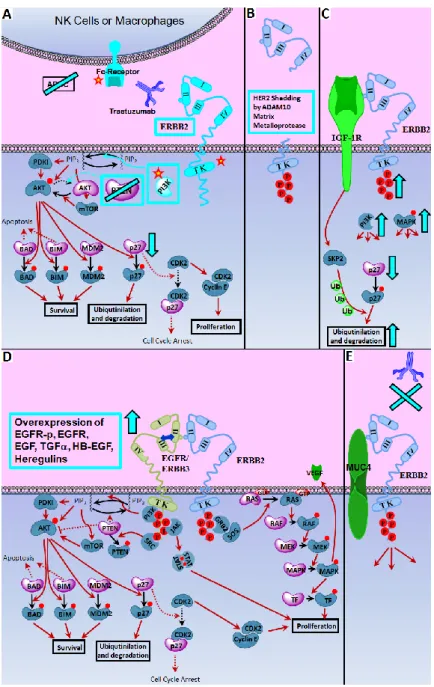
Figure 1.5 Mechanisms of Trastuzumab Resistance. Potential culprits of anti-ERBB2 resistance are shown. Mutations on intracellular region of ERRB2 receptor (mainly at Tytosine Kinase region), activating PI3K mutations and PTEN deletion is shown to cause constitutive activation of Akt induced signaling, leading to p27 degradation (A). Mutations on antibody-binding epitope (A), receptor shedding by ADAM proteases (B) and receptor masking through ERBB2-MUC4 interaction (E) prevents antibody binding. Mutations on Fc receptor hamper the ADCC response (A). In addition cross-talk between ERBB2 and other growth factor receptors (C and D) activate the downstream proliferative pathways. Effectors shown in dark blue are the molecular forms demonstrated or suggested to be active in Trastuzumab resistance, participating in proliferation and survival pathways. Effectors shown in pale purple are the molecular forms demonstrated or suggested to be decreased in Trastuzumab resistance, participate in anti-proliferative pathways. /: molecular transitions/signal transductions in action. : inhibited signal transductions.
:
upregulation, activation or stabilization. : downregulation, inactivation or degradation.X
: inhibition of receptor binding. : mutation or polymorphisms. : phosphorylated residues. TK: Tyrosine kinase domain.20
1.3.3.1 Variations in ERBB2-ECD
Since Trastuzumab binds to a juxtamembrane position on the extracellular domain of ERBB2 receptor tyrosine kinase, any variation in that region will interfere with Trastuzumab-response. Missense mutations and in-frame deletions at the juxtamembrane domain of ERBB2 were found in transgenic mice overexpressing ERBB2/NEU and shown to be capable to oncogenic transformation when introduced into wild type ERBB2/NEU [55]. Yet, common point mutations in ERBB2-ECD have not been documented in human breast tumors (Nahta et al. 2006). A rare His-559-Ala point-mutation identified on the Trastuzumab-binding site on the juxtamembrane domain of ERBB2 might account for some resistance observed in ERBB2-overexpressing invasive human breast cancers, but far from explaining the high frequency of Trastuzumab-resistance observed in clinic [75].
Truncated ERBB2 receptors lacking extracellular N-terminal region were detected in human breast tumors. Since they lack the Trastuzumab binding extracellular domain IV, it is not surprising that they are irresponsive to Trastuzumab treatment. ERBB2 Carboxy-Terminal-Fragments (CTFs) were suggested to be produced either through proteolytic receptor shedding (as in the case of p95) [121– 124] or by alternative ERBB2 translation (either starting from methionine-611 located upstream of the transmembrane domain as in the case of 611-CTF, or from methionine-687 downstream of the transmembrane domain) [167, 168]. CTFs were detected in a subset of breast cancer patients that developed nodal metastasis, leading to worse prognosis and poor clinical outcome [129, 130]. In addition, ERBB2-overexpressing patients were shown to be less responsive to Trastuzumab if they expressed p95; the response rate was 51.4% for tumors expressing full-length ERBB2, while it was 11.1% for patients expressing p95 [169]. HSP90 inhibitors which block downstream signalling induced by p95-ERBB2, were suggested as a therapeutic option for p95-overexpressing, Trastuzumab resistant breast cancer types [170].
ERBB2 N-terminal fragments (p100 and p110) circulating in the serum that are produced either through the cleavage of matrix metalloproteases [119, 120] or by alternative splicing also participate in Trastuzumab resistance [171, 172]. Identical to full-length ERBB2 sequence at its 5’ end, the alternative transcript diverged from the wild type sequence due to an in frame stop codon 61 nucleotides upstream of the
21
transmembrane domain. The truncated ERBB2 of 100 kDa is found both intracellularly at the perinuclear cytoplasm and secreted into the conditioned media [171]. It has been reported that, p110 competed with full-length ERBB2 present on cell surface for binding to anti-ERBB2 antibodies [123, 173], and elevated levels of p110 circulating in the serum was shown to be associated with poor prognosis in advanced breast cancer patients [125–128].
Another alternative splicing event deleted the entire exon 16 which is coding for amino acids immediately preceding the transmembrane domain of the receptor on ERBB2 ECD-IV. Associated with constitutive ERBB2 activation [174] and stronger transformation capacity [175], this ERBB2 variant (16ERBB2) with a 16 amino acid in-frame deletion (C634-S649del) was detected in human breast tumors [174, 176]. A later report indicated that, the oncogenic ERBB2Δ16 was found in 89% of the lymph-node positive breast tumors with ERBB2-overexpression [177]. Although C634-S649 is not involved in Trastuzumab-ERBB2 interaction [17], tumor cell lines with this variant were found to be resistant to Trastuzumab [174, 177].
One other alternative splicing event produces a 68 kDa Herstatin which only retains the total extracellular domain I (ECD-I) and most of extracellular domain II (ECD-II); replacing the remaining sequence [178]. Through its unique C-terminal tail which substitutes the extracellular domain III (ECD-III) of the wild type ERBB2, Herstatin was shown to bind ERBB2 ECD-II [179], EGFR, Her4, IGF-IR [180], prevent ERBB2 dimerization with Her3 and EGFR [181] and inhibit TGF-α, EGF and heregulin mediated growth of breast cancer cells [182, 183] similar to the dimerization inhibitor antibody Pertuzumab [99, 184]. On the contrary to other alternative splicing products of ERBB2 which are suggested to be associated with worse prognosis, Herstatin which acts as an endogenous pan-HER inhibitor was suggested as a therapeutic regimen to prevent ERBB2 mediated growth [185]
1.3.3.2 Activating Mutations on ERBB2 Transmembrane and Kinase Domain
Being the earliest detected mutation on NEU; Val664Glu was commonly observed in Neu transgenic mice and was shown to constitutively activate NEU favouring its homo- and hetero-dimerization [51–53, 186]. While the human